
Abstract
Acute paracetamol over dose-induced hepatotoxicity is considered an important medical hazard especially among women. Omega-3 long-chain polyunsaturated fatty acids (Omega-3 PUFAs) daily doses are nowadays recommended for their antioxidant and anti-inflammatory potentials. Fourier transform infrared (FTIR) spectroscopy is considered a reliable method in analyzing cellular alterations and is now efficiently used to diagnose several diseases and the efficacy of drugs even in the early stages. The aim of our study was to evaluate the hepatoprotective effect of Omega-3 PUFAs against paracetamol-induced hepatotoxicity in rats confirmed through measuring protein alterations in hepatocytes by FTIR. Rats were pretreated with Omega-3 PUFAs (50 and 100 mg/kg) for 21 days prior to oral ingestion of paracetamol. FTIR results revealed that Omega-3 PUFAs (50 mg/kg) limited the toxic effects of paracetamol by restoring the hepatic amide I to amide II ratio. In addition; biochemical analyses demonstrated that serum ALT, AST, Cholesterol, LDL-cholesterol and Il-6 levels as well as hepatic TNF-α, MDA, NOx levels were decreased. Besides; serum HDL-cholesterol level and hepatic GSH level were increased. Histopathological examinations of hepatic sections validated the hepatoprotective potential. The overall effect of this dose was comparable to those of the usual recommended hepatoprotective supplement; silymarin. In conclusion; it would be recommended to use Omega-3 PUFAs in low doses on daily bases as a hepatoprotective agent.
Introduction
Paracetamol is a safely and commonly prescribed analgesic and antipyretic particularly among women; yet acute overdose could lead to hazardous and life threatening hepatotoxicity. 1,2 The main reason for development of such medical complication is the production of N-acetyl-p-benzoquinoneimine (NAPQI), resulting from the oxidation of paracetamol by the cytochrome P450 enzyme family. In normal conditions this metabolite binds to glutathione but in cases of overdoses, glutathione stores are depleted and as a result; NAPQI binds to cellular proteins. 3,4 Overproduction of reactive oxygen species, inflammatory cascade and consequent cell death follows. Therefore; the availability of glutathione and antioxidant moieties could have a marked impact in the prevention of acute paracetamol-induced hepatic insult. 5,6
Marine derived Omega-3 long-chain polyunsaturated fatty acids (Omega-3 PUFAs) especially, eicosapentaenoic acid (EPA) and docosahexaenoic acid (DHA) have long been reported for their valuable hepatoprotective effects and their ability to decrease hepatic injury and steatosis. 7 –9 They have a variety of proposed mechanisms of action; the most significant of which would be; modulating cell proliferation, regulating fatty acid metabolism, inhibiting lipogenesis as well as suppressing inflammation and oxidative stress. 10,11
Silymarin is a well-known traditional herbal medicine extracted from Milk thistle (Silybum marianum L. Gaertn) fruits. Previously it has been reported to exert hepatoprotection by several mechanisms such as; scavenging free radicals, raising the glutathione content, inhibiting lipid peroxidation, generating membrane stabilization and restoring enzymes levels. 12,13
Fourier transform infrared (FTIR) spectroscopy is considered a reliable method in providing discernments into disease progression at the molecular level by monitoring the vibration modes of functional groups present in proteins, lipids, polysaccharides, and nucleic acids in the tissue. Due to its numerous advantages; being cheap, accurate and less time consuming than the traditional pathological measurements that use stains and immunohistochemical markers; it is becoming an increasingly powerful tool to diagnose several diseases even in the early stages. 14,15
Therefore; our study aimed to explore the hepato-protective potential of Omega-3 PUFAs against paracetamol-induced toxicity in rats. The different mechanisms by which Omega-3 PUFAs exert their activity were investigated; specially their effect on liver protein secondary structural alterations; confirmed by FTIR technique. Female rats were used to in our study to evaluate whether it would be useful to use Omega-3 PUFAs as a supplement in women to overcome the possibility of development of paracetamol-induced liver injury.
Materials and methods
Animals
Adult Female Wistar albino rats, 200–250 g were obtained from the animal house colony, National Research Centre NRC, Giza, Egypt. All animals were housed in a well-ventilated environment (8/cage, 22 ± 3°C, 55 ± 5% humidity and 12 h dark & light cycles); received standard rat food pellets and water was provided ad libitum throughout the experimental period. The animals were treated according to the national and international ethics guidelines stated by the ethics committee of NRC. The study’s approval No. is 13/111.
Drugs
Paracetamol (GSK co, EGYPT) provided as powder. Omega-3 PUFAs (Vitamin World, Inc. Ronkonkoma, NY11779 U.S.A.) provided as oily solution. Silymarin (MADAUS GmbH 51101, Koln, Germany) purchased as tablets every tablet contains 70 mg of silymarin. Due to poor water solubility and the physical properties of the treatments; they were carefully daily freshly prepared in distilled water as a suspension prior to administration with continuous shaking before ingestion to assure homogenate suspension.
Experimental design
Study groups (eight female rats each) were treated as follows: Group (1) Control group (untreated group): Receiving oral distilled water ingestion (5 ml/kg). Group (2) Paracetamol group: Receiving oral distilled water ingestion (5 ml/kg). Group (3) Standard group: Receiving silymarin at dose of 100 ml/kg/day p.o. 16 Group (4): (O-50): Receiving Omega-3 PUFAs at dose of 50 mg/kg/day p.o. Group (5): (O-100): Receiving Omega-3 PUFAs at dose of 100 mg/kg/day p.o. 17 All groups received the corresponding drug treatments for 21 days. On day 21 all groups except the first group were administered paracetamol (600 mg/kg p.o.). 18 Twenty-four hours later blood samples were collected by orbital puncture of the retro-orbital plexus 19 ; under phenobarbital anesthesia using special glass capillary tubes. The collected samples were allowed to clot, then centrifuged for 20 min at 3000 r.p.m. Serum was separated and stored into Eppendorf tubes at –20°C to be used for determination of liver function parameters including (AST), (ALT), (HDL-Cholesterol), (LDL-Cholesterol), Total Cholesterol, (IL-6) and (TNFα). After collection of blood samples, rats were sacrificed by decapitation and their livers were immediately removed. Each liver was divided into three parts; the first part was preserved in saline and used for FTIR examination. The second part was kept at −80°C for determination of (MDA), (GSH) and (NOx). The third part was preserved in phosphate buffered formalin 10% for further histopathological investigation.
FTIR micro-spectroscopical analysis of biological tissues
At postmortem, liver was immediately excised from rats, trimmed of connective tissue and a part of the liver was immediately placed in liquid nitrogen then storage in at −80°C for FTIR measurements the samples were freeze dried in lyophilizer then dried powder were analyzed using VERTEX 70 FTIR (Fourier Transform Infrared Spectrometer) and the IR Spectra were recorded in a spectral range of 4000–400 cm−1, resolution 2 cm−1 and scan speed 2 mm/s with using ATR unit with Diamond crystal. 20
Determination of liver function parameters (ALT and AST)
Serum activities of (ALT) and (AST) were determined spectrophotometrically at wave length 546 nm. 21
Determination of lipid profile parameters (total cholesterol, high and low densities lipoproteins
Serum total cholesterol, HDL-Cholesterol and LDL-Cholesterol were measured spectrophotometrically at wave length 500 nm. 22 –24
Determination of inflammatory markers
Determination of serum interleukein-6
This test was performed using Rat Interleukin 6 enzyme-linked immunosorbent assay (ELISA) kit (Glory Science Co.) for the quantitative determination of rat IL-6 concentration at wave length 450 nm. 25
Determination of tissue tumor necrosis factor-alpha
Serum levels of TNF-α were quantified as performed by enzyme-linked immunosorbent assay (ELISA) kit (Glory science Co.) and read at 450 nm. 26
Determination of anti-oxidant activity, oxidative state and nitrosative stress
Reduced glutathione, malondialdehyde and nitric oxide in liver tissue
Supernatant of rat liver homogenate (20%) was used for the spectrophotometric determination of reduced glutathione (GSH) at wave length 405 nm, 27 (MDA) at wave length 534 nm, 28 and nitric oxide (NOx) at wave length 540 nm. 29
Histopathological examination
For histopathological studies, autopsy samples were taken from the liver of rats from different groups and fixed in 10% formal saline for 24 h. Washing was done in tap water then serial dilution of alcohol (methyl, ethyl and absolute ethyl) were used for dehydration. Specimens were cleared in xylene and embedded in paraffin at 56°C in hot air oven for 24 h. Paraffin bees wax tissue blocks were prepared for sectioning at 4 microns thickness by sledge microtome. The obtained tissue sections were collected on glass slides, deparaffinized, stained by hematoxylin and eosin stain for routine examination then examination was done through the light electronic microscope. 30
Immunohistochemistry
Demonstration of Bax and activated Caspase-3 immunostaining in liver sections of normal and treated rats, as apoptotic markers, was performed according to the method described by Ibrahim et al. 31 Rat anti-caspase-3 (diluted to 1:1000, Abcam, Ltd., USA) and Bax (1:200, Abcam, Ltd., USA) were used as biotinylated primary antibodies. Color intensity of positive immune-reactive cells was determined in 10 random low microscopic field (X10) using Image analyzer (Leica Qwin 500, Cambridge, England). The image was transformed into a gray image [a grid of pixels each representing the intensity or brightness at that point by a range of numbers, typically from 0 (black) to 255 (white)]. A grayscale image is a color mode that displays image using 256 shades of gray, referred to as 8-bit grayscale image. Each color was defined as a value between 0 and 255, where 0 is the darkest (black) and 255 is the lightest (white). Cells that are positively stained showed brown color while negative staining is indicated by blue color
Ten different fields were randomly selected under high-power magnification (x100), and calculations were performed according to a semi-quantitative method. The average value was considered as the percentage value of apoptosis. The Bax and caspase-3 staining was evaluated as follows: 0 points: Absent hepatocytes staining. 1 point: <25% hepatocytes staining. 2 points: 25–49% hepatocytes staining. 3 points: 50–74% hepatocytes staining. 4 points: ≥75% hepatocytes staining.
Statistical analysis
All results were expressed as mean ± standard error of the mean. Data analysis was achieved by one-way analysis of variance (ANOVA) followed by Tukey’s multiple comparison test using software program Graph Pad Prism (version 8.00). Difference was considered significant at P < 0.05. Pearson’s correlation study was conducted for FTIR vs. ALT and AST using the same software program where difference was considered significant at P < 0.0332.
Results
Result of FTIR
Paracetamol (600 mg/kg) induced significant decrease in the ratio of amide I to amide II when compared to the normal control group. Pretreatment of rats with Omega-3 (50 and 100 mg/kg) resulted in a significant protein ratio arousal as compared to paracetamol group respectively. Results obtained from the Omega-3 50 group were superior over those of silymarin (Figures 1 and 2).
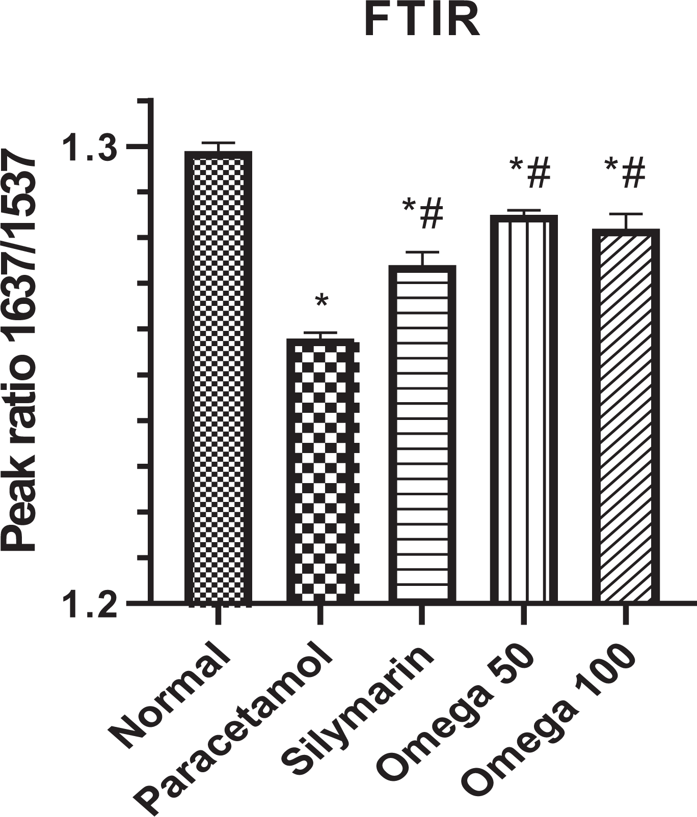
Effect of oral administration of Omega-3 on tissue proteins level (ratio between amide I to amide II waves. *Significant from normal control, #significant from paracetamol (P < 0.05).

The representative FTIR spectra of the experiment, amide 1 appeared at 1637 cm−1 and amide II appeared at 1537 cm−1.
Effect on serum biochemical parameters
Effect on liver functions
Effect on serum ALT and AST
Paracetamol (600 mg/ kg) induced acute significant increase in serum ALT and AST levels as compared to the normal control group. Pretreatment of rats with Omega-3 (50 and 100 mg/kg) significantly lowered both ALT and AST as compared to the paracetamol group and the results were comparable to those obtained from silymarin (Figure 3(a) and (b)). It worth mentioning that the lower dose of Omega-3 showed better outcomes.
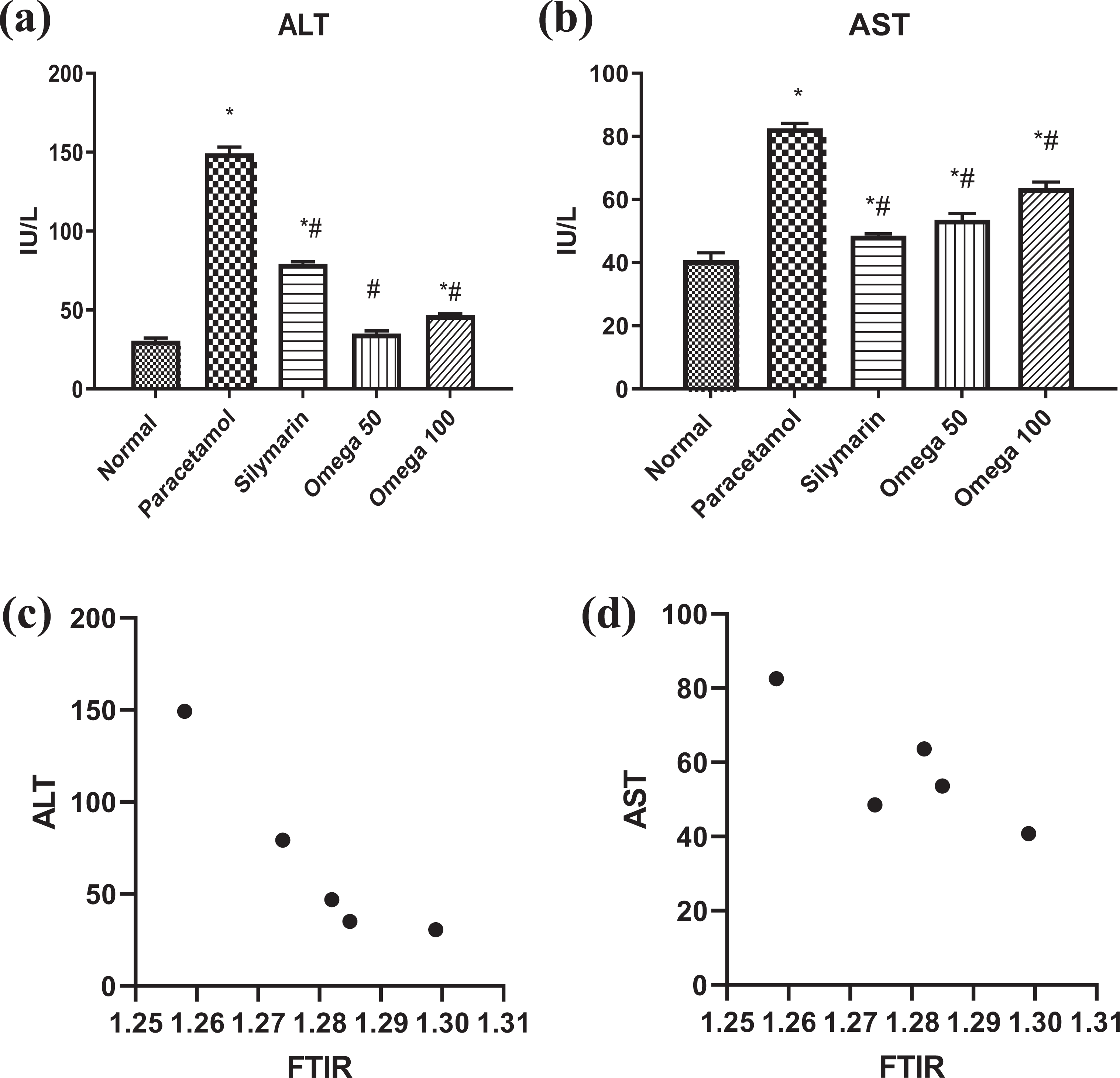
(a, b) Effect of oral administration of Omega-3 on serum ALT and AST. *Significant from normal control, #significant from paracetamol (P < 0.05). (c, d) Correlation study: FTIR vs. ALT and AST.
Correlation study: FTIR vs. ALT and AST
Comparing the means of groups; correlation analysis revealed the existence of a negative correlation between FTIR and serum ALT level only (R2 = 0.8755) at P < 0.0332 (Figure 3(c) and (d)).
Effect on serum lipid profile
Effect on serum HDL, LDL and total cholesterol
Paracetamol (600 mg/kg) significantly decreased HDL level and prompted a significant elevation in total cholesterol and LDL levels. Silymarin ingestion resulted in a significant decrease in serum total cholesterol and LDL levels as compared to paracetamol group. Meanwhile; the HDL level was normalized. Only pretreatment of rats with Omega-3 (50 mg/kg) lowered LDL level as compared to paracetamol group while both doses lowered the total cholesterol level as compared to paracetamol group. In addition; both doses normalized HDL level (Figure 4(a) to (c)).

(a–c) Effect of oral administration of Omega-3 on serum lipid profile. *Significant from normal control, #significant from paracetamol (P < 0.05).
Effect on serum inflammatory markers
Effect on serum (IL-6)
Paracetamol (600 mg/ kg) significantly elevated serum IL-6 level. Pretreatment of rats with Omega-3 (50 and 100 mg/kg) displayed anti-inflammatory activity lowering serum level of IL-6 (Figure 5). The results were comparable to those of silymarin and it worth mentioning that Omega-3 at the lower dose level showed better outcomes.

Effect of oral administration of Omega-3 on serum IL-6. *Significant from normal control, #significant from paracetamol (P < 0.05).
Effect on tissue (TNF-α)
Paracetamol (600 mg/kg) resulted in a significant elevation in hepatic tissue TNF-α level. Pretreatment of rats with Omega-3 at doses of (50 and 100 mg/kg) induced a significant reduction in the elevated tissue level of TNF-α (Figure 6). The results were comparable to those of silymarin and it worth mentioning that Omega-3 at the lower dose level showed better outcomes.

Effect of oral administration of Omega-3 on tissue TNFα. *Significant from normal control, #significant from paracetamol (P < 0.05).
Effect on tissue anti-oxidant activity, oxidative state and nitrosative state
Effect on tissue (GSH), (NOx) and (MDA)
Paracetamol (600 mg/kg) prompted a significant reduction in tissue (GSH) level; in addition to a significant elevation in tissue (MDA) and (NOx). Pretreatment of rats with Omega-3 at doses of (50 and 100 mg/kg) showed a pronounced anti-oxidant activity represented by the significant elevation of GSH level compared to the normal control along with the normalization of NOx level at the group ingesting the lower Omega-3 dose and the results were comparable to those of silymarin. Meanwhile the MDA level was only affected at the lower dose level (Figure 7(a) to (c)).

(a–c) Effect of oral administration of Omega-3 on tissue GSH, NOx and MDA. *Significant from normal control, #significant from paracetamol (P < 0.05).
Histopathological examination and immunohistochemical study
Histopathological examination of hepatic sections in the paracetamol intoxicated group revealed extensive inflammatory cells infiltrating the portal tract and extending in between hepatocytes. Pyknotic nuclei of some hepatocytes are observed all over the section which indicates hepatocellular degeneration. Groups treated with either silymarin or omega 50 mg/kg showed scattered inflammatory cells in between hepatic cords and hepatocytes. On the other hand the group treated with omega 100 mg/kg displayed extensive inflammatory cellular infiltration and degenerated cells all over examined fields. In conclusion hepatocytes induced apoptosis associated with the activation of caspase and Bax, revealed by their positive brown immunostaining, expresses that ingestion of Omega-3 at dose level of 50 mg/kg was safer than 100 mg/kg on liver tissue with minimal side effects and the results were comparable to those of silymarin (Figure 8).

Histopathological examination and immunohistochemical study.
Normal sections revealed negative staining of both caspase-3 and Bax point of “0” range. Control positive revealed point “4” range positivity for both stains according to the used semi quantitative assessment. 32,33
Omega 50 as well as silymarine Bax staining revealed point “1” range. Omega 100 Bax showed point “2” scoring level which was higher than silymarine but less than control positive.
Omega 50 caspase-3 revealed point “1” range and was superior over silymarine which showed point “2” scoring. Omega 100 caspase-3 showed point “3” scoring.
Discussion
The present study aimed to evaluate the diverse means by which Omega-3 PUFAs could hinder acute paracetamol-induced toxicity in female rats; with special attention to the effect on liver protein secondary structural alterations; confirmed by FTIR technique.
Fourier Transform Infrared (FTIR) micro-spectroscopy is now considered as a trustable technique for the biochemical analysis of tissues and cellular materials providing quick, accurate and objective information and has been applied in many areas of medical research. 20,34 Analysis of liver tissue protein alterations by FTIR micro-spectroscopy could be used as a reliable marker on the holistic biochemistry of liver tissue. Previous studies revealed that diseased tissues showed a reduced ratio of amide I to amide II when compared with normal tissue revealing a change in protein structure in diseased liver. 35 –37
Acute hepatotoxicity induced by paracetamol represents a well-known medicinal complication especially among women. 38 –40 Our study revealed that amide I and amide II peak ratio was significantly decreased in paracetamol intoxicated group compared to the normal control. Moreover; ingestion of a paracetamol single acute over dose (600 mg/kg) resulted in a significant increase in serum ALT, AST, LDL and total cholesterol as well as a significant reduction in the HDL level. In addition; liver tissue oxidative stress was significantly increased indicated by the elevated levels of MDA and NOx and decreased GSH level. Inflammatory markers represented by serum IL-6 and tissue TNF-α were significantly increased. Finally both histopathological and immunohistochemical examinations certified the existence of paracetamol-induced severe hepatotoxicity.
Former investigators stated that; acute overdose of paracetamol resulted in massive destruction to hepatocytes, leading to elevation of serum ALT, AST, total cholesterol and triglycerides levels; in addition to a generalized hepatic oxidative stress status represented by accumulation of MDA as well as NOx and depletion of GSH. In addition; inflammatory response represented by elevated level of TNF-α was recorded. Besides; a significant increase in the levels of apoptosis-related proteins such as Bax and caspase-3 was observed suggesting a vital role of apoptosis in paracetamol-induced hepatic insult. 41 –47
Previously, it was reported that; dietary fish oil supplementation rich in Omega-3 fatty acids (Omega-3 PUFAs) possessed several medicinal beneficial actions; suppressing inflammatory response and oxidative stress as well as modulating cell proliferation. 10 The mechanisms underlying the hepatoprotective effects of Omega-3 PUFAs includes its ability to increase GSH along with its capability to scavenge free radicals and consequently inhibit lipid peroxidation. 9,43 Meanwhile; Maksymchuk, 2014, previously demonstrated that there was more than two-fold increase in the content of cytochrome P450 2El (CYP2E1) in the liver of rats receiving omega-3 PUFAs for 4 weeks in the standard daily diet. In another study; consumption of omega-3 PUFAs led to a 3-fold (p < 0.05) increase in CYP2E1 content. Such changes in the enzyme expression did not have an impact on the level of lipid peroxidation and on the prooxidant/antioxidant balance in the liver. 48,49 In addition; it has been previously reported that Omega-3 PUFAs inhibit the conversion of arachidonic acid into the pro-inflammatory eicosanoids through cyclooxygenase-2 and 5-lipoxygenase pathways either via competing with the substrate or via inhibiting the activity of eicosanoid-generating enzymes. Moreover; their metabolites including resolvins and protectins are involved in hindering inflammation and steatosis for example; “Resolvin D1, resolvin E1, and protectin D1” inhibit transendothelial migration of neutrophils into sites of inflammation. Resolvin D1 inhibits IL-1beta production. Protectin D1 attenuates the production of TNF-alpha and IL-1beta. Besides, Omega-3 fatty acids were previously reported to activate peroxisome proliferator-activated receptors alpha, which up regulate several genes associated with fatty acid and lipid metabolism that stimulate fatty acid oxidation, decreasing liver fats and thus reducing hepatic lipogenesis and steatosis. Both ALT and AST levels were reported to be reduced by Omega-3 PUFAs in several models of hepatic injury. Finally; it was reported that there was a direct link between docosahexaenoic acid (DHA) treatment and suppression of apoptosis represented by a reduction in active caspase-3. 9,17,50 –52 Several doses have been proposed for the daily intake of Omega-3 fatty acids with some reports highlighting that in some cases lower doses would result in favorable outcomes with less side effects. 17,53 In our research, (50 mg/kg) of Omega-3 fatty acids (Omega-3 PUFAs) showed pronounced protection against paracetamol induced liver damage represented by increased amide I to amide II peak ratio compared to the paracetamol intoxicated group. Furthermore; antioxidant as well as anti-inflammatory actions and improved serum lipid profile along with declined levels of serum liver enzymes were detected. Finally histopathological and immunohistochemical investigations revealed hepatic tissue protection. Results were generally comparable to those of silymarin.
Conclusion
Omega-3 fatty acids could be a very useful supplement in the prevention of liver injury especially in women but only in low doses.
Footnotes
Declaration of conflicting interests
The author(s) declared no potential conflicts of interest with respect to the research, authorship, and/or publication of this article.
Funding
The author(s) disclosed receipt of financial support for the research article: This research was conducted as a part of a master degree funding program granted by the National Research Centre (NRC) of Egypt.