
Abstract
Bisphenol A (BPA) is one of the widely used chemical as a plasticizer and regarded as endocrine disruptor because of its ability to derail body metabolic functions and adverse effect on the vital organs. The present work outlined the subchronic effect of low-dose BPA (10 mg/kg) on histology of spleen, level of hepatic trace metals, and hepatic protein profile of Wistar rats. To conduct the research work, animals were grouped into two categories (n = 5). Group 1 was labelled as the control group and group 2 was taken as an experimental group. Experimental group was exposed to low-dose BPA for 12 weeks. Histopathology of spleen highlighted dilation in splenic sinuses, follicle activation, followed by depopulation in the area of white pulp and red pulp in the experimental group. Iron staining revealed significant hemosiderosis in the experimental group when compared with the control group. Statistically significant decrease was noted in zinc and copper concentrations, while nonsignificant change was observed for magnesium concentration through atomic absorption spectroscopy. Sodium dodecyl sulfate–polyacrylamide gel electrophoresis was run for hepatic protein profiling, and as compared to control, elevated levels of different proteins were observed in the experimental group. It can be concluded from the above results that even low dose of BPA causes changes in the major organs of the body. Hence, it is suggested that BPA alternative should be used, so that public health status can be secured.
Introduction
Endocrine disruptors are chemicals that inhibit or induce change in normal hormonal function of the body, and occurrence of these chemicals in environment is perilous for both the environment and the living organism in many respects. Among the reported endocrine disruptors, bisphenol A (BPA) is one with over six billion pounds production globally each year as a plasticizer. 1 BPA has vast applications in numerous products particularly in the synthesis of polycarbonate, adhesive resins, electrical gadgets, optical glass, paint, thermal papers, food cans, medical equipment, dental sealants, and so on. 2 In the production of vinyl chloride, it is also extensively used as a stabilizer and antioxidant. 3 Anthropogenic activities are the major source of BPA migration into environment. Manufacturing, treatment, and recycling of plastic and epoxy resins contaminate the ecosystem and food web by releasing its monomers. 4 Fu and Kawamura in 2010 reported that urban areas of Asia are highly contaminated with BPA monomers ranging up to 170–880 pg/m3. 5 It has been reported that dust samples of indoor air has BPA concentration approximately up to 0.2–17.6 mg/kg. 6 Surface water and industrial sewage are highly contaminated, that is, Elba river of Germany is contaminated with 4–92 µg/dm3 concentration of BPA, and 36.7 mg/kg BPA concentration was detected in the raw sewage wastes of Canada. 7 Human population is significantly exposed to BPA and its monomers through canned food and beverages. 8 It was reported that high BPA concentration is present in canned vegetables and fruits approximately ranging from 3.7 µg/kg to 959 µg/kg, while from 1.0 µg/kg to 223.91 µg/kg was detected in seafood. 8
Numerous health-related issues have been reported due to consumption of BPA and its migration into ecosystem, namely mutagenicity, 9 teratogenicity, 10 obesity, 11 hepatotoxicity, 12 neurotoxicity, 13 and immunotoxicity. 14 Its bioaccumulation has been reported in the trophic level of food chain that can even cause death of fresh water and marine fauna. 15 It has been reported that exposure of rodents to BPA results in derailed estrogenic level which likely acts through the estrogen receptor (ER). 16 Early reproductive aging, increase in prostate body weight, and estrogenic activity were reported in mice due to administration of BPA. 17
Animal data revealed that BPA can induce damage in the nervous system by disrupting the function of dopamine system and hippocampus. 13 Mild doses of BPA induces changes in the development of mammary gland and increased frequency of tumors. 18 Hassan et al. in 2012 reported the hepatotoxicity due to BPA administration characterized by the formation of reactive oxygen species and oxidative stress. 19 Destruction in hepatic cells particularly in cell line HepG2 was also observed due to administration of low-dose concentration of BPA and significantly raised level of liver enzymes has been investigated in rats with high dose of BPA (50 mg/kg). 20 Industrial application of BPA is parallel with its damaging effects. To the best of our knowledge and reported data, low-dose subchronic effect of BPA had not been studied in Wistar rats, thus the current work has been conducted with the objective to investigate the effect of BPA on histopathology of spleen, hepatic trace elements, and protein profile.
Materials and methods
Animals
Previously reared adult male Wistar rats (Rattus norvegicus) of 200–220 g were used and maintained in the animal house of Department of Zoology, University of the Punjab, Lahore, Pakistan. Standard rat chow and normal drinking water were provided. All the animals were taken care according to the standard guidelines for animal handling and care.
Animal groups
Animals were categorized into two groups: the control group and the experimental group (n = 5).
Subchronic inflammation
Subchronic inflammation was induced by administration of low-dose BPA (10 mg/kg of body weight) dissolved in 1 ml of corn oil for 12 consecutive weeks. Dose of BPA was given orally by oral gavage to each rat of the experimental group and the rats in the control group received 1 ml of corn oil for 12 consecutive weeks. After the decided time duration, the animals were anesthetized with 1% ketamine for dissection. Dissection of animals was performed under sterile conditions. After the completion of surgical procedure, liver and spleen were excised out. Liver samples were stored in Eppendorf at −80°C for further analysis, while spleen tissues were fixed in formalin for histopathological examination.
Histological processing and staining of spleen
For tissue processing and hematoxylin and eosin (H&E) staining, standard protocol was followed with minor modification as described. 21
Iron staining was performed for determination of hemosiderosis. For iron staining, standard protocol 22 has been used with slight modification. Briefly, slides having sections were deparaffinized with xylene and then rehydrated with distilled water for about 5 min. After rehydration, slides were dipped in the working solution (2% hydrogen chloride + 2% potassium ferrocyanide) for 30 min, followed by three washes in distill water for about 3 min. For counterstaining, slides were retained in nuclear fast red (200 ml distilled water + 10 g aluminum sulfate + 0.2 g nuclear fast red) for 10 min. After retaining, slides were again washed with distilled water and dehydrated using ascending grade of alcohol. For tissue clearance, slides were again dipped in xylene for 5 min. Canada balsam was mounted, and slides were examined under microscope.
Hepatic trace metals assessment
Hepatic trace metals assessment was done according to the mentioned protocol 23 with slight modification. Briefly, wet digestion of tissue was carried out, and trace metals level was estimated through atomic absorption spectroscopy (AAS). For wet digestion, liver tissue (0.25 gram) was taken in a china dish followed by the addition of 2 ml conc. 70% HNO3. It was placed on a hot plate for 7–10 min and was refluxed at the hot plate without boiling. China dish was covered with watch glass to avoid fumes after complete tissue digestion in nitric acid, then it was removed from the hot plate, followed by addition of 500 µl deionized water and 750 µl hydrogen peroxide (30%), respectively. The mixture was again placed on China dish until effervescence stopped. Total volume of solution was brought up to 30 ml, after removal of China dish from the hot plate. Prepared solution was then filtrated with Whatman filter paper 1 and stored at room temperature for trace metals analysis through atomic absorption spectroscopy.
Hepatic protein profile
Modified protocol of Abbasi et al. 24 has been used for protein extraction from liver tissue. The samples were prepared by incubating 0.20 g liver tissue in 1% sodium dodecyl sulfate (SDS) buffer for 45 min, followed by centrifugation for 20 min at 7000 × g. After centrifugation, supernatant was pipetted out for sodium dodecyl sulfate–polyacrylamide gel electrophoresis (SDS-PAGE), and pellet was discarded.
To resolve proteins, 12% polyacrylamide gel was prepared and proceeded as described elsewhere. 25
Results
Histopathological examination
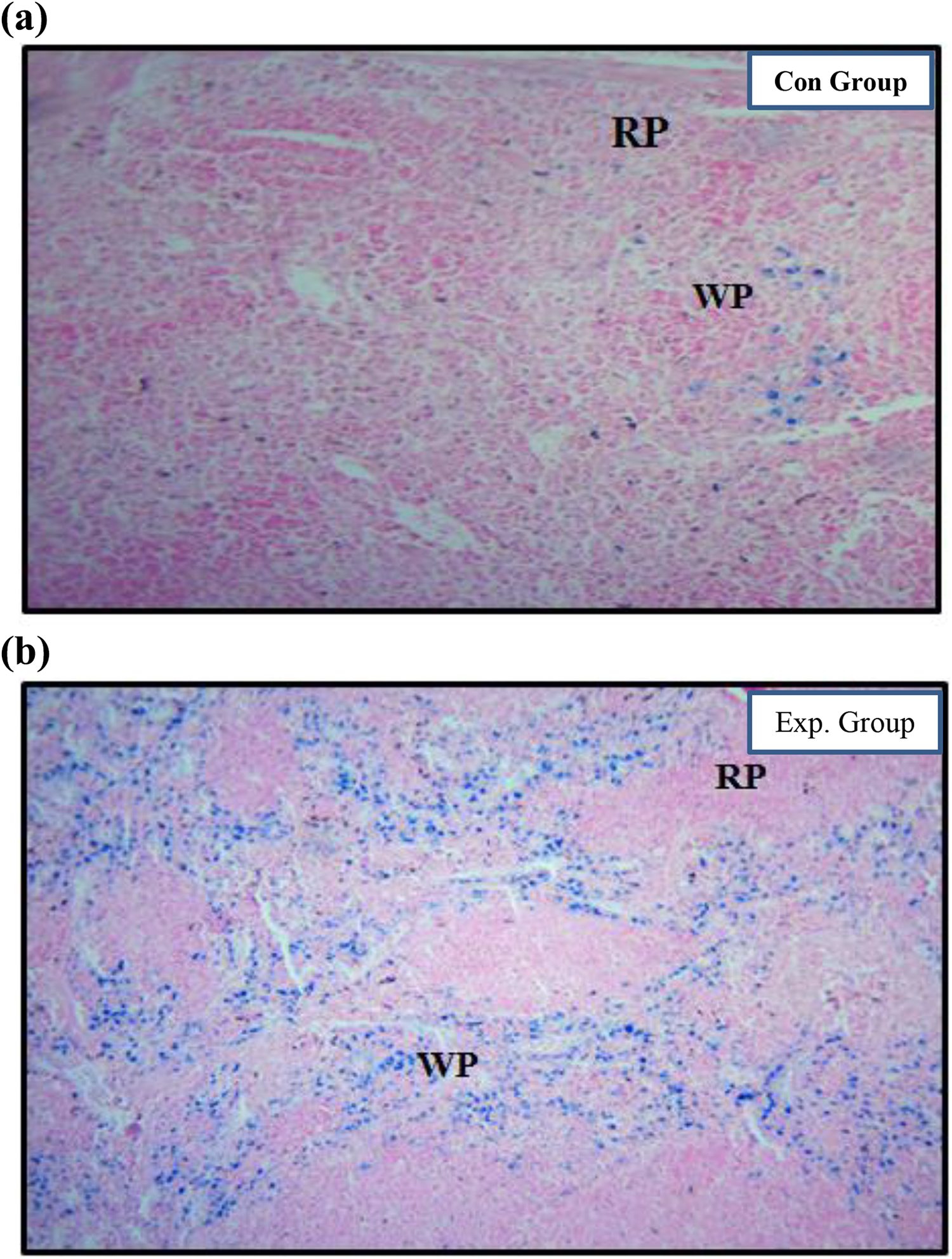
Histologically, spleen is distributed into red and white pulp. Splenic sinuses and splenic cords constitute red pulp, while structure of white pulp is composed of periarterial lymphoid sheath, follicles, and marginal zone. Splenic sections of the control group showed normal architecture with distinguished red and white pulp with normal morphological features (Figure 1(a)).

(a) Control splenic H&E-stained sections of rats; normal structure with normal RP and WP can be seen (magnification ×100). (b) H&E-stained splenic sections of rats exposed with low-dose BPA. Experimental group showed dilation and vacuolization in RP. Significant depopulation and follicle activation was also observed in WP (magnification ×100). BP: bisphenol A; H&E: hematoxylin and eosin; RP: red pulp; WP: white pulp.
BPA-administrated sections showed severe changes in splenic architecture. The experimental group showed dilation of splenic sinuses, apparent reduction in the count of macrophages, and vacuolization in the area of red pulp. Depopulation, follicle activation, dilation in the area of marginal zone, and destroyed follicles are seen in the area of white pulp. Accumulation of lymphocytes surrounding the central arteries and erythrocytes in red pulp were also noted (Figure 1(b)).
In Prussian blue-stained splenic sections of control group sections, no significant pigmentation was evident (Figure 2(a)), while BPA-exposed group showed hemosiderosis (blue pigmentation) which indicate excessive iron accumulation in red and white pulp. In red pulp; in particular splenic sinuses were overloaded with excessive iron accumulation, while in the area of white pulp, marginal zones showed excessive deposition of iron (Figure 2(b)).
(a) Control splenic Prussian blue-stained sections of rats. No pigmentation in the form hemosiderosis was observed (magnification ×100). (b). Prussian blue-stained splenic sections of rats exposed with low-dose BPA. Significant hemosiderosis particularly in the area of red and white pulp (magnification ×100). BP: bisphenol A.
Hepatic trace metal level
Results of hepatic metal estimation through atomic absorption spectroscopy showed that as compared to the control group, there was a significant decrease (p value = 0.0204) in zinc (Zn) metal content (Figure 3(a)). Significant decrease (p value = 0.0338) in copper (Cu) metal content was also observed (Figure 3(b)), while nonsignificant decrease (p value = 0.2281) in magnesium (Mg) metal level was noted (Table 1)(Figure 3(c)).
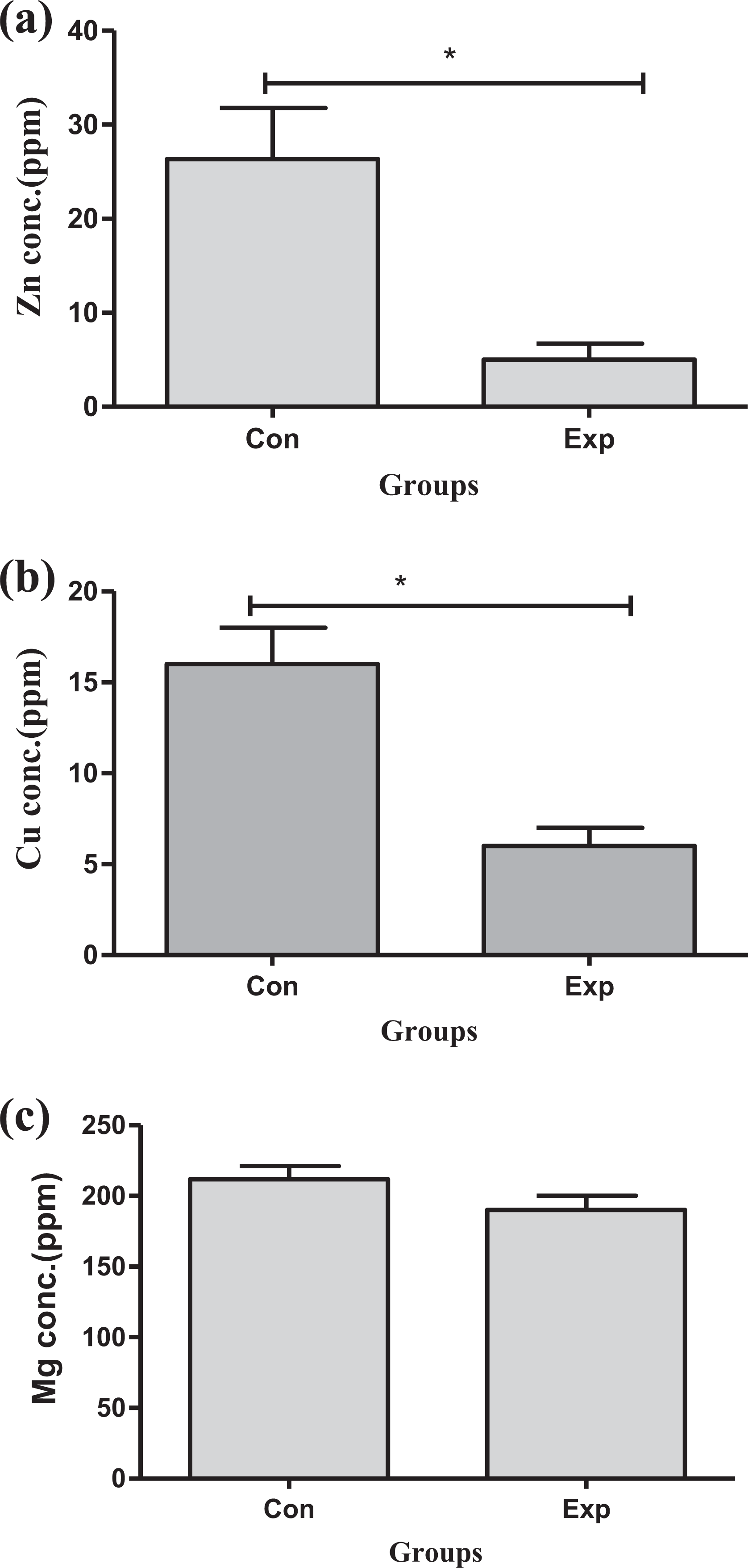
Comparison of atomic absorption spectrophotometric analysis for the concentration of hepatic trace metal (ppm) of low-dose BPA treated rats with the control group. (a) Statistically significant decrease in zinc concentration was observed in the experimental group. (b) Decrease in copper concentration was observed in the experimental group as compared to control. (c) Decrease in magnesium concentration was observed in the experimental group as compared to control ( *= P≤ 0.05).
Variation in hepatic metal content of control and low-dose BPA-exposed experimental groups.a

BP: bisphenol A; Zn: zinc; Cu: copper; Mg: magnesium; SEM: standard error of the mean.
a Values represent mean ± SEM.
Hepatic protein profile
SDS-PAGE was run for the analysis of proteins. To analyze the molecular weight of protein, Thermo Scientific unstained protein ladder in the range of 10–220 kDa was used and to analyze the gel, software (TotalLab Quant; version 5) was used. On SDS-PAGE, different molecular weight of proteins including 72 kDa, 60 kDa, 55 kDa, 50 kDa, 40 kDa, 30 kDa, 22 kDa, and 14 kDa demonstrated increase in their expression after the use of BPA as compared to the control group (Figure 4).

Electropherogram showing changes in expression of protein in low-dose experimental group as compared to the control group.
Discussion
BPA known as endocrine-disrupting chemical has worldwide application in different plastic products of domestic use. 1 The extensive use of plastic products produce alarming situations and give scientists and environmental protection agencies a new research attention area by understanding the BPA mechanism and its adverse effects particularly on the mammalian body. 26 Accordingly, keeping in view of previous research perspective, current work was designed by taking the objective that how BPA affects splenic architecture and hepatic trace elements and protein profile in mammals.
In our study, long-term exposure of BPA has induced severe changes in spleen architecture. From the results of H&E and iron staining, histopathological changes were observed, characterized by dilation in the area of red pulp, that is, dilation in splenic sinuses, depopulation in the area of white pulp, activation of lymphoid follicles, and significant hemosiderosis in red and white pulp areas. Ahmed et al. in 2015 reported similar changes in spleen with 70 days exposure of BPA (150 mg/kg). 27 In another study, it was reported that splenic tissues showed multiple focal necrosis, destruction of follicles, dilation of central arteries due to 6-week exposure of 2.5 g/kg of BPA administration in basal diet. 28
Xu et al. in 2016 reported significant splenocytic proliferation, degeneration in T and B cells, and apparent reduction in the count macrophages were observed in male mice treated with long-term exposure of BPA. 29 These changes might be due to severe oxidative stress by the formation of reactive oxygen species (ROS) induced by low-dose BPA. ROS are formed due to inhibition of several antioxidant enzymes activity and stimulation of lipid peroxidation activity. 30 Oxidative stress is considered as one of the key factors that play crucial role in numerous histopathological conditions, that is, fibrosis and follicle necrosis. It was also reported that oxidative stress produced by BPA impairs mitochondrial function in spleen. 31
Liver is one of the target tissues for the metabolism of endocrine-disrupting chemicals. Specific ERs are present in the liver, and cellular responses concerning estrogen interactions have been identified in the liver. 32
In the current research work, hepatic trace elements were investigated to determine the systematic toxic effect of BPA. Zn, Cu, and Mg were considered for the determination of hepatic trace metals. The levels of Mg, Zn, and Cu have been widely used to investigate the pathogenic risk factors. Our results showed that there is a decrease in hepatic Zn and Cu metal contents in BPA-treated group, while on the other hand, there is no significant difference in hepatic Mg metal content. So the present research work revealed that BPA impairs hepatic trace metal content.
Prasad in 2013 reported that hepatic Zn deficiency is one of the significant factor in the development and activation of malignancy. 33 It was also reported that even low-dose administration of BPA can activate oxidative stress by decreasing hepatic Zn metal content and induce dysfunctioning of various antioxidant enzymes involved in Zn metabolism. 34 It has been reported that decrease in Cu metal content is one of the key factors that induce hepatic injury and disease by inducing oxidative stress. 35 According to our findings, the decrease in Zn and Cu metal contents might be due to oxidative stress. Mg has significant role in the release of insulin. 36 Deficiency of Mg can also prompt negative impact on liver metabolism. 37
Hepatic protein profile showed variations in the expression of different proteins ranging from 72 kDa to 14 kDa. These proteins might be heat-shock proteins (HSPs; 72 kDa), tyrosine protein kinase (60 kDa), transthyretin protein (55 kDa), actin-binding proteins (50 kDa), fatty acid-binding protein (40 kDa), heme-binding protein (30 kDa), insulin-like growth factor (22 kDa), and selenium binding protein (14 kDa).
HSPs showed increased expression in the experimental group that provides evidence that BPA exposure has induced stress in hepatic tissues. HSP is also known as stress protein because these proteins are expressed particularly in response to toxicant administration, heavy metal accumulation, and free radical formation. 38 Previous reported studies also showed that exposure to xenoestrogens results in the activation of HSP. 39
Tyrosine protein kinase plays significant role in calcium ion channels of membranes, and besides, it also plays role as a neurotransmitter. Increased expression of tyrosine protein kinase in the experimental group provides clue that BPA induction might have led to some neurotoxic effect and decrease in communication cells binding to receptor of tyrosine protein kinase. Heme-binding protein is associated with biliverdin metabolism and provides cellular protection against ROS. 40 Its expression indicates that BPA has induced effect on biliverdin metabolism.
Previous reported data showed that insulin-like growth factor induces expression during sudden and programmed cell death. 41 The increased expression of insulin-like growth factor indicates that BPA administration induces necrosis of hepatocytes. 42 It has been reported that increased expression of insulin-like growth factor stimulates β amyloid.Mao et al. 43 reported that in rats BPA exposure at early stage of life leads to glucose intolerance and dysfunction of β cells. It can be concluded that BPA might have caused glucose intolerance. It has been reported that selenium-binding protein is involved in xenobiotic metabolism. 44 Selenium-binding protein is also involved in immunological responses, and Schott et al. in 2018 reported that pathological conditions lead to lower expression of selenium-binding protein. 45 From our study it can be concluded that decreased expression of selenium-binding protein provides indication of change in immunological responses.
Concluding remarks
All the present findings leads us to the conclusion that oral use of BPA has induced splenic toxicity, derailed level of hepatic trace metals, and variation in expression of hepatic proteins. Hence, it is concluded that even the low dose of BPA can be hazardous to health if extensively used.
Future suggestion
The consumption of BPA should be limited and specious handling of plastic containers should also be limited to reduce health risks. Canned food container should not be lined with BPA as the BPA transformation in mammalian body induces severe health issues and syndromes. BPA used in baby bottles and toys should be completely banned to avoid toxicity of BPA. Government and nongovernmental organizations should create awareness to the public to use plastic containers free of BPA. Furthermore, researchers should extend the investigation based upon the toxicity of xenoestrogens so that worldwide use of BPA and other likely xenoestrogens would be replaced by nontoxic chemicals.
Footnotes
Declaration of conflicting interests
The author(s) declared no potential conflicts of interest with respect to the research, authorship, and/or publication of this article.
Funding
The author(s) received no financial support for the research, authorship, and/or publication of this article.