
Abstract
The limited effectiveness of the conventional methods for cancer treatment makes the researchers to find novel safe and effective therapeutic strategies. One of these strategies is to use small interfering RNAs (siRNAs). A major challenge here is the siRNA delivery into the cells. The purpose of this study was to design and prepare a biocompatible, biodegradable, and safe nanosized particle for siRNA delivery into human breast cancer MCF-7 and leukemia K562 cells. Chemically synthesized magnetic nanoparticles containing polyethyleneglycol-lactate polymer (PEG-LAC), chitosan, and polyethyleneimine (PEI) were successfully prepared and used as a gene delivery vehicle. The nanoparticles were characterized by Fourier transform infrared spectroscopy and zeta potential. The Fe3O4-PEG-LAC-chitosan-PEI nanoparticle showed efficient and stable survivin siRNA loading in gel retardation assay. The cytotoxicity of the prepared nanoparticle was studied using 3-(4,5-dimethyl-2-thiazolyl)-2,5-diphenyl-2H-tetrazolium bromide assay and was compared with that of mitoxantrone (MTX) in combination with the prepared siRNA delivery system to evaluate the possible synergic effect of MTX and survivin siRNA. The nanoparticles with and without noncomplementary siRNA showed low toxicity against both cell lines; however, a twofold decrease was observed in cell survival percent after MTX addition to MCF-7 cells treated with either nanoparticle itself or complexed with noncomplementary siRNA. While survivin siRNA nanoplex caused threefold decrease in the cell survival percent, its combination with MTX did not result in a significant increase in the cytotoxic effect. Therefore, Fe3O4-PEG-LAC-chitosan-PEI nanoparticle should be considered as a potential carrier for enhanced survivin siRNA delivery into MCF-7 and K562 cells.
Introduction
RNA interference (RNAi) is a powerful aspect of gene therapy which has attracted much attention in the last decades. 1 –4 However, delivery of small interfering RNAs (siRNAs) into specific tumor tissues and cells followed by their intracellular release from the endosomal/lysosomal compartments into the cytoplasm still remains a major problem that needs to be solved before this experimental technique could become a clinically viable therapeutic option. 5,6
Cell death and survival are of the most important pathways playing key role in cancer progression. Inhibitor of apoptosis proteins are anti-apoptotic genes who directly bind to caspases and block apoptosis. One family member, survivin, has multiple functions including cytoprotection, cell death inhibition, and cell cycle regulation, all of which favor cancer cell survival. It has been shown that inhibition of survivin can restore the sensitivity of cancer cells to anticancer agents. 7 Therefore, survivin may be a potential target for siRNA-based anticancer therapy due to its higher levels of expression in carcinoma cells. 8
Naked siRNAs are unfavorable for systemic delivery because of their inherent limitations such as negative charge, large molecular weight, size, and instability. They are easily degraded by serum endonucleases and are quickly eliminated by renal excretion. 9 In this regard, engineered nanocarriers that can stably encapsulate/complex, protect, and selectively deliver siRNAs to target tumor tissues and cells are highly promising as next-generation gene delivery vehicles. 10 The ideal system should preferably be nontoxic, biodegradable, nonimmunogenic, and able to efficiently deliver siRNAs to target tumors. More importantly, after tumor tissue-specific delivery, the nanosystem should facilitate intracellular uptake and promote endosome release/escape of the payload siRNAs into cytoplasm allowing their interaction with the endogenous RNA-induced silencing complex. 11 Although viral vectors can serve as efficient gene delivery vehicles, the apparent toxicity and safety concerns limit their utility for human applications, necessitating the development of safer delivery technologies. 3 Accordingly, the Food and Drug Authority has not yet approved any viral vector-based gene delivery system. Nonviral gene vectors have gained significant interest because of their safety, nonimmunogenicity, and easy manufacture. Several polycations, including polyethyleneimine (PEI), 12 polyamidoamine, 13 cationic polysaccharide, 14 poly (2-dimethyl amino)ethyl methacrylate, 15 and cyclodextrin cationic carriers, 16 have been reported to be capable of delivering nucleic acids. Chitosan and polyethyleneglycol (PEG) are also generally used in cationic particle synthesis for oligonuleotide delivery. 17 –21 Among them, PEI is one of the most efficient and widely studied gene carriers in vitro and in vivo because of its “proton sponge effect” which can buffer the endosome environment and cause the release of the complexes into the cytoplasm. 4
PEI increases transfection efficiency because of its high positive charge which provides a means of escaping from the late endosome through proton sponge effect. PEI increases the loading amount of the negatively charged oligonucleotide strand due to its hydrophilicity. It also leads to greater degradation of poly lactic-co-glycolic acid and siRNA release. 22 Chitosan is a natural biocompatible polysaccharide which conserves as an effective coating to stabilize the nanoparticle, preventing particle agglomeration. 23 PEG is commonly used to decrease the toxicity of PEI, to increase the colloidal stability of the particle through steric hindrance, and to provide nonfouling properties beside its high biocompatiblity. 23
In the present research, we designed and synthesized novel positive charged nanoparticles used for siRNA transfection into cancerous cells. After characterizing the nanoparticle itself and in complex with the siRNA, we have studied the efficacy of the treatment with survivin siRNA carried by synthesized nanoparticles individually and in combination with chemotherapeutic agent, mitoxantrone (MTX), on MCF-7 and K562 cells.
Materials and methods
Materials
SiGENOME MOUSE Birc5 (11799) siRNA (5′-GAACUAACCGUCAGUGAAU-3′) was purchased from Thermo Scientific (Thermo Fisher Scientific Inc., Waltham, Massachusetts, USA). Mitoxantrone (20 mg/10 mL) was purchased from Kocak Farma (Istanbul, Turkey). RPMI-1640 medium, reduced serum medium 1X (Opti-MEM®), and fetal bovine serum (FBS) were obtained from Gibco (Life Technologies, Thermo Fisher Scientific Inc.). Penicillin/streptomycin and 3-(4,5-dimethyl-2-thiazolyl)-2,5-diphenyl-2H-tetrazolium bromide (MTT) were purchased from Sigma-Aldrich (St Louis, Missouri, USA). RNase A was purchased from CinnaGen (Tehran, Iran).
Nanoparticle synthesis
Magnetic core
The aqueous FeCl3 (1.66 g in 20 mL deionized water) and FeCl2·4H2O (1.00 g in 20 mL deionized water) were mixed together with stirring. Black precipitant was observed once NH4OH solution (25%, 20 mL) was added into the solution, indicating the formation of magnetite nanoparticles. The dispersion was continuously stirred for another 30 min to complete the reaction. Then, it has been placed on a magnet to let the magnetite absorb to that surface. After pouring out the reaction fluid, oleic acid solution in hexane (2.5–10 wt% in 20 mL hexane) was introduced into the magnetite dispersion under stirring. The dispersion was concentrated by evaporating hexane to obtain a black thick liquid of concentrated magnetite in hexane. 24,25
PEG-LAC polymer layer
Polyethylene glycol-lactide (PEG-LAC) copolymerization was done in the presence of stannous octoate catalyst. In this reaction, 1.00 g lactide and 0.2 g PEG 2000 were mixed in 9.00 mL toluene containing 0.005 g stannous octoate. The reaction balloon was placed in hot oil bath (140°C) for 6 h to complete the reaction. After letting the reaction mixture cool down at room temperature, the obtained copolymer was precipitated using ice-cold diethylether liquid and kept in vacuum oven to dry. 26
To cover the PEG-LAC layer on the magnetite core surface, 20 mL of the magnetite dispersion in hexane was introduced into 20 mL of aqueous copolymer solution (1 wt% copolymer). The mixture was then ultrasonicated for 4 h to transfer the particles from hexane top layer to aqueous bottom layer. The remaining dispersions in aqueous phase were then dialyzed using dialyze bag (1200) and lyophilized. 25
Chitosan layer
To cover the chitosan layer on the previously synthesized particle’s surface, 0.05 g of the magnetite particle (previous step’s product) was dissolved in 1 mL dichloromethane and added to the aqueous solution of 0.05 g chitosan containing 0.2 mL acetic acid, drop wise. After stirring for about an overnight, it was passed through a 0.22 µM filter and lyophilized. 23,25
PEI layer
A total of 0.02 g PEI branched (25 kDa) was dissolved in 10 mL deionized water and the same amount (0.02 g) of the previously prepared product was added while stirring. The mixture was placed on stirrer for 24 h. 25
FTIR analysis
FTIR spectral analysis was carried out for the structural characterization of the synthesized materials. The FTIR spectra were recorded on a FTIR spectrophotometer (Bruker Tensor 27 spectrometer, Billerica, Massachusetts, USA) in the range of 400–4000 cm−1. The sample (2 mg) and 200 mg potassium bromide were mixed and ground for 3 min. The mixture was pressed into pellets for measurements.
siRNA loading
Nanoplexes with different polymer/siRNA ratios were prepared by adding siRNA solution in Opti-MEM (20 nM) to the polymer solutions (various concentrations) in distilled deionized water. The resulting solutions were vortexed for 10 s and incubated for 30 min at room temperature.
Cell culture
Human breast cancer MCF-7 cells and human chronic myelogenous leukemia K562 cells as adherent and suspension cells, respectively, were both obtained from Pasteur Institute Cell Culture Collection (Tehran, Iran). The cells were grown in RPMI 1640 medium (pH 7.4) supplemented with 10% FBS and 100 units mL−1 penicillin/streptomycin and incubated at 37°C in a humidified 5% CO2-containing incubator.
Characterization
The particle size and size distribution of the prepared nanoparticles with and without siRNA loading were determined using the laser diffraction particle size analyzer (Shimadzu, Japan) equipped with the Wing software (version 2101). The mean diameter and size distribution of the resulted homogeneous suspension were assessed. Each value was determined in triplicate.
The zeta potential of the polyplexes prepared in deionized water (0.5 µg mL−1, pH 7.4) was determined at 25°C in a DTS5001 cell with a Zeta sizer 2000 unit (Malvern, UK).
Cytotoxicity assay
The cytotoxicity of the particles was evaluated via MTT assay in 96 well plates. MTT is a colorimetric assay in which the MTT dye changes to insoluble formazan inside the living cells by NAD(P)H-dependent cellular oxidoreductase enzymes. The absorbance measured for the obtained purple color is in direct relation with the viable cell count.
27
MTX was used (1 µM)
28,29
beside to determine the effect of siRNA designed for survivin gene and MTX on MCF-7 and K562 cancer cells in combination with naoparticle, siRNA-containing nanoplexes, and individually. The obtained results were reported as survival percent using the following equation:

Gel electrophoresis for retardation and stability assays
The nanoplexes were all prepared as previously mentioned at siRNA loading section. Gel electrophoresis assays were carried out on a 3% agarose gel using a constant siRNA amount (100 ng) and a range of particle concentration in the w/w ratios 0.1:1, 1:1, 5:1, 10:1, and 20:1 (nanoparticle: siRNA). A total of 90 mV was applied to the gel for about 45 min. Following electrophoresis, the gel was stained in ethidium bromide solution and observed under GelDoc (Uv Doc-008.XD). The stability measurement was done just at the same conditions mentioned above, but the ratios used were 0.1:1, 1:1, 5:1, and 20:1 in the presence and absence of RNase A (20 mg mL−1). RNase A was added in the same concentration as siRNA after the complex formation and incubated for another 30 min at room temperature. 22
Statistical analyses
All statistical analyses were performed using SPSS 17.0 software. Data obtained from at least three separate experiments were expressed as mean ± standard deviation. Data were analyzed by one-way analysis of variance and t-test. p-Values less than 0.05 were considered statistically significant.
Results
Characterization of the synthesized nanoparticles
The synthesized nanoparticle’s surface structure modifications were checked after each step by FTIR spectroscopy (Figure 1). The FTIR spectrum of Fe3O4 MNPs (a), PEG-LAC (b), Fe3O4-PEG-LAC-chitosan (c), and Fe3O4-PEG-LAC-chitosan-PEI (d) confirms the presence of characteristic functional groups of components. Fe3O4 MNPs spectrum (Figure 1(a)) shows a characteristic absorption band at 570 cm−1 which is referred to Fe–O bond indicating the successful synthesis of the Fe3O4 magnetic nanoparticles (MNPs). 30 Figure 1(b) indicates the FTIR spectrum of PEG-LAC sample. The absorption bands at 900–1200 cm−1 refer to C–O stretching, where the bands between 1200 and 1500 cm−1 show the esteric bond formation. The strong band at 1753 cm−1 shows the carbonyl group (C=O) presence and the bands indicated at around 3000 and 3500 cm−1 refer to C–H and O–H stretching, respectively. The FTIR spectrum of the Fe3O4-PEG-LAC-chitosan is illustrated in Figure 1(c) where the Fe3O4 characteristic band is re-appeared at 569 cm−1. The bands present here are the same as Figure 1(b). The exception is the presence of two N–H stretching absorptions (NH2) at 2860 and 2925 cm−1. As shown in Figure 1(d), although the Fe3O4-PEG-LAC-chitosan-PEI represents the same bands, there is an increased intensity of NH2 band while the intensity was decreased for the other bands. This seems to be the proof of PEI coating on the nanoparticle surface which provides high positive charge through its amine groups. The final chemical structure and the nanoparticle preparation steps were demonstrated in Figure 2 where a schematic picture of the nanoparticle structure is shown as well (Figure 2 inset).

FTIR spectra of the synthesized nanoparticle obtained at each step. (a) Fe3O4 core, (b) Fe3O4-PEG-LAC, (c) Fe3O4-PEG-LAC-chitosan, and (d) Fe3O4-PEG-LAC-chitosan-PEI. PEG: polyethylene glycol; LAC: lactide; PEI: polyethylene imine; FTIR: Fourier transform infrared.

Schematic depiction of the nanoparticle preparation. Inset: schematic picture of the synthesized layer by layer nanoparticle. PEG: polyethyleneglycol; LAC: lactide; PEI: polyethyleneimine.
The size and zeta potential values of the nanoparticles before and after making nanoplexes with siRNA strands are shown in Table 1. As shown in Table 1, the nanoparticle diameter obtained individually or even in complex with siRNA strand was lower than 100 nM confirming their susceptibility to enter the cells. It has been reported that particles with the size ≤100 nM can be used for medical purposes. 31
Size (diameter, nM) and zeta potential (mV) values of the synthesized nanoparticle before and after complexation with siRNA.
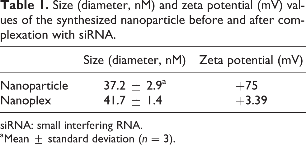
siRNA: small interfering RNA.
aMean ± standard deviation (n = 3).
The nanoparticles positive charge was an insurance of electrostatic nanoplex formation, where the nanoplex showed lower positive charge.
Gel retardation assay of the nanoparticle and siRNA
The gel retardation assay was done on nanoplexes prepared by adding different nanoparticle amounts to a constant amount of siRNA (100 ng). Naked siRNA was used to show the unbound siRNA location on gel. The nanoparticle: siRNA weight ratios used were 0.1:1, 1:1, 5:1, 10:1, and 20:1. The results showed that the prepared nanoparticles can form nanoplexes with siRNA in 5:1 ratio and above, where the unbound siRNA bond has disappeared from the gel surface (Figure 3(a)).

Gel electrophoresis of the nanoparticle and siRNA. (a) Gel retardation assay on various nanoparticle amounts complexed with a constant amount of siRNA (100 ng) preparing the 0.1:1, 1:1, 5:1, 10:1, and 20:1 (nanoparticle: siRNA) ratios (lanes 2–6) for 30 min. The first lane represents the uncomplexed siRNA bond. (b) Stability evaluation assay on various nanoparticle amounts complexed with constant amount of siRNA (100 ng) preparing the 0.1:1, 1:1, 5:1, and 20:1 (nanoparticle: siRNA) ratios 30 min after complex formation (lanes 2–5), and after incubation with RNase A enzyme for 30 min (lanes 6–9). The first lane represents the uncomplexed siRNA bond. siRNA: small interfering RNA.
The stability test was carried out following the same pattern on agarose gel (nanoparticle: siRNA ratios were 0.1:1, 1:1, 5:1, and 20:1), but the difference was the addition of RNase A to the nanoplexes after their formation lasting for about 30 min. The results showed that the formed nanoplexes were stable in the presence of the enzyme and the siRNA degradation did not occur (Figure 3(b)). So, the 2:1 nanoparticle: siRNA ratio was selected for other investigations.
Cytotoxicity of the nanoparticle
The toxicity of the nanoparticle itself and conjugated with different siRNA strands in the presence/absence of the anticancer agent, MTX, on MCF-7 and K562 cells was evaluated using MTT assay.
The results shown in Figure 4(a) indicated the survival percent of the MCF-7 cells 24–72 h after receiving the nanoparticle in different concentrations as 0, 1, 5, 10, 100, and 250 µg mL−1. Although all groups showed significant decrease in survival percent compared to the untreated cell (p < 0.05), the 200 µg mL−1 concentration was chosen for next assays due to gel retardation assay results to supply cells with sufficient amount of siRNA. Figure 4(b) shows the survival percent of K562 cells in the presence of nanoparticle in various concentrations (0, 0.01, 0.1, 1, 10, 50, 100, and 500 µg mL−1) in three time intervals after exposure (24, 48, and 72 h). The obtained results showed the approximate constant survival percent while increasing the dose higher than 10 µg mL−1 (p > 0.1). In both cell lines, the lowest survival percent (the maximum effect) was obtained after 48 h, therefore, this period of time was used for further assays.

The inhibitory effect of Fe3O4-PEG-LAC-chitosan-PEI nanoparticle in different concentrations on MCF-7 (1–250 µg mL−1) (a) and K562 (0.01–500 µg mL−1) cells (b). The survival percent was measured by MTT assay after 24, 48, and 72 h treatment with the nanoparticle. Values are means ± standard deviation of triplicate. PEG: polyethylene glycol; LAC: lactide; PEI: polyethylene imine; MTT: 3-(4,5-dimethyl-2-thiazolyl)-2,5-diphenyl-2H-tetrazolium bromide.
Figure 5 indicates the MTT assay results done on the same cell lines after 24–72 h exposure to various treatments including nanoparticle (200 µg mL−1), nanoplexed with survivin siRNA, and nanoparticle complexed with noncomplementary siRNA strand, in the presence and absence of MTX on MCF-7 (a) and K562 cell lines (b). The results showed approximate similar survival percent on both cells in 24 h (p > 0.05) and an approximate threefold decrease in cell survival percent (p < 0.01) after cells exposure to survivin siRNA containing nanoplexes in comparison with nanoparticle.

MTT assay performed 24, 48, and 72 h after exposure to various treatments including nanoparticle (200 µg mL−1), nanoplexed with survivin siRNA, nanoparticle complexed with noncomplementary siRNA strand, in both conditions of presence and absence of MTX on MCF-7 (a) and K562 (b) cell lines. Each value represents the mean ± standard deviation of three independent assays. MTX: mitoxantrone. Sur: survivin siRNA. Non: noncomplementary siRNA strand; siRNA: small interfering RNA; MTT: 3-(4,5-dimethyl-2-thiazolyl)-2,5-diphenyl-2H-tetrazolium bromide.
Discussion
Although many attempts have been made to design suitable carriers for intracellular delivery of siRNA strands into cells, 4,32 –35 this issue still remains as a major challenge for researchers. In the current study, we aimed at designing and preparation of novel cationic polyplexes that can be used as a vehicle in siRNA delivery.
Chitosan is a biocompatible polysaccharide that has been extensively studied for gene and siRNA delivery, mainly due to its cationic nature and mucoadhesive property. Different modifications of chitosan have been proposed for gene and/or siRNA delivery. 36 Among them, the carrier constituting chitosan and branched PEI has immerged as a potential candidate for siRNA delivery due to its remarkable in vitro and in vivo gene delivery success. 37 These complexes have shown efficient and safe cellular entry of siRNA and high perinuclear distribution which may immerge as a viable approach for lung cancer treatment. 17
As discussed in results section, the FTIR spectra were all verifications of successful nanoparticle preparation. Referred to Table 1, high positive charge and small nanoscaled size of the synthesized particle make it an appropriate candidate for siRNA delivery into cancerous cells. As shown in Table 1, the slight increase in particle size and decrease in zeta potential of the nanoparticle seem to be the reason of the complex formation between the siRNA and the synthesized nanoparticle, which was then proved by gel retardation assay. High stability of nanocomplex against time (30 min) and RNaseA may be the result of nanoparticle’s ability in siRNA protection and efficient binding to the siRNA strand, which seems to be beneficial in its easy handling. Similar protective effect against RNaseA enzyme have been reported after nanoplex formation by Han et al. 38 Interactions of siRNA and positive-charged nanoparticles will be electrostatic. Weak polymer-siRNA binding could be easily disrupted in the presence of ionic molecules and proteins, where the strong binding may lead to structural stability and no siRNA release in cytoplasm. Near the tumor cells, positively charged nanoplexes would bind to cell surface and be internalized via endocytosis. 38
The MTT results confirmed the low toxicity of Fe3O4-PEG-LAC-chitosan-PEI particles against MCF-7 and K562 cells. The results showed in Figure 4 were helpful in choosing the nanoparticle concentration of 200 µg mL−1 for further investigations. According to the results indicated in Figure 5, the nanoparticles with and without noncomplementary siRNA showed low toxicity against both cell lines; however, a twofold decrease was observed in the cell survival percent after MTX addition to MCF-7 cells treated with either nanoparticle or noncomplementary siRNA. While survivin siRNA nanoplex caused threefold decrease in the cell survival percent, its combination with MTX did not result in a significant increase in the cytotoxic effect. MTX cytotoxicity was more effective on MCF-7 cells than on K562 cells, which might be because of the differences between these two cell types. The MCF-7 cells are adhesive where the K562 cells are in suspension form. The survivin expression level is higher in MCF-7 cells compared with K562 cells, 39 which leads to lower cell survival percent in nanoplex treated group. The decrease showed for nanoplex-treated group was so sharp that it seems to cover the cytotoxicity effect of MTX. Attacking the cancer cells via two different strategies seems to be more useful than one. MTX addition leads to decrease in cell survival percent approximately at all groups specially at 48 h time interval. In accordance with the research done by Wu et al., the induced cell survival shown after MTX addition to each group seems to be in relation with the survivin gene expression induction by MTX. 40 Reduced cell viability has been reported previously by other researchers after chemical medicine and siRNA simultaneous exposure to the cancerous cells such as etoposide, 41 doxorubicine, 22 mitoxantrone, 35 and cisplatin. 42
Zhu et al. designed biodegradable cationic micelles for green flourescent protein (GFP) siRNA and paclitaxel co-delivery into MDA-MB-435-GFP cells. 33 The micelles prepared by Zhu and coworkers were easy to prepare with controlled molecular characteristics, biodegradable and had low cytotoxicity. Those biodegradable micellar carriers were promising in combination cancer therapy with therapeutic siRNA and chemotherapeutics. 33 Suh and coworkers 34 synthesized a new cationic lipid by linking negatively charged glutamic acid to oleoylamine via a peptide bond. The system was able to transport siRNA to various cancer cells in vitro with reduced cytotoxicity. 34 Chang et al. reported various compositions of palmitoleyl (Pal) for combined delivery of a chemical anticancer compound, mitoxantrone, and an anticancer siRNA (siMcl-1). 35 The system showed efficient cellular delivery and also had effective anticancer activity following intratumoral administration. Highly engineered nanoparticles have also been designed and used by Hossain et al. for intracellular delivery of siRNA using pH-sensitive carbonate apatite taking the advantage of endocytosis-mediated siRNA entry and its subsequent escape from the endocytic vesicle in a time-dependant manner. The nanoparticles showed excellent siRNA complexation with a desirable nanosize level suitable for endocytosis (Hossain et al. 43 ). The RNAi effect of carbonate apatite was higher and comparable to that of cytotoxic Lipofectamine 2000 in Hela cells. Furthermore, nanoparticle-facilitated delivery of validated siRNA against cyclin B1 resulted in significant growth inhibition of cancer cells. 43
Also, targeted siRNA polyplexes consisting of two molecularly precise components, a covalent siRNA conjugate equipped with a folic acid receptor targeting ligand and PEG shielding function, together with a defined three-arm polycation for siRNA packaging and nanoparticle formation has been prepared by Dohmen et al. 44 Combination of this structure with PEI-containing unit leads to efficient and specific reporter gene silencing in folic acid receptor positive KB cells. Other researchers have reported biodegradable methacrylamide- and chitosan-based cationic polymers as promising nonviral vectors for siRNA delivery. 45 Importantly, the polyplexes preserve their gene-silencing activity in the presence of serum proteins, while their cytotoxicity remained low.
In accordance with the report published by Yang et al., poly(2-dimethylaminoethylamine 2-(2-aminoethyoxy) ethoxy) phosphazene a new cationic compound with multiple amino groups showed a great potential in gene expression and gene silencing. 22 The level of Enhanced Green Fluorescent Protein (EGFP) expression and survivin gene silencing mediated by the prepared carrier were more evident than PEI carriers in MCF-7 cells. The siRNA-complexed particle induced an apparent apoptosis as well as greatly enhanced the sensitivity of MCF-7 cells to adriamycin compared with PEI-based nanoparticles. 22
Conclusion
Fe3O4-PEG-LAC-chitosan-PEI carrier had sufficient positive charge to form efficient complexes with survivin siRNA and its nanoscaled size makes it an appropriate candidate for gene delivery. Fe3O4-PEG-LAC-chitosan-PEI-mediated survivin siRNA delivery was found to be safe and specific as indicated by cell viability assays. Above findings suggest that our biocompatible Fe3O4-PEG-LAC-chitosan-PEI carrier composed of chitosan and branched PEI has a great potential in siRNA-based cancer studies.
Footnotes
Declaration of Conflicting Interests
The author(s) declared no potential conflicts of interest with respect to the research, authorship, and/or publication of this article.
Funding
The author(s) disclosed receipt of the following financial support for the research, authorship, and/or publication of this article: This project was supported by Iran National Science Foundation (INSF, which was a part of PhD thesis no: 83).