
Abstract
This study investigated the gastroprotective effects of diallyl disulfide (DADS), a secondary organosulfur compound derived from garlic (Allium sativum L.) on experimental model of ethanol (EtOH)-induced gastric ulcer in rats. The antiulcerogenic activity of DADS was evaluated by gross/histopathological inspection, pro-inflammatory cytokines, and lipid peroxidation with antioxidant enzyme activities in the stomach. DADS (100 mg/kg) was administered by oral gavage 2 h prior to EtOH treatment (5 ml/kg). The animals were killed 1 h after receiving EtOH treatment. Pretreatment with DADS attenuated EtOH-induced gastric mucosal injury, as evidenced by decreased severity of hemorrhagic lesions and gastric ulcer index upon visual inspection. DADS also prevented histopathological alterations and gastric apoptotic changes caused by EtOH. An increase in tumor necrosis factor-α (TNF-α) and inducible nitric oxide synthase was observed in the gastric tissues of EtOH-treated rats that coincided with increased serum TNF-α and interleukin 6 levels. In contrast, DADS effectively suppressed production of pro-inflammatory mediators induced by EtOH. Furthermore, DADS prevented the formation of gastric malondialdehyde and the depletion of reduced glutathione content and restored antioxidant enzyme activities, such as catalase, glutathione peroxidase, and glutathione reductase in the gastric tissues of EtOH-treated rats. These results indicate that DADS prevents gastric mucosal damage induced by acute EtOH administration in rats and that the protective effects of DADS may be due to its potent antioxidant and anti-inflammatory activities.
Introduction
Gastric ulcer is a common disease affecting many people worldwide. 1 The pathophysiology of gastric ulcer is resulted from an imbalance between gastric aggressive factors and mucosal defensive factors in gastric mucosa. 2 Some etiologies of imbalance occurring in gastric ulcer include stress, liver diseases, Helicobacter pylori infection, cigarette smoking, steroidal and nonsteroidal drugs, and ethanol (EtOH) consumption. 3 Among various etiologies, intake of EtOH is an important contributor to gastric ulceration. 4,5 EtOH-induced gastric ulcer model in rats are considered to be a reliable tool for studying the pathogenesis of acute gastric injury and screening the gastroprotective effects of antiulcerogenic agents. 6
The mechanisms of gastric damage caused by EtOH are still not fully understood; however, EtOH increases generation of reactive oxygen species (ROS), leading to oxidative stress concurrent with decreased antioxidant enzyme activities, which is believed to be responsible for gastric mucosal injuries including ulceration, erosion, hemorrhage, and edema. 7,8 Besides, infiltrated and activated neutrophils in gastric mucosa induce the production of pro-oxidative and pro-inflammatory enzymes during the initial process of gastric ulcer caused by EtOH. 9,10 In particular, the high levels of tumor necrosis factor α (TNF-α) and interleukin 6 (IL-6) in the gastric lesions were provoked by EtOH, suggesting that these pro-inflammatory cytokines play an important role in the pathogenesis of EtOH-induced acute gastric damage. 10–12 Nitric oxide (NO) is implicated in various physiological functions such as dilating blood vessels, increasing blood flow, and stimulating gastric angiogenesis during the ulcer healing process. 13 Under pathological conditions, an increase in inducible NO synthase (iNOS) activity in gastric mucosa causes the development of gastric ulcer. 10 To date, many studies have demonstrated that several compounds possessing antioxidant properties and anti-inflammatory activities may confer gastroprotective effects on EtOH-induced gastric mucosal damage in various experimental animals. 14,15
Garlic (Allium sativum L.) has diverse biological activities, including anticarcinogenic, antiatherosclerotic, antidiabetic, antioxidant, and anti-inflammatory effects. 16 Garlic oil contains more than 20 organosulfur compounds (OSCs), which are believed to play a major role in the biological activities. 17 Diallyl disulfide (DADS), a major component of the secondary OSCs derived from garlic, has potent antioxidant 17–19 and anti-inflammatory activities. 20,21 This component also downregulates the expression of numerous genes involved in oxidative stress and pro-inflammatory response. 22–24 Based on these considerations, we hypothesized that DADS could attenuate gastric damage induced by EtOH. Therefore, the aim of this study is to evaluate the possible activity of DADS on EtOH-induced gastric ulcer in rats, and to further assess its antiulcer mechanism. This is the first report regarding the antiuclerogenic property of DADS contributing to increase the knowledge on natural compound-derived substances and offer therapeutic alternatives for the treatment of gastric ulcer.
Materials and methods
Animals and environmental conditions
A total of 24 female Sprague-Dawley rats aged 7 weeks were obtained from a specific pathogen-free colony at Samtako Co. (Osan, Korea) and used after 1 week of quarantine and acclimatization. Two animals per cage were housed in a room maintained at a temperature of 23 ± 3°C and a relative humidity of 50 ± 10% with a 12-h dark/12-h light cycle and with 13 to 18 air changes per hour. Commercial rodent chow (Samyang Feed Co., Wonju, Korea) sterilized by radiation and sterilized tap water were available ad libitum. The Institutional Animal Care and Use Committee of Chonnam National University approved the protocols for the animal study (CNU IACUC-YB-2012-24), and the animals were cared for in accordance with the guidelines for animal experiments of Chonnam National University.
Test chemicals and treatment
Absolute EtOH (CAS No. 64-17-5) was purchased from Sigma Aldrich Co. (St Louis, Missouri, USA). DADS was purchased from Tokyo Kasei Chemical Co. (Tokyo, Japan). All other chemicals were of the highest grade commercially available. DADS was dissolved in corn oil and prepared prior to treatment. The application volume of DADS (2 ml/kg body weight) was calculated in advance based on the most recently recorded body weight of the individual animal. The animals were fasted overnight before the experiment, and DADS was administered orally to rats at the dose of 100 mg/kg body weight. Two hours after the DADS treatment, the rats were treated with a single oral dose of absolute EtOH (5 ml/kg body weight, equivalent to 3.945 g EtOH) to induce acute gastric mucosal injury. 10,15,25 All animals were killed 1 h after receiving the EtOH treatment.
Experimental groups
A total of 24 healthy female rats were assigned randomly to the following 4 experimental groups: (1) vehicle control, (2) DADS, (3) EtOH, and DADS + EtOH groups (n = 6 per group). The effective dose of DADS was based on earlier studies. 26,27
Necropsy, blood collection, and serum cytokine measurement
All treated animals were anesthetized by carbon dioxide inhalation for blood sample collection 1 h after EtOH treatment. Blood samples were drawn from the posterior vena cava under anesthesia, and all rats were euthanized by exsanguination after bleeding. The samples were centrifuged at 800×g for 20 min, within 1 h after collection, and stored at −80°C before analysis. Serum IL-6 and TNF-α concentrations were measured using commercial kits according to the manufacturer’s protocols (Thermo Scientific, Waltham, Massachusetts, USA).
Gastric lesion index and stomach weight
Stomach was removed, opened along the greater curvature, and washed with cold phosphate-buffered saline (PBS). The stomach was stretched on a clean paper with the mucosal surface facing upward. Photographs of hemorrhagic erosions in the stomach were taken with a photometric digital camera (C-4000, Olympus, Tokyo, Japan). The degree of gastric mucosal damage was graded according to ulcer score scales as described previously 28 and presented in Table 1. Mean assessments for each group were calculated and expressed as an ulcer index (UI). After visual inspection, the absolute and relative (organ/body weight ratio) weights of the stomachs were measured in all rats. The stomach tissue was cut in half and stored at −80°C for biochemical analysis.
The gastric mucosal ulcer scoring system.

Histopathological examination
The remaining half of the stomach was fixed in 10% neutral-buffered formalin solution for 2 weeks. The fixed stomachs were processed routinely and were embedded in paraffin, sectioned to 4 μm thickness, deparaffinized, and rehydrated using standard techniques. The extent of EtOH-induced gastric mucosa injury and the protective effects of DADS were evaluated by assessing histopathological changes in stomach sections stained with hematoxylin and eosin. All lesions were examined manually with a light microscope with 10× and 20× objective lenses in a totally blinded manner.
TUNEL assay
The fixed gastric tissues were embedded in paraffin, sectioned to 4 μm thickness, deparaffinized, and rehydrated using standard techniques. The level of DNA fragmentation was detected using a terminal deoxynucleotidyl transferase (TdT) deoxyuridine triphosphate nick-end labeling (TUNEL) assay, which was performed according to the manufacturer’s instructions (ApopTag Peroxidase In Situ Apoptosis Detection kit; Chemicon, Billerica, Massachusetts, USA). In brief, the slides were applied proteinase K (Chemicon) for 15 min at room temperature, washed twice with distilled water, and then treated with 3.0% hydrogen peroxide (H2O2) in PBS (pH 7.2) for 5 min at room temperature. The sections were washed with PBS, treated with equilibrium buffer for 10 s. Working strength TdT enzyme was applied and incubated for 1 h at 37°C. After incubation, the slides were treated with working-strength stop/wash buffer for 10 min, incubated with anti-digoxigenin for 30 min at room temperature, and then visualized with chromogen 3,3′ diaminobenzidine (DAB). The sections were counterstained with Harris’s hematoxylin before being mounted. Each slide was examined manually with a light microscope (Leica, Wetzlar, Germany) with 10× and 20× objective lenses in a totally blinded manner.
Immunohistochemical examination
The fixed gastric tissues were processed routinely using standard techniques. The sections were heated for 30 min in a sodium citrate buffer (0.01 M, pH 6.0) and then treated with 0.3% (v/v) H2O2 in water for 15 min. The slides were washed with 0.01 M PBS (pH 7.2) and then blocked with 1% bovine serum albumin (BSA), 0.2% Tween 20 in PBS for 1 h at room temperature. The sections were then incubated overnight with monoclonal antibody against caspase 3 (1:200; Cell Signaling Technology, Danvers, Massachusetts, USA) at 4°C. The second antibody biotinylated goat anti-rabbit immunoglobulin G (1:500; Cell Signaling Technology) was applied followed by streptavidin peroxidase and then visualized with the DAB with its substrate buffer. The sections were counterstained with Harris’s hematoxylin before being mounted. Each slide was examined manually with a light microscope (Leica) with 10× and 20× objective lenses in a double-blinded manner.
Western blotting analysis for TNF-α and iNOS expression
Frozen stomach tissues were lysed with a radio-immunoprecipitation assay lysis buffer (Cell Signaling Technology) and were centrifuged at 12,000×g at 4°C for 10 min to obtain the cellular proteins in the supernatant. Equal amounts of proteins from each sample were resolved by sodium dodecyl sulfate–polyacrylamide gel electrophoresis, transferred to polyvinylidene difluoride membranes (Whatman, Maidenstone, UK) and blocked in blocking buffer (150 mM sodium chloride (NaCl) in 10 mM Tris, pH 7.5 containing 5% nonfat dry milk) for 1 h at room temperature. The membranes were incubated with primary rabbit antibodies against TNF-α and iNOS (1:100; Abcam, UK) and β-actin (1:1,000; Cell Signaling Technology) for 18 h at 4°C, washed three times (20 mM tris(hydroxymethyl)aminomethane (Tris)–hydrochloric acid, pH 7.5, 137 mM NaCl, and 0.1% Tween 20), incubated with horseradish peroxidase-conjugated secondary antibodies (1:2000) for 1 h at room temperature, washed three times, and then detected by enhanced chemiluminescence. Protein concentration was determined with a BCA protein assay kit (Pierce, Rockford, Illinois, USA). Protein expression was quantified based on band density using TINA 20 Image software (Raytest Isotopenmessgeraete GmbH, Straubenhardt, Germany). Relative intensity was calculated by dividing the densities of respective loading control density value. Values are presented as mean ± SD.
Determination of lipid peroxidation and GSH, and antioxidant enzyme activities
A portion of the frozen stomach was homogenized in a glass–Teflon homogenizer with 50 mM phosphate buffer (pH 7.4) to obtain 1:9 (w/v) whole homogenate. The homogenates were then centrifuged at 11,000×g for 15 min at 4°C to discard any cell debris, and the supernatant was used to measure malondialdehyde (MDA), glutathione (GSH) concentrations, and antioxidant enzyme activities. The concentration of MDA, an index of lipid peroxidation, was assayed by monitoring thiobarbituric acid reactive substances formation using the method of Berton et al. 29 In brief, stomach homogenates were mixed with 35% perchloric acid. The mixture was centrifuged (8000 rpm, 20 min at 4°C) and 0.6% thiobarbituric acid was added to the supernatants, followed by boiling at 100°C for 30 min. After cooling the supernatant, absorbance was determined at 535 nm. The results are expressed as μmol/mg protein. GSH content was measured by the method followed by Moron et al. 30 In brief, samples were mixed with 100 mM potassium phosphate buffer (pH 7.0) containing 1 mM ethylenediaminetetraacetic acid, 0.2 mM nicotinamide adenine dinucleotide phosphate, and 0.6 mM 5,5′-dithio-bis (2-nitrobenzoic acid). After shaking the reaction mixture for 5 min, its absorbance was measured at 412 nm. The absorbance values were extrapolated from a GSH standard curve and expressed in nmol/mg protein. The antioxidant enzyme activities, including glutathione peroxidase (GPx), glutathione reductase (GR), and catalase were also determined using commercial assay kits according to manufacturer’s protocols (Cayman, Ann Arbor, Michigan, USA). Results are expressed in units per milligram protein. Total protein content was determined by the method described by Lowry et al. 31 using BSA as the standard.
Statistical analysis
Results are expressed as mean ± SD, and all statistical comparisons were made by one-way analysis of variance followed by Tukey’s multiple comparison test. The statistical analysis was performed comparing the treatment groups with the control group using GraphPad InStat v. 3.0 (GraphPad Software Inc., La Jolla, California, USA). Differences with the value of p ≤ 0.05 were considered statistically significant.
Results
Effects of DADS on body weight and stomach weight
No significant differences were observed in the body weights between the groups (data not shown). The absolute (1.35 ± 0.409) and relative (0.49 ± 0.102, p < 0.05) stomach weights in the EtOH group were slightly or significantly increased compared with those in the control group (Table 2). Although the absolute (1.25 ± 0.183) and relative (0.47 ± 0.051) stomach weights in the DADS + EtOH group were also slightly lower than those in the EtOH group, no significant differences were detected compared with those in the control group.
Absolute and relative stomach weights of female rats treated with DADS and/or EtOH.
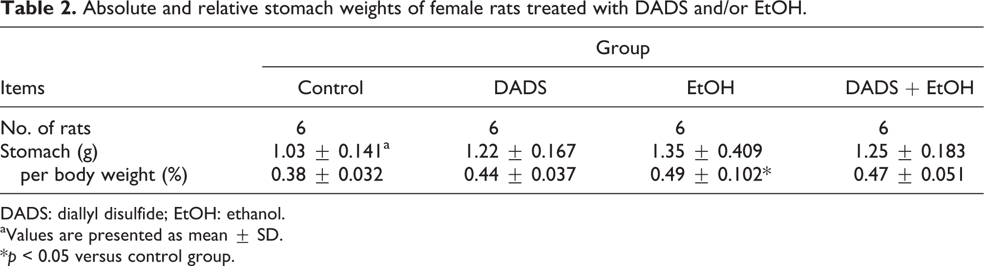
DADS: diallyl disulfide; EtOH: ethanol.
aValues are presented as mean ± SD.
*p < 0.05 versus control group.
Effects of DADS on EtOH-induced acute gastric injury
The gastric UI was scored by visual inspection according to the method of La Casa et al. 28 The control and DADS groups presented stomachs with normal appearance (Figure 1(a) and (b)). In the EtOH group, no visible lesions were observed in the nonsecretory part of the stomach, whereas severe hemorrhagic longitudinal linear lesions with hyperemia were observed in the gastric body of the stomach (Figure 1(c)). The gastric UI score in the EtOH group (6.3 ± 2.25, p < 0.01) also increased significantly compared with that in the control group (Figure 1(e)). Although these findings were also observed in the DADS + EtOH group, the incidence and severity of hemorrhage and hyperemia lesions were lower than that in the EtOH group (Figure 1(d)). The UI score in the DADS + EtOH group (2.0 ± 1.79, p < 0.01) also decreased significantly compared with that in the EtOH group (Figure 1(e)).

Effects of DADS on EtOH-induced acute gastric injury. Representative photographs of gastric mucosa treated with DADS (100 mg/kg) and/or EtOH (5 ml/kg). Images showing normal appearance of stomach for (a) vehicle control and (b) DADS-treated rats. (c) Stomach of EtOH-treated rat shows severe hemorrhagic longitudinal linear lesions with hyperemia. (d) Stomach of DADS + EtOH-treated rat shows mild hemorrhagic lesions. (e) Image showing gastric mucosal ulcer index determined by visual inspection. Values are presented as mean ± SD (n = 6). **p < 0.01 versus control group; †† p < 0.01 versus EtOH group. DADS: diallyl disulfide; EtOH: ethanol.
Effect of DADS on EtOH-induced histopathological alterations
The antiulcer activity of DADS observed in gross findings was confirmed by the histopathological examination. The control and DADS groups presented gastric mucosa with normal architecture (Figure 2(a) and (b)). However, gastric mucosa of all EtOH-treated rats showed extensive histopathological changes, characterized by severe hemorrhage, infiltration of inflammatory cells, loss of epithelial cells, submucosal edema, and vascular congestion (Figure 2(c)). Although these findings were also observed in the DADS + EtOH group (Figure 2(d)), the incidence and severity of gastric histopathological lesions decreased compared with those in the EtOH group.
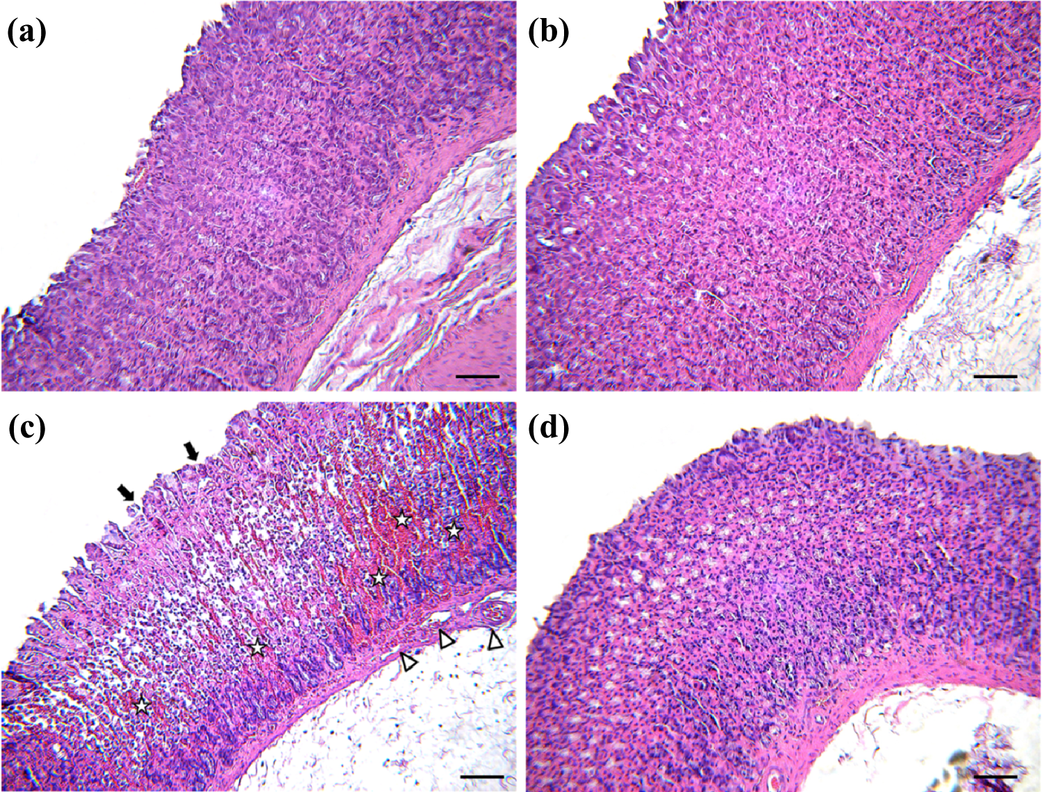
Effects of DADS on EtOH-induced histopathological alterations. Representative H&E-stained histological sections of stomach of rats treated with DADS (100 mg/kg) and/or EtOH (5 ml/kg). Gastric tissues of (a) vehicle control and (b) DADS-treated rats show normal appearance. (c) Gastric tissue of EtOH-treated rat shows severe hemorrhage, infiltration of inflammatory cells (asterisk), loss of epithelial cells (arrows), and submucosal edema and vascular congestion (arrow head). (d) Gastric tissue of DADS + EtOH-treated rat shows nearly normal appearance. H&E stain. Bar = 50 μm (×400 magnification). H&E: hematoxylin and eosin; DADS: diallyl disulfide; EtOH: ethanol.
Effect of DADS on EtOH-induced gastric mucosal cell apoptosis
The apoptotic changes observed in gastric mucous are presented in Figure 3. The control and DADS groups showed few apoptotic changes in gastric mucosal cell (Figure 3(a) and (b)). An increased number of gastric apoptotic cells was observed in the upper epithelial mucosal layer in the EtOH group (Figure 3(c)). In contrast, pretreatment with DADS resulted in a decrease in the number of TUNEL positive cells compared with that in the EtOH group (Figure 3(d)).

Effect of DADS on EtOH-induced gastric mucosal cell apoptosis. Representative photographs of TUNEL assay performed on stomach sections of rats treated with DADS (100 mg/kg) and/or EtOH (5 ml/kg). (a) Vehicle control and rats treated with (b) DADS, (c) EtOH, and (d) DADS + EtOH. The brownish-colored cells represent TUNEL positive cells. Bar = 50 μm (×400 magnification). TUNEL: terminal deoxynucleotidyl transferase deoxyuridine triphosphate nick-end labeling; DADS: diallyl disulfide; EtOH: ethanol.
Effect of DADS on caspase 3 immunopositivity
To further confirm the gastroprotective effect of DADS on EtOH-induced gastric mucosal cell apoptosis, we conducted immunohistochemical analysis for caspase 3, and the results are presented in Figure 4. Caspase 3 positive cells were seldom seen in the control and DADS groups (Figure 4(a) and (b)). However, the number of caspase 3 positive cells increased in the EtOH group compared with those in the control group (Figure 4(c)). In contrast, caspase 3 positive cells in the DADS + EtOH group decreased compared with those in the EtOH group (Figure 4(d)).

Effect of DADS on EtOH-induced caspase-3 immunopositivity. Representative photographs of caspase-3 immunohistochemical analysis on stomach sections of rats treated with DADS (100 mg/kg) and/or EtOH (5 ml/kg). (a) Vehicle controls and rats treated with (b) DADS, (c) EtOH, and (d) DADS + EtOH. The brownish-colored cells represent caspase-3-positive cells. Bar = 50 μm (×400 magnification). DADS: diallyl disulfide; EtOH: ethanol.
Effects of DADS on EtOH-induced inflammatory mediators TNF-α, iNOS, and IL-6
To determine whether DADS elicits its effects on the inflammatory mediators TNF-α, iNOS, and IL-6, we confirmed the protein and serum levels of TNF-α, iNOS, and IL-6. As shown in Figure 5, gastric tissue treated with EtOH showed a significant increase in the protein levels of TNF-α (2.04 ± 0.394, p < 0.01) and iNOS (4.05 ± 0.242, p < 0.01) compared with those in the control group (Figure 5(a) and (b)). Similarly, serum levels of TNF-α (22.04 ± 1.734, p < 0.01) and IL-6 (28.82 ± 4.564, p < 0.01) in the EtOH group also increased significantly compared with those in the control group (Figure 5(c) and (d)). In contrast, gastric TNF-α (1.18 ± 0.228, p < 0.01) and iNOS (2.29 ± 0.12, p < 0.01) protein levels in the DADS + EtOH group decreased significantly compared with those in the EtOH group. Moreover, pretreatment with DADS exhibited a significant decrease in the serum TNF-α (20.35 ± 1.241, p < 0.01) and IL-6 (17.49 ± 4.459, p < 0.01) levels compared with those in the EtOH group.

Effect of DADS on EtOH-induced pro-inflammatory mediators. (a) Western blot analysis of TNF-α and iNOS expression in the gastric tissue of rats treated with DADS (100 mg/kg) and/or EtOH (5 ml/kg; loading control: β-actin). (b) Bar graph showing relative intensity levels of TNF-α and iNOS for vehicle, DADS, EtOH, and DADS + EtOH-treated rats, respectively. Serum (c) TNF-α and (d) IL-6 levels were determined using enzyme-linked immunosorbent assay. Values are presented as mean ± SD (n = 6). **p < 0.01 versus control group; †† p < 0.01 versus EtOH group. DADS: diallyl disulfide; EtOH: ethanol; TNF-α: tumor necrosis factor α; iNOS: inducible nitric oxide synthase; IL-6: interleukin 6.
Effects DADS on EtOH-induced lipid peroxidation and depletion of GSH
The concentration of MDA (1.28 ± 0.067, p < 0.01), an end product of lipid peroxidation, in rats treated with EtOH increased significantly, whereas GSH content (0.63 ± 0.029, p < 0.01) decreased significantly compared with that in the control group (Figure 6(a) and (b)). However, the level of MDA (1.15 ± 0.045, p < 0.05) in the DADS + EtOH group decreased significantly compared with that in the EtOH group. Gastric GSH content (0.73 ± 0.025, p < 0.01) increased significantly in the DADS + EtOH group compared with that in the EtOH group.

Effects DADS on EtOH-induced lipid peroxidation and depletion of glutathione. (a) Malondialdehyde and (b) glutathione concentrations in gastric tissues of rats treated with DADS (100 mg/kg) and/or EtOH (5 ml/kg). Values are presented as mean ± SD (n = 6). **p < 0.01 versus control group; † p < 0.05, †† p < 0.01 versus EtOH group. DADS: diallyl disulfide; EtOH: ethanol.
Effects of DADS on EtOH-induced suppression of antioxidant enzyme activities
As shown in Figure 7, GPx (0.39 ± 0.158, p < 0.01), GR (0.63 ± 0.129, p < 0.01), and catalase (1.42 ± 1.795, p < 0.01) activities in the EtOH group decreased significantly compared with those in the control group (Figure 7(a) to (c)). In contrast, pretreatment with DADS resulted in a significant increase in GPx (1.44 ± 0.329, p < 0.01), GR (1.36 ± 0.228, p < 0.01), and catalase (5.94 ± 1.973, p < 0.01) activities compared with those in the EtOH group.
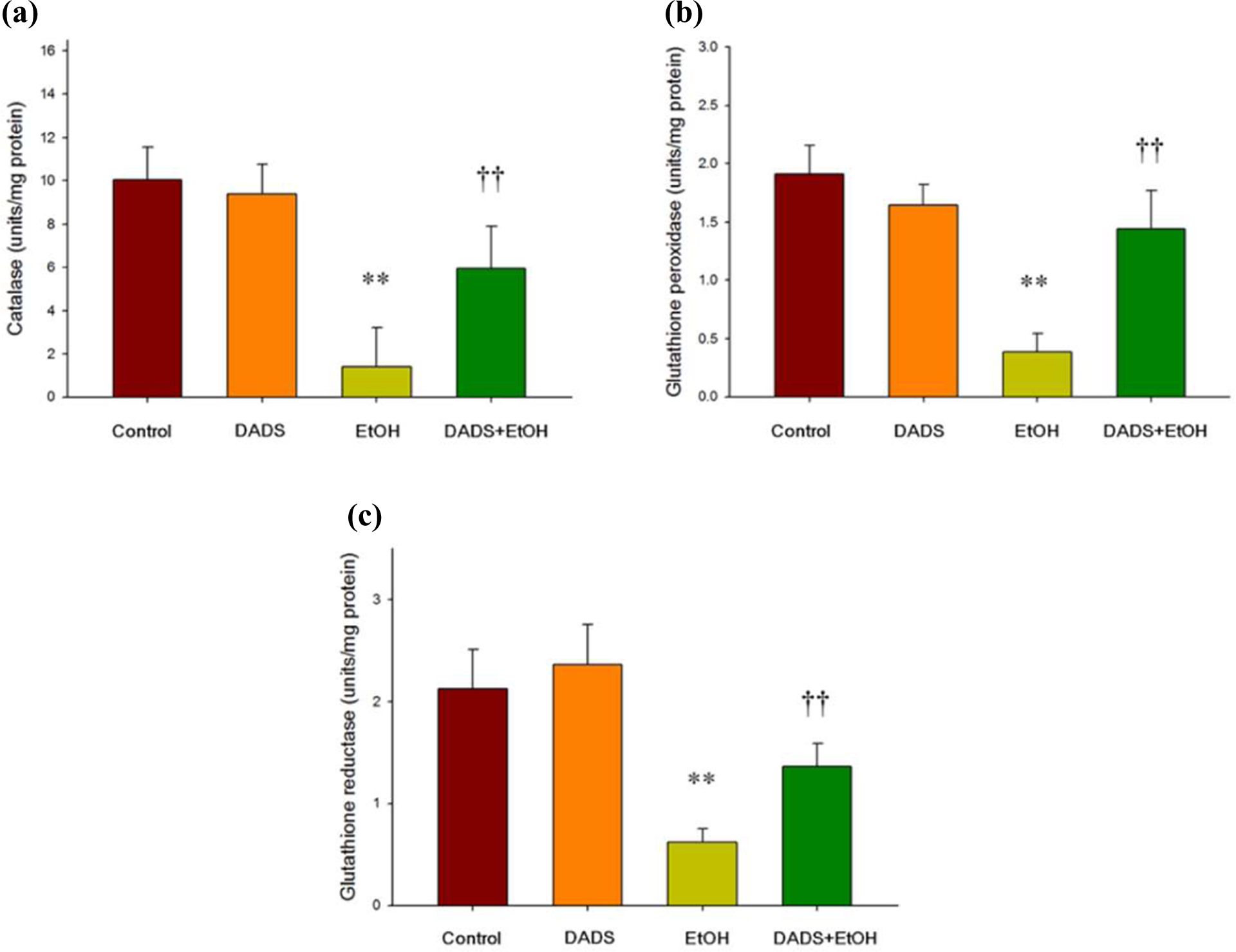
Effects of DADS on EtOH-induced suppression of antioxidant enzyme activities. (a) Catalase, (b) glutathione peroxidase, and (c) glutathione reductase activities in the gastric tissues of female rats treated with DADS (100 mg/kg) and/or EtOH (5 ml/kg). Values are presented as mean ± SD (n = 6). **p < 0.01 versus control group; †† p < 0.01 versus EtOH group. DADS: diallyl disulfide; EtOH: ethanol.
Discussion
Various etiologies, such as stress, cigarette smoking, sepsis, alcohol, and steroidal and nonsteroidal drugs, cause an imbalance between gastric aggressive and protective factors that play a role in gastric ulcerogenesis. 3 The current therapeutic treatments of gastric ulcers generally dependent on the inhibition of gastric acid secretion by histamine H2-antagonists, proton pump inhibitors, and acid-independent therapy provided by sucralfate and bismuth cholinergics. 32 However, nowadays, most of these therapies induce the development of tolerance and adverse effects that make the efficacy of these treatments questionable. 33 In contrast, natural compounds can provide a safe and comfortable approach for prevention of EtOH-induced gastric ulcer. 34–36 In this study, the gastroprotective activity of DADS was evaluated on EtOH-induced gastric ulcer model in rats. EtOH treatment resulted in extensive gastric mucosal injury in rats, and DADS pretreatment conferred protection on the EtOH-induced gastric mucosal damage and oxidative stress via its ability to enhance antioxidant enzyme activities and suppress inflammatory response.
EtOH-induced gastric damage is one of the most intensively used models for studying the pathogenesis of acute gastric injury. 1,6 EtOH-induced acute gastric lesions are characterized by various pathological alterations including hemorrhage, inflammatory cell infiltration, edema, and loss of epithelial cells. 15,25 In this study, EtOH treatment to rats caused an elevation in severity of hemorrhagic and hyperemic lesions concurrent with increasing gastric UI. These alterations in the gross findings were well correlated with extensive histopathological changes such as mucosal edema, vascular congestion, inflammatory cell infiltration, loss of epithelial cells, and increased gastric epithelial cell apoptosis. Our findings are also in accordance with the results of previous studies. 15,25,37 However, administration of DADS significantly attenuated the EtOH-induced acute gastric mucosal damage and apoptotic change, as evidenced by a decrease in the gastric UI and the incidence and severity of the gross/histopathological changes. These results indicate that DADS confers antiapoptotic activity and gastroprotection against EtOH-induced gastric mucosal injury.
The inflammatory process is a major component of mucosal defense against exogenous and endogenous factors in the gastrointestinal tract. Impairment of this response can lead to mucosal injury and an impaired healing process. 38 The production of inflammatory mediators, such as TNF-α and IL-6, is important for the mechanisms of EtOH-induced gastric lesions. 11 TNF-α is one of the pleiotropic inflammatory cytokines and plays a key role in tissue injury and inflammation. 39 IL-6, one of the key pro-inflammatory cytokines, is commonly produced in local tissues and released into circulation. Elevated IL-6 level is responsible for local tissue damage by activating neutrophils and macrophages at the inflammatory site. 40,41 Previous studies have reported that TNF-α and IL-6 levels were remarkably increased in EtOH-induced gastric ulcer lesion, and its reduction effectively reversed gastric mucosal damage caused by EtOH. 10,41 In this study, EtOH caused an increase in gastric and serum TNF-α and IL-6 levels. In contrast, pretreatment with DADS remarkably inhibited the production of TNF-α and IL-6 level in gastric mucosa and serum, indicating that the reduction of pro-inflammatory cytokines is one of the major factors involved in the gastroprotective action on EtOH-induced gastric ulcer.
NO is involved in the modulation of the gastric mucosal integrity and the role of endogenous NO in physiological processes has been established in many tissues, including gastrointestinal tract. However, excessive NO, mainly produced by iNOS, enhances gastric ulcerogenic response. 42 Ahn et al. 14 demonstrated that reducing iNOS in gastric mucosa attenuates EtOH-induced gastric damage. In the present study, we investigated the effect of DADS on the gastric iNOS expression. Our results demonstrated that pretreatment with DADS decreased gastric iNOS expression in EtOH-induced gastric ulcer, suggesting that a reduction of iNOS expression is probably involved in the gastroprotective action of DADS on EtOH-induced gastric mucosal damage.
The pathogenesis of gastric mucosal damage includes generation of ROS, which play a vital role in lipid peroxidation of cellular components, accompanied by impairment of the antioxidative enzyme system. 7,43 In contrast, antioxidants can confer protection on gastric mucosal injury induced by various ulcerogenic agents including EtOH. 2,15,44 In the present study, EtOH treatment caused high levels of oxidative stress, as evidenced by a significant elevation in the MDA concentration and a significant decrease in GSH content with decreased GR, GPx, and catalase activities, suggesting a role for oxidative stress in EtOH-induced gastric ulcer. However, DADS treatment attenuated the EtOH-induced increased MDA concentration and effectively improved EtOH-induced depletion of GSH content and suppression of antioxidant enzyme activities in gastric tissues. According to some reports, garlic oil possesses antioxidant properties that provide protection against EtOH-induced gastric injury. 45 Furthermore, antioxidant activity of DADS exhibits a protective effect on various pathological conditions induced by oxidative stress, including gentamicin-induced nephrotoxicity and carbon tetrachloride-induced hepatotoxicity. 26,46 Therefore, this apparent ameliorative effect may be due to the ability of DADS to inhibit lipid peroxidation and enhance antioxidant enzyme activities.
In summary, the results of this study demonstrated that DADS can provide a marked gastroprotective effect on EtOH-induced gastric mucosal damage in rats. These gastroprotective effects of DADS may be due to its antioxidant properties, as evidenced by increased GSH content and enhanced antioxidant enzyme activities, and decreased lipid peroxidation. DADS also showed an anti-inflammatory activity, as demonstrated by a decrease in pro-inflammatory mediators. Although further studies are still needed to better elucidate the mechanisms underlying these protective effects, DADS may be a promising gastroprotective agent against various gastric injuries caused by oxidative stress and the inflammatory response.
Footnotes
Authors’ Note
Authors I-C L and H-S B contributed equally to this work as co-first authors.
Conflict of interest
The authors declared no conflicts of interest.
Funding
This research was supported by Basic Science Research Program through the National Research Foundation of Korea funded by the Ministry of Education, Science and Technology (NRF-2013R1A1A2010835). The animal experiment in this study was supported by the Animal Medical Institute of Chonnam National University.