
Abstract
Reactive oxygen species (ROS) such as
Introduction
Reactive oxygen species (ROS) are formed during partial reduction of molecular oxygen in the mitochondrial electron transport chain. Presence of unpaired electron on the outer orbital of oxygen makes ROS highly toxic. 1 Many antineoplastic agents are known to induce oxidative stress in biological systems. During cancer chemotherapy, oxidative stress-induced lipid peroxidation generates numerous electrophilic aldehydes and oxygen free radicals that are capable of damaging compounds of all biochemical classes including nucleic acid, proteins, lipids, lipoproteins, carbohydrates, and connective tissue macromolecules. 1,2 This product of oxidative stress may even interfere with the ability of anticancer drugs to kill cancer cells by inhibiting caspase activity or death receptors leading to inhibition in drug-induced apoptosis and also cause severe toxicity to healthy cells. These toxicities would also diminish the efficacy of the treatment and are major contributing factors to dose reduction, delays, and cessation of cancer treatment. 2
Camptothecin (CPT) is a class I topoisomerase inhibitor that stabilizes topo I DNA specifically binding at the top1-DNA interface and forms ternary cleavable complex, thus preventing DNA religation step. Stabilization of top1-cleavage complex by top 1 inhibitors arrests the replication fork which generates bulky DNA lesions. 3 CPT is a broad spectrum antinoeplastic agent that has received Food and Drug Administration approval for use against ovarian, lung, breast, and colon cancers, but occurrence of leukopenia, thrombocytopenia gastrointestinal malignancies, melanoma, fever, alopecia, vomiting, diarrhea, and hemorrhagic cystitis were observed to be its dose-limiting toxicities. 4,5 Although CPT has shown promising antineoplastic effects, its use has been limited due to several toxicities because of its pro-oxidant nature. 4
Vitamin E is an effective antioxidant that is known to stop the production of ROS formed when fat undergoes oxidation. 6 It has been theorized by researchers that vitamin E blocks lipid peroxidation-mediated toxicity and increases the efficacy of drug. Pretreatment with vitamin E is also known to induce apoptosis in experimental tumor cell line. 7 It has also been reported to increase survival time in patients with terminal cancer. An increase in serum vitamin E levels has been reported to inhibit protein kinase C, 5-lipoxygenase, smooth muscle cell proliferation, platelet aggregation, and oxygen burst in neutrophils. 8,9 In the present study, we investigated the effect of CPT administration in myocardial tissue of Wistar rats and influence of vitamin E pretreatment in order to assess whether exogenous supplementation of supra dietary dose of vitamin E can enhance the efficacy of standard and experimental cancer therapies by decreasing the upsurge in CPT-induced toxicities.
Materials and methods
Drugs and chemicals
(S)-(+)-CPT, α-tocopherol actetate, glutathione reductase (GRD), epinephrine, and reduced glutathione (GSH) were purchased from Sigma Chemicals (St Louis, Missouri, USA). All other chemicals used were of high analytical grade, and the solvents were of Qualigen grade.
Animal model
Adult male albino rats of Wistar strain (weighing 120 ± 20 g) were obtained from Bharat Serum Pvt. Ltd (Thane, Navi Mumbai, Maharashtra, India). The animals were maintained under standard conditions of humidity, temperature (25 ± 2°C) and light (12-h light/12-h dark). They were fed standard rat pellet diet obtained from Lipolin, India and water ad libitum. Experimental animals were handled according to the institutional legislation, regulated by the Committee for the Purpose of Control and Supervision of Experiments on Animals (CPCSEA) after institutional ethical clearance.
Experimental design
Following the acclimatization period, the rats were randomly divided into four groups consisting of six animals each. Group I: Control rats were given drug vehicle dimethyl sulfoxide (DMSO) in saline solution (0.9% sodium chloride) for four consecutive days, intravenously. Group II: Rats were injected CPT (6 mg/kg body weight) dissolved in DMSO for four consecutive days, intravenously. Group III: Rats served as control group for vitamin E and received α-tocopherol (6 mg/kg body weight) orally daily for a period of 30 days. Group IV: Rats received α-tocopherol prior to CPT injection as described for groups II and III rats.
At the end of the experimental period, the animals were sacrificed by decapitation. Heart was excised immediately, washed with ice-cold saline, and processed for further analysis.
Biochemical analysis
The heart tissue was homogenized with the help of a glass–teflon homogenizer in 0.01 M tris(hydroxymethyl)aminomethane)-hydrochloric acid buffer (pH 7.4) for enzyme assays and in 0.15 M potassium chloride for lipid peroxidation studies. The aliquots of the homogenate were suitably processed for the assessment of following biochemical parameters.
Lipid peroxidation
Lipid peroxidation was determined by the procedure followed by Okhawa et al. 10 Malondialdehyde (MDA), formed as an end product of lipid peroxidation, served as a measure of the intensity of oxidative stress. MDA reacts with thiobarbituric acid to generate a colored product that can be measured optically at 532 nm. Tetramethoxy propane was used as the standard, and the results were expressed in nanomoles of MDA per milligram protein.
Estimation of antioxidant enzymes
Estimation of SOD activity
Superoxide dismutase (SOD) was assayed by monitoring the oxidation of epinephrine according to the procedure followed by Mishra and Fridovich. 11 The method is based on the ability of SOD to inhibit autoxidation of adrenaline to adrenochrome at alkaline pH. The reaction mixture contained 0.3 M carbonate buffer (pH 10.2) and 0.6 mM of ethylenediaminetetraacetic acid (EDTA) solution. Then, 0.1 ml of the homogenate was added, and the absorbance was monitored after adding 1.8 mM of epinephrine at 420 nm for 3 min at 30 s interval. Autoxidation of epinephrine was also monitored in the reaction mixture without adding the homogenate. The final activity was expressed in terms of units per milligram protein. One unit of enzyme activity is defined as the enzyme required for 50% inhibition of adrenaline autoxidation.
Estimation of CAT activity
Catalase (CAT) activity was determined according to the method described by Clairborne. 12 The method relies on decomposition of hydrogen peroxide (H2O2), which is indicated by a decrease in absorbance at 240 nm. 0.1 ml of the homogenate and 4.0 ml of phosphate buffer (pH 7.0) were added to 30 mM H2O2, and the decrease in optical density (OD) due to decomposition of H2O2 was monitored at 240 nm for 3 min at 30 s interval. The enzyme activity was expressed as micromoles of H2O2 decomposed per minute per milligram protein.
Estimation of GST activity
Glutathione S-transferase (GST) activity was assayed by the method followed by Habig et al. 13 The enzyme-catalyzed condensation of GSH with the model substrate, 1-choloro, 2,4-dinitrobenzene (CDNB), leads to a product formed (2,4-dinitrophenyl-glutathione) that absorbs light at 340 nm, which facilitated the analysis of enzyme activity based on product formation. The reaction mixture contained phosphate buffer (pH 6.5), 30 mM CDNB, and 0.1 ml of the homogenate. Tubes were incubated at 37°C for 5 min, then 30 mM GSH was added, and the change in OD was monitored at 340 nm for 3 min at 30 s interval. The enzyme activity was expressed as micromoles of CDNB conjugated per minute per milligram protein.
Estimation of GPx activity
Glutathione peroxidase (GPx) activity was determined by the method described by Paglia and Valentine, by monitoring nicotinamide adenine dinucleotide phosphate (NADPH) oxidation in the presence of GRD, which reduces glutathione disulfide (GSSG) using hydro peroxide as substrate. 14 To 0.1 ml of the homogenate, 0.1 M phosphate buffer (pH 7.0), 1 mM of EDTA, GRD (2.4 units), 10 mM of GSH, and 1 mM of sodium azide were added, and the tubes were incubated at 37°C for 10 min. Then, 1.5 mM NADPH and 1.5 mM of H2O2 were added, and the absorbance was monitored at 340 nm for 5 min at 30 s interval. The activity was expressed in nanomoles of NADPH oxidized per minute per milligram protein.
Estimation of GRD activity
Glutathione reductase (GRD) catalyzes the NADPH-dependent reduction of GSSG to GSH. The oxidation of NADPH to NADP+ is accompanied by a decrease in absorbance at 340 nm, thus providing a spectrophotometric means of detection by Carlberg and Mannervik method. 15 The reaction mixture consisting of 50 mM phosphate buffer (pH 7.5), 1 mM of EDTA, 0.67 M of GSSG, and 0.1 ml of homogenate were incubated at 37°C for 10 min. Then, 0.1 mM of NADPH was added to the tubes, and the change in absorbance was read immediately at 340 nm for 3 min at 30 s interval. The enzyme activity was expressed as nanomoles of NADPH oxidized per minute per milligram protein.
Assessment of nonenzymic antioxidant
Assessment of total GSH levels
Free endogenous GSH was assayed by the method followed by Moron et al. using 5,5′-dithio-bis 2-nitrobenzoic acid (DTNB) as the coloring reagent. 16 Homogenate proteins were precipitated by the addition of 5% trichloroacetic acid (TCA) and centrifuged at 3000 r/min for 10 mins. Then, 0.6 mM of DTNB reagent and 0.2 M phosphate buffer were added to an aliquot of clear supernatant. The absorbance was read at 412 nm against a blank containing TCA. The amount of GSH was expressed as nanomoles of GSH per milligram protein.
Assessment of vitamin E levels
Vitamin E level was estimated using the spectrophotometric method of Bieri et al. in which tocopherol is oxidized to tocopheryl quinone by ferric chloride, and the resultant ferrous ion forms a red-colored complex with dipyridyl reagent. 17 To the sample were added 0.5% of 2, 2′-dipyridyl and 0.2% ferric chloride in ethanol and kept for incubation. Red color was diluted with distilled water and then read at 520 nm. Absorbance was recorded within 30 min, and the result was expressed in milligram of vitamin E per gram tissue.
Estimation of diagnostic marker enzymes
Using King’s method, both aspartate aminotransferase (AST) and alanine aminotransferase (ALT) were estimated only with a difference that the former uses
Histopathological studies
Samples of heart were fixed in 10% formalin, processed routinely for paraffin embedding, and sectioned at 4 μm thickness. Sections were stained with hematoxylin–eosin and mounted. The sections were examined by light microscope to detect the histological changes induced by the treatments.
Statistical analysis of data
Data are presented as mean ± SD. The significance of difference among the groups was assessed using one-way analysis of variance test followed by Tukey’s honest significant difference post hoc test between all group means. The value of p < 0.05 was considered statistically significant.
Results
No distinctive clinical signs, mortality, or morbidity was observed in any of the experimental groups during the study period. Treatment of rats with CPT caused no significant change in body weight or tissue weight. In the present study, the content of MDA, a product of lipid peroxidation, in heart tissue was significantly (p < 0.001) increased in the CPT-treated group as compared to control. Pretreatment with vitamin E markedly reduced the MDA content (p < 0.05) suggesting inhibition of lipid peroxidation and protection of tissue against free radical damage (Figure 1). Myocardial tissue of CPT-treated rats showed 26% depletion in vitamin E than that of the control group. Groups III and IV showed about 65% and 36% increase in the level of vitamin E, respectively, indicating a good accumulation of orally administered vitamin E (Figure 2). There was significant (p < 0.001) loss of GSH in group II animals when compared with the control (Figure 3). Table 1 represents alterations in cardiac marker enzymes. CPT administration caused significant decrease in enzymes like AST, ALT (p < 0.001) and ALP (p < 0.01). In group IV, these drug-induced alterations were reduced, and enzyme levels were resumed to normal indicating ameliorative nature of vitamin E. When compared with control, LDH increased significantly (p < 0.01) in group II, whereas group IV showed significant reversion in the levels of LDH against group II. Table 2 represents the altered activity of antioxidant enzymes SOD, CAT, GRD, GPx, and GST in the heart tissue of CPT-injected rats and protection rendered by vitamin E. In comparison with control, CPT-treated rats showed increase in the activities of SOD, CAT, GPx, and GST, whereas significant decrease (p < 0.05) in the activity of GRD was observed. Treatment of group IV with vitamin E significantly prevented CPT-induced alteration in the levels of these antioxidant enzymes. The restoration of enzymic and nonenzymic antioxidant levels toward control range by vitamin E indicates its protective effect against oxidative stress-induced changes in antioxidant molecules. The normal rats receiving vitamin E alone (Group III) did not show any significant change when compared with the control rats (Group I), indicating that vitamin E does not per se have any adverse effects. Histopathological observation also showed severe cardiac damage following intravenous CPT treatment which was averted in rats preadministered with vitamin E (Plate A to D).

Change in lipid peroxides level after CPT and vitamin E administration. Values are expressed as mean ± SD for six rats in a group. Comparisons are made between (a) groups I and II; (b) groups I and IV; and (c) groups II and IV. ***p < 0.001; *p < 0.05: statistical significance and NS: nonsignificant. CPT: camptothecin.

Change in vitamin E level in all groups after CPT and vitamin E administration. Values are expressed as mean ± SD for six rats in a group. Comparisons are made between (a) groups I and II; (b) groups I and IV; and (c) groups II and IV. ***p < 0.001; *p < 0.05: statistical significance. CPT: camptothecin.

Effect of CPT and vitamin E on the levels of GSH. Values are expressed as mean ± SD for six rats in a group. Comparisons are made between (a) groups I and II; (b) groups I and IV; and (c) groups II and IV. ***p < 0.001; *p < 0.05: statistical significance and NS indicates nonsignificant. CPT: camptothecin; GSH: glutathione.
Effect of CPT and vitamin E on the levels of diagnostic marker enzymes.a
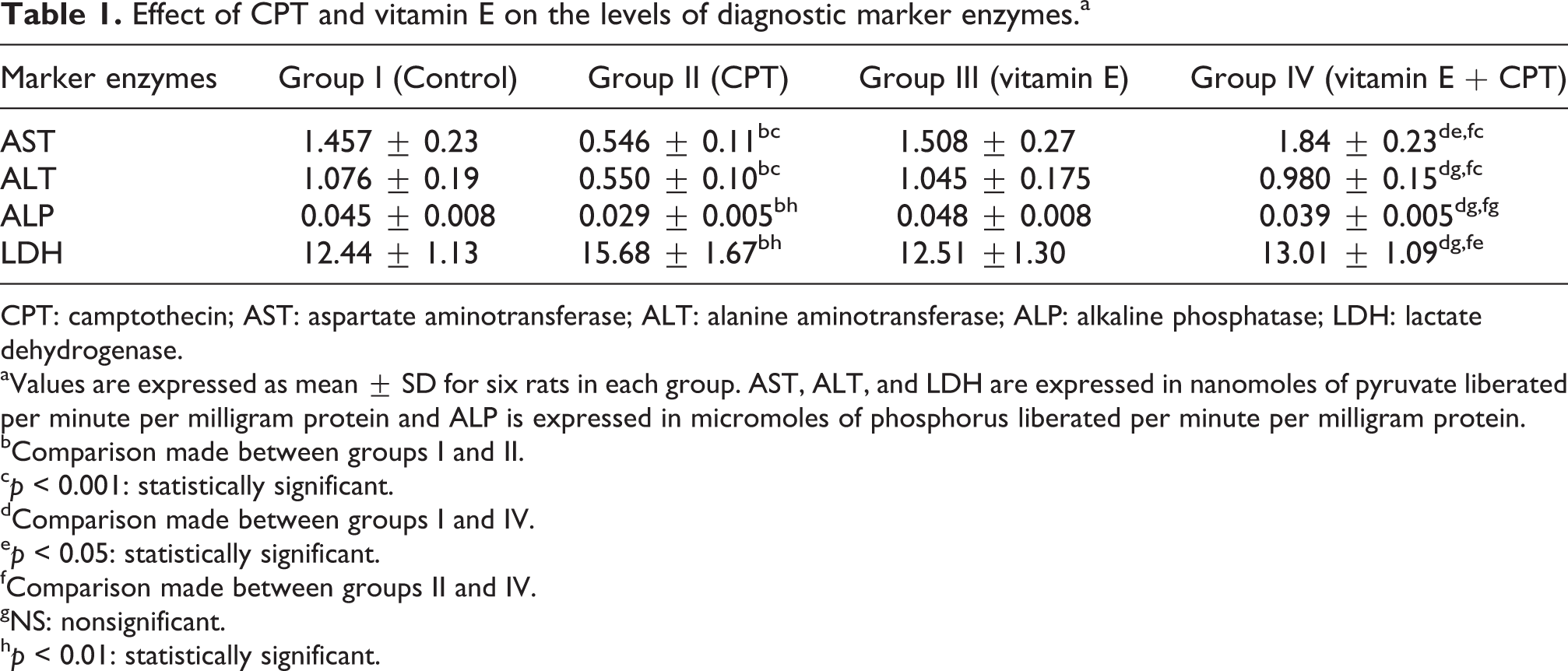
CPT: camptothecin; AST: aspartate aminotransferase; ALT: alanine aminotransferase; ALP: alkaline phosphatase; LDH: lactate dehydrogenase.
aValues are expressed as mean ± SD for six rats in each group. AST, ALT, and LDH are expressed in nanomoles of pyruvate liberated per minute per milligram protein and ALP is expressed in micromoles of phosphorus liberated per minute per milligram protein.
bComparison made between groups I and II.
c p < 0.001: statistically significant.
dComparison made between groups I and IV.
e p < 0.05: statistically significant.
fComparison made between groups II and IV.
gNS: nonsignificant.
h p < 0.01: statistically significant.
Effect of CPT and vitamin E on the activities of antioxidant enzymes.a
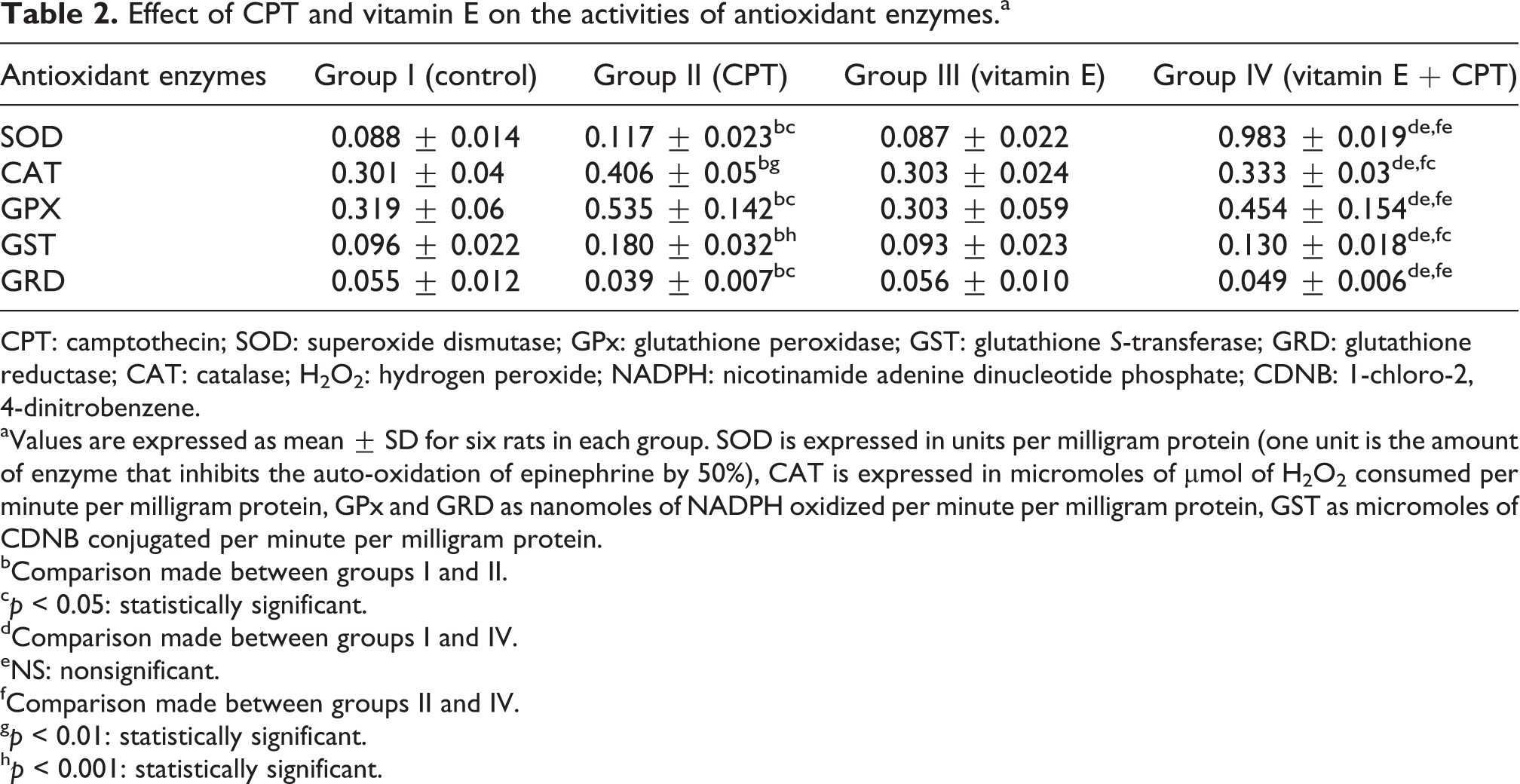
CPT: camptothecin; SOD: superoxide dismutase; GPx: glutathione peroxidase; GST: glutathione S-transferase; GRD: glutathione reductase; CAT: catalase; H2O2: hydrogen peroxide; NADPH: nicotinamide adenine dinucleotide phosphate; CDNB: 1-chloro-2,4-dinitrobenzene.
aValues are expressed as mean ± SD for six rats in each group. SOD is expressed in units per milligram protein (one unit is the amount of enzyme that inhibits the auto-oxidation of epinephrine by 50%), CAT is expressed in micromoles of μmol of H2O2 consumed per minute per milligram protein, GPx and GRD as nanomoles of NADPH oxidized per minute per milligram protein, GST as micromoles of CDNB conjugated per minute per milligram protein.
bComparison made between groups I and II.
c p < 0.05: statistically significant.
dComparison made between groups I and IV.
eNS: nonsignificant.
fComparison made between groups II and IV.
g p < 0.01: statistically significant.
h p < 0.001: statistically significant.
Discussion
During chemotherapy, oxygen radicals are generated which catalyze the oxidative modification of lipids. The presence of double bond adjacent to a methylene group makes the methylene C–H bonds of polyunsaturated fatty acid weaker, and therefore, hydrogen becomes more prone to abstraction. Accumulation of lipid peroxides, alkoxy and peroxy radicals result in initiation of lipid peroxidation and introduction of hydrophilic moiety in hydrophobic phase. This is a self-perpetuating process since peroxy radicals are both reaction initiators as well as the products of lipid peroxidation. 21 The present data revealed that CPT administration produced a marked oxidative impact as evidenced by the significant increase (p < 0.001) in lipid peroxides that might result from increased production of free radicals. Use of antioxidants can interfere in the process of lipid peroxidation by scavenging free radical species which is comparable to our result of decreased levels of lipid peroxides in the group pretreated with tocopherol in the presence of CPT (Figure 1).
ROS can attack vital cell components and can alter intrinsic membrane properties like fluidity, ion transport, loss of enzyme activity, protein cross-linking, inhibition of protein synthesis, DNA damage, ultimately resulting in cell death. 22 Serum aminotransferase activities have long been considered to be sensitive indicators of tissue injury. 23 ROS-mediated injury to the cell alters their membrane permeability leading to leakage of enzymes from the cells. 24 Therefore, the marked release of AST and ALT into the circulation and diminished levels of enzymes in tissue, as per our report, indicates severe damage to tissue membranes and loss of functional integrity during drug intoxication. ALP is a hydrolase enzyme responsible for removing phosphate groups from many types of molecules including nucleotide, protein, and alkaloids. 25 The observed significant decrease in ALP levels of CPT-treated animals may result from the increased need of energy through glycolytic and oxidative pathways of glucose-6-phosphate, rather than ALP activity. 26
LDH is an essential enzyme involved in anerobic glycolysis and is responsible for the anerobic transformation of pyruvate to lactate. Increased expression of LDH under hypoxic conditions has been demonstrated in various cell lines. 27 –29 Increased LDH production in cardiomyocytes of CPT-treated rats could also be attributable to anerobic conditions. The cardiocytes are expected to increase the production of LDH under anerobic conditions, until they become necrotic. Animals that received vitamin E prior to CPT treatment (group IV) showed significant increase in the levels of transaminases and phosphatase and decrease in LDH when compared with enzyme levels of CPT-treated rats (group II). The reversal of decreased levels of enzymes by vitamin E may be due to prevention of the leakage of intracellular enzymes by its membrane-stabilizing activity. This is in agreement with the commonly accepted view that changed levels of transaminases return to normal with the healing and regeneration of cardiomyocytes. These results are consistent with our previous reports of CPT-induced hepatotoxicity. 30 –32
Under physiological conditions, the damaging effects of ROS are prevented by endogenous antioxidant enzymes. The present work shows changes in anti-peroxidative enzyme profile. CAT, SOD, GPx, and GST are class of antioxidant enzymes responsible for scavenging H2O2 and organic hydroperoxides. These enzymes together are called cellular antioxidant defense system. The damaging effect of superoxide radicals are prevented by SOD enzyme that catalyzes the dismutation of two superoxide radicals producing H2O2 and oxygen. Further, antioxidative factors like CAT and GPx have a predominant role in controlling the concentration of H2O2 and in elimination of ROS. CAT along with GPx, converts H2O2 generated after dismutation of superoxide radicals by SOD to water. 33,34 Amidst of these enzymes, GST catalyzes the conjugation of GSH to electrophilic centers on a wide variety of substrates. These activities detoxify endogenous compounds such as lipid peroxides and bring about breakdown of xenobiotics. 35 Enzymes like GST and GPx are GSH-dependent enzymes; therefore, in the process of radical detoxification they generate GSSG. GRD, an enzyme crucial for regeneration of GSH, ensures its availability to GSH-dependent enzymes for their proper functioning. 36 Thus, these enzymes are interdependent and perform their function in harmony. The present investigation shows significant increase in GPx, CAT, GST, and SOD when compared with the control group (Table 2). Elevated levels of peroxides cause increased rate of lipid peroxidation, 37 which justifies inducible synthesis of these antioxidant enzymes 38 as an adaptive mechanism to detoxify the reactive species in order to curtail tissue damage. The activity of GRD was significantly less in CPT-treated groups than that of control (Table 2). This enzyme contains sulfydryl group residues that are essential for catalytic activity and vulnerable to free radical-mediated inactivation. 36 The reduced GRD activity may be justified by the fact that under high oxidative stress GSSG can be effluxed out of the cell. 39 In our study, pretreatment with an antioxidant significantly reversed the altered antioxidant enzymes status when compared with only CPT-treated rats to near about control range. Similar to our findings, Selvakumar et al. have reported that use of lipoic acid can ameliorate tissue peroxidative damage and abnormal antioxidant levels in drug-induced toxicity. 40

The tripeptide thiol GSH has facile electron-donating capacity, linked to its sulfhydryl (–SH) group. It is an important water-phase nonenzymic antioxidant and essential cofactor for antioxidant enzymes which provides protection against intracellular oxygen radicals. 41 GSH serves both as a nucleophile and as an effective reductant by interacting with many oxidizing and electrophilic compounds. In the present study, level of GSH was decreased significantly in myocardial tissue of CPT-administered rats. But the level of GSH in vitamin E-administered rats was restored to near normal (Figure 3). Depletion of GSH content may be attributed to the direct conjugation of drug and its metabolites with free or protein-bound SH groups, 42 thereby interfering with the antioxidant functions. Moreover, when GSH is utilized by free radical detoxifying enzymes, it causes increase in GSSG content of the cell. 43 In order to maintain the redox potential, the GSSG formed should be converted to GSH by GRD. Decrease in GRD levels can be the reason for decrease in the GSH found in the present report.
Vitamin E supplementation before CPT injections significantly protected these alterations in enzymic and nonenzymic antioxidant defense system. The decrease in vitamin E clearly indicates overproduction of ROS and other free radicals in CPT-treated animals, whereas the levels of vitamin E in groups III and IV increased more than control due to vitamin E treatment for 30 days (Figure 2). When compared, group IV illustrates decreased amount of vitamin E than group III that signify the involvement of vitamin E in detoxification of ROS and free radical intermediates. The cytosolic defenses are undoubtedly important in preventing free radical-mediated damage to cellular and subcellular target molecules. α-Tocopherol administration appears to maintain α-tocopherol reserve in cell and cellular antioxidant status of cell. 44
Light microscopy observation reveals that histology of control and vitamin E control rats showed normal architecture thereby confirming that vitamin E supplementation does not per se have any adverse effects. Heart tissue of control rats showed normal myocardial fiber and myocytes with no evidence of cellular injury and vacuolated cells (Plate A and C). In contrast, groups treated with CPT at doses of 6 mg/kg showed cardiotoxicity. The most pronounced cardio-histopathological abnormalities observed in rats treated with CPT involved degenerative changes like congestion, necrosis, fragmentation of fibers, and hemorrhages (Plate B). Our histopathological studies along with biochemical findings confirm the possibility of membrane damage and cellular leakage caused by CPT. The tissue histology of animals treated with vitamin E before CPT showed small amount of pathological alterations when compared with the CPT alone-treated rats (Plate D). Vitamin E did not completely ameliorate the tissue damage but certainly reduced the adverse effects of the drug and maintained structural integrity to some extent. This is possibly by decreasing the upsurge in oxidative stress after drug treatment that resulted in disturbance to normal architecture of tissue.
Vitamin E is a fat-soluble compound that includes both tocopherols and tocotrienols. The most potent and useful form of vitamin E is α-tocopherol. It is an effective antioxidant that is known to stop the production of ROS. 6 It also protects cell membranes from oxidation by reacting with lipid radicals produced in the lipid peroxidation chain reaction. 45 This would remove the free radical intermediates and prevent the oxidation reaction from continuing. The present study thus validates vitamin E as a potentially useful candidate against CPT-induced tissue injury and oxidative stress during chemotherapy. Therefore, we propose that vitamin E should be added to the list of pharmacological preconditioning agents as it introduces a novel and protective strategy in drug-mediated cardiotoxicity.
Footnotes
Acknowledgment
The authors gratefully acknowledge Dr A Ingle, Scientist, Advanced Centre for Treatment, Research and Education in Cancer (ACTREC), Tata Memorial Centre, Mumbai, Maharashtra, India, for his help in histopathological studies.
Conflict of interest
The authors declared no conflicts of interest.
Funding
This research received no specific grant from any funding agency in the public, commercial, or not-for-profit sectors.