
Abstract
In carbon monoxide (CO) poisoning, CO affects the oxygen-carrying capacity of the hemoglobin molecule. Nucleolar-organizing regions (NORs) are genetic loci on chromosomes that are composed of ribosomal DNA and proteins. NORs can be stained with silver. A total of 18 rats were exposed to CO in three different concentrations (1000, 3000, and 5000 ppm) with 6 rats as controls. The animals were euthanized 7 days after CO intoxication. Lung tissues were taken, embedded in paraffin blocks, and sectioned at 5 μm thickness. Argyrophilic nucleolar-organizing region (AgNOR) staining was carried out. One hundred nuclei per individual were evaluated, and total AgNOR number per total nuclear number and total AgNOR area per nuclear area (TAA/NA) for each nucleus were analyzed. The CO exposure groups had significantly higher TAA/NA values and AgNOR numbers than the control group (p < 0.05). Although the differences between 1000 ppm and the other two CO-exposed groups were meaningful (p < 0.05) in the TAA/NA values, there were no differences among the CO exposure groups for the AgNOR number (p > 0.05). The increase in TAA/NA value depends on the increase in the CO exposure. Significant correlations between both the AgNOR values and histopathological scoring methods were found. Therefore, AgNOR staining method may be used as an indirect indicator for evaluating the degree of cell damage rate.
Introduction
Carbon monoxide (CO) is a colorless, odorless, tasteless, and nonirritating gas. CO is released from fuels that are not lit highly enough and can cause acute and chronic poisoning deaths. 1,2 CO gas is heavier than room air and can accumulate quickly, even in well-ventilated indoor environments. The most common sources of CO include gas exhaust from motor vehicles, fumes from fires, gas-powered engines, forest fires, and paints containing methylene chloride. 3,4 CO poisoning is known to be the cause of more than half of all fatal poisonings in many countries worldwide. 5
In CO poisoning, inhaled CO gas quickly passes through alveolar capillary membranes and enters the intravascular space, which is the primary location for connecting to the hemoglobin (Hb). Most CO becomes stable after accumulating in the erythrocytes, and the affinity of Hb for CO is 200–250 times higher than for oxygen (O2). Shifting the dissociation curve of oxyhemoglobin to the left explains the decrease in arterial O2 transport and the hypoxic symptoms of CO poisoning. The toxic effects of CO cannot be explained with this mechanism alone. Other mechanisms are thought to be involved. 6,7 CO intoxication creates structural changes in the Hb (by entering into the erythrocyte, CO inhibits heme proteins such as myoglobins), which makes it difficult to transfer O2 into the tissues. CO also impairs cellular respiration by inactivating reduced cytocrome-a3. 5,8 For these reasons, CO affects the O2-carrying capacity of the Hb molecule by binding to Hb.
CO poisoning causes a variety of symptoms in different organs of the human body due to hypoxia. 9 The symptoms caused by CO poisoning can occur in the early period, or they can be seen weeks after exposure. In addition to the main symptoms that affect the neuropsychiatric and cardiovascular systems, other systems and organs are also affected. One of the affected systems is the respiratory system, where pulmonary edema, pneumonia, hemorrhage, and unilateral diaphragmatic paralysis may occur. 10
The nucleoli are prominent structures within cell nuclei. In the nucleolus, ribosomal RNAs are synthesized, processed, and assembled with ribosomal proteins. The size and organization of the nucleolus are directly related to ribosome production and reflect the functional compartmentalization of the nucleolar machinery. When this activity is blocked or disrupted, the nucleolar proteins can interact independently from the processing activity. Nucleolar-organizing regions (NORs) are genetic loci on chromosomes that are composed of ribosomal DNA (rDNA) and proteins, some of which are argyrophilic. NORs are loops of DNA, tandemly repeated with intergenic spacers and transcribed into ribosomal RNA, which is processed into preribosomes in the nucleoli, eventually becoming part of mature ribosomes in the cytoplasm. 11
In humans, the NORs (sites of the ribosomal genes) are located on the secondary constrictions of the five pairs of acrocentric chromosomes 13, 14, 15, 21, and 22 and can be stained with silver when they are active. Silver binds with those rDNA sites that are transcriptionally active or have already been transcribed and still retain residual rRNA nonhistone-associated proteins. Therefore, argyrophilic NOR (AgNOR)-associated proteins are one of the most reliable methods used to visualize nucleoli in interphase nuclei. 12
Numerous studies have been performed concerning the importance of the interphase quantity of AgNOR in tumor pathology, 13,14 hair root cells of humans, 15,16 developmental stages of Down syndrome in infants, 17 and so on. However, to the best of our knowledge, there are no studies in the literature about the relationship between AgNOR proteins and CO exposure in lung cells. Therefore, we carried out the current study in order to detect any possible effects of CO exposure on the NOR protein synthesis of lung cells.
In this study, we aimed to show hypoxia-induced cell proliferation, due to CO intoxication, using the AgNOR method, and investigate any possible relationship between the AgNOR staining method and histopathological evaluation as prognostic factors.
Materials and methods
In our study, 24 male albino Wistar rats, weighing 150–180 g, were used. The experimental protocols were in compliance with the Atatürk University Ethics Committee on Research in Animals as well as the internationally accepted principles for laboratory animal use and care. Three steel tubes, with each of 10 L in volume, had full of a CO–air mixture at three concentrations (1000, 3000, and 5000 ppm) were provided by Habaş Industrial and Medical Gases Production Industries Inc., Kocaeli, Turkey. CO exposure was applied in an enclosed transparent jar with a dimension of 20 cm × 40 cm × 60 cm. There were inlet and outlet openings with 2 cm diameter at opposite sides of the jar.
The rats were randomly divided into four groups: control group, 1000 ppm, 3000 ppm, and 5000 ppm groups, consisting of six animals each. The experimental groups were exposed to CO one time, each group at a different density (1000, 3000, and 5000 ppm) with a flow rate of 4 L/min for 30 min in the transparent jar. After intoxication, the rats were housed in standard plastic cages on sawdust bedding under a 12-h light/12-h dark cycle in an air-conditioned room at 20°C and provided food and water ad libitum. One week after CO exposure, the animals were anesthetized intraperitoneally with urethane at a dosage of 1.25 g/kg and euthanized by intracardiac perfusion. Fixation in a 10% formaldehyde solution was achieved. These experimental procedures were performed in the Atatürk University Pharmacology Laboratory.
The lung tissue samples (approximately 1 × 1 × 1 cm3 in size) were obtained from the front-bottom edge of the animals’ lower lobes of their left lungs. After routine histological follow-ups, the pieces of lung tissue in the paraffin blocks were cut to 5 μm thick. Before AgNOR staining, the tissue sections were deparaffinized in xylene and then rehydrated in graded alcohol solutions. The slides were air-dried at room temperature for 15 min before being fixed in absolute methanol for 5 min. AgNOR staining was carried out according to the Benn and Perle protocol and Lindner protocol, with a slight modification for all groups. 18,19
Slides containing stained tissue sections were viewed using a light microscope (Eclipse 80i; Nikon, Tokyo, Japan) and photographed using a digital camera (Digital Sight DS-fi1; Nikon). The captured images were transferred to image processing software (ImageJ ver. 1.47t, National Institutes of Health, Bethesda, Maryland, USA), and the measurements were obtained using the “freehand selections” tool. One hundred nuclei per animal were assessed, and the mean AgNOR number and total AgNOR area per nuclear area (TAA/NA) for each nucleus were estimated.
For the histopathological examination, another series of sections were obtained and stained with hematoxylin and eosin (H&E). The sections were evaluated for the degrees of alveolar hemorrhage, vascular congestion, inflammation, and edema. Each lung slide was examined, and the severity of observed changes was scored using a scale of none (0), mild (1), moderate (2), and severe (3) damage. Histopathological analyses were made by an experienced pathologist. Statistical evaluations (Kruskal–Wallis/Dunn and one-way analysis of variance) were performed using NCSS software (Ver. 9; NCSS, LCC, Kaysville, Utah, USA).
Results
The mean AgNOR number and TAA/NA values of the rats from the control and CO-exposed groups and statistical values are given in Table 1. CO-exposed groups significant at p = 0.027 had higher mean AgNOR numbers (1.643 for 1000 ppm, 1.603 for 3000 ppm, and 1.628 for 5000 ppm groups) and TAA/NA (0.052 for 1000 ppm, 0.081 for 3000 ppm, and 0.080 for 5000 ppm groups) than the control group (1.237 and 0.0255, respectively, p = 0.0007; Figure 1).
TAA/NA and mean AgNOR number values of groups.

TAA: total argyrophilic nucleolar organizing region area; NA: nuclear area; AgNOR: argyrophilic nucleolar organizing region.
aIf the median values of the groups have the same letter, it said that they are not significantly different.

Box plots showing TAA/NA and mean AgNOR number values of groups. TAA: total argyrophilic nucleolar-organizing region area; NA: nuclear area.
While the differences between the 3000 ppm group and 5000 ppm group were not meaningful, the differences were statistically significant among the other groups for the TAA/NA value. Additionally, for the mean AgNOR numbers, there were no statistically significant differences among the CO-exposed groups (p > 0.05; Table 1). Figure 2 shows the demonstrative examples of silver-stained NORs in the lung cells of CO-exposed groups.

Silver-stained NORs (arrows) in lung cells: (A) control group, (B) 1000 ppm group, (C) 3000 ppm group, and (D) 5000 ppm group (×1000 magnification). NOR: nucleolar-organizing region.
According to the histopathological scoring method, the differences between the control and CO-exposed groups were statistically significant for congestion, inflammation, hemorrhage, and edema (p < 0.001; Table 2). Figure 3 shows the demonstrative examples of H&E-stained lung tissue images, which were used for histopathological scoring.
The scores of histopathological evaluations.

aComparison of control group versus other groups; the differences were statistically significant in congestion and inflammation (p < 0.001) and hemorrhage and edema (p < 0.0001), respectively.
bComparison of 5000 ppm group versus 1000 ppm group and 3000 ppm group; the differences were not statistically significant in congestion, inflammation, and hemorrhage (p > 0.05); only difference was meaningful in edema (p < 0.001).
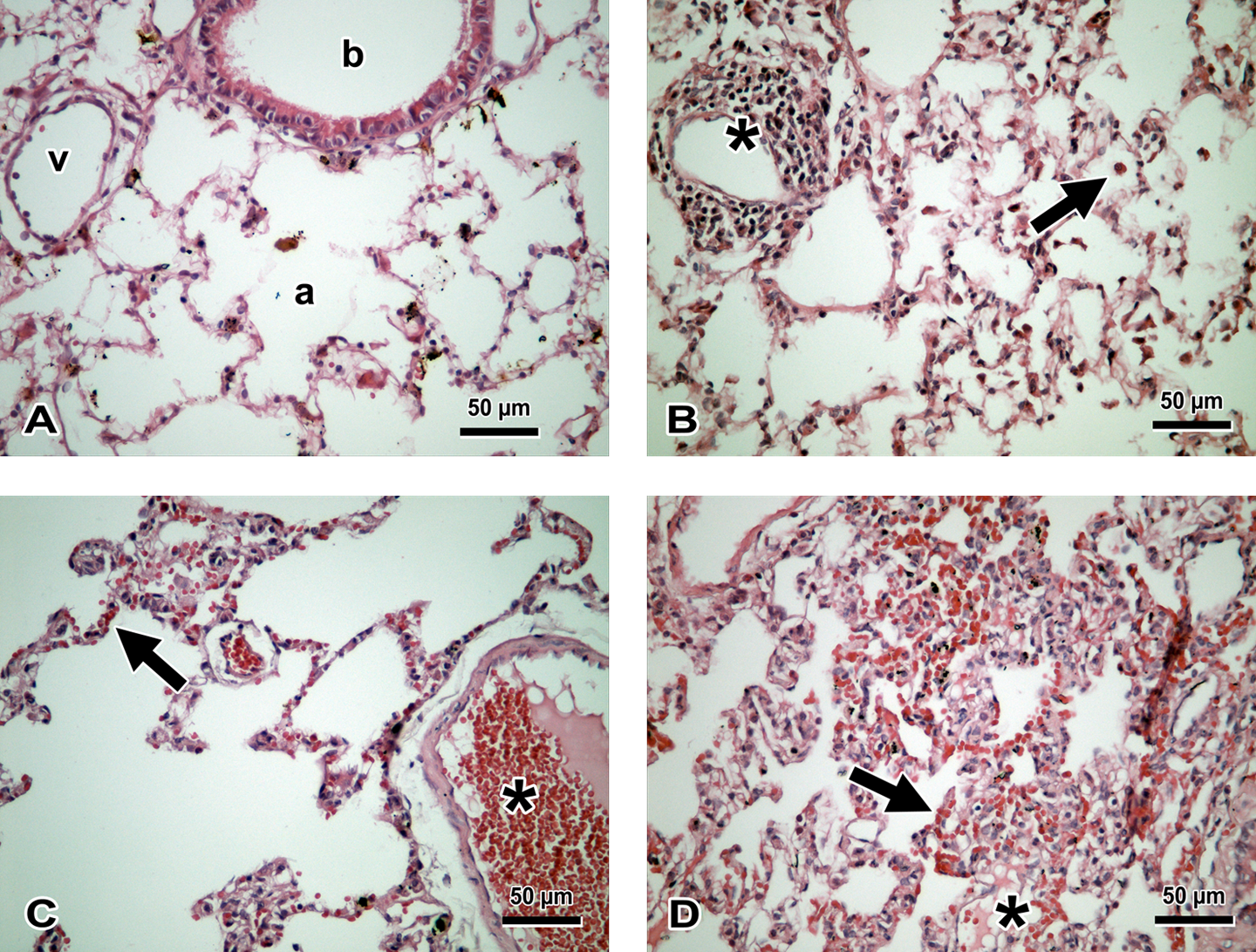
H&E-stained lung tissue. (A) control group: a: alveoli, b: bronchiole, v: vascular structure; (B) 1000 ppm group: macrophage in alveoli (arrow); *lymphoid aggregate, mild inflammation; (C) 3000 ppm group: moderate alveolar hemorrhage (arrow); *vascular congestion; and (D) 5000 ppm group, severe alveolar hemorrhage (arrow); *edema. H&E: ×400. H&E: hematoxylin and eosin.
When the correlations between the TAA/NA and histopathological scoring methods were evaluated, a significant relationship was found (p < 0.001; R 2 = 68.9%). Additionally, the correlation was statistically meaningful for the mean AgNOR number (p = 0.015; R 2 = 22.8%; Figure 4).
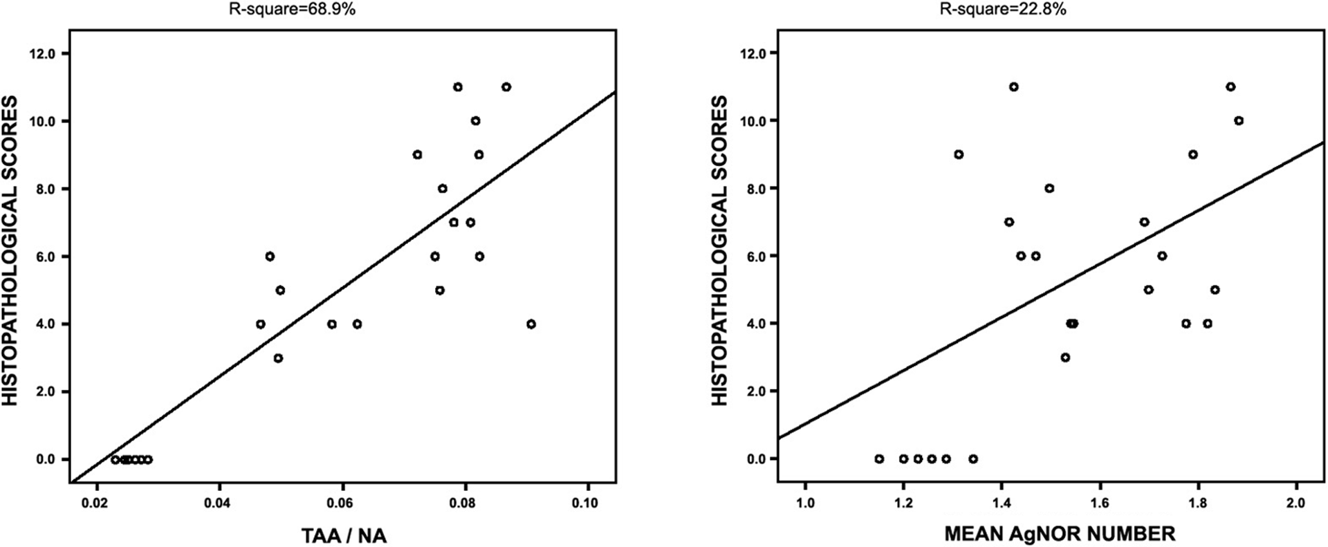
Correlations between histopathologic scoring and AgNOR values. AgNOR: argyrophilic nucleolar-organizing region.
Discussion
The physiological amounts of endogenous CO act as neurotransmitters. Low levels of CO appropriately influence direct inflammation, cell proliferation and apoptosis, and increase mitochondrial biogenesis. 5 With increased exposure to CO, poisoning signs begin to emerge in the organs. 20 CO exposure affects the O2-carrying capacity of Hb in two ways. First, CO competitively inhibits the binding of O2 to Hb, and it prevents the transport and release of O2 to the tissues. CO leading to relative anemia is caused by tissue hypoxia or asphyxia. 21,22 Second, by making structural changes in the Hb, CO prevents releasing O2 to the tissues, thus impairing cellular respiration. 8 Lung injury after CO poisoning is rare; however, when it exists, it has been thought that it is not caused by the histotoxicity of CO, but probably because of the hypoxia resulting from impaired O2 transfer. 10
In CO poisoning, prolonged hypoxia can affect capillary permeability and may cause pulmonary edema. 23,24 Pulmonary edema, pneumonia, and adult respiratory distress syndrome may occur after CO poisoning. 25,10 Pulmonary lesions are cardiogenic damage, which may develop due to CO poisoning. Additionally, pulmonary edema can be seen as cardiogenic damage. 26 –28
It was difficult to point out the damage to the lung tissue after CO poisoning. To the best of our knowledge, the literature contains no reports that showed the relationship between AgNOR protein synthesis and lung damage caused by CO intoxication. It is known that the low levels of CO direct cell proliferation, and the AgNOR staining method can be used as a proliferation marker. 5,13,14,29
The nucleolus is connected with the regulation of major physiological cellular processes, including mitosis, ribosome assembly, stress response, and the generation of ribonucleoprotein complexes. 30 More than 700 nucleolar proteins, most of which seem to be involved in the processing and maturation of ribosomal RNAs, and the generation of mature ribosomes, have been described in humans. 30 –32 NORs are functional subunits of the nucleolus in which actively transcribed rDNA is surrounded by a large number of regulatory proteins in interphase. 33
The number, shape, and distribution of AgNOR reactive sites counted in the cells provide important knowledge about the behavior of those cells. In tumor cells, the relationship between cell proliferation and AgNOR amounts has been investigated widely by comparing AgNOR values with kinetic data obtained by applying a panel of proliferation markers. 13,14,34
We have performed several studies using the AgNOR method, 13 –17,29 which supply an index of cellular activity in connection with proliferation, differentiation, and secretory activities. Our study showed that rRNA gene activity, as determined by total AgNOR number per total nuclear number or TAA/NA, increased depending on the increase of CO exposure in the lung cells. In lung cells, AgNOR protein synthesis tends to increase during CO exposure, and all cells tend to protect their situation toward dangerous agents. Perhaps these proteins occur against to CO exposure or trigger the synthesis of some other proteins that have protective roles in the lung cells. Both the AgNOR values may be used as indirect indicators for evaluating the degree of cell damage (occurring hypoxia) rate. Additional studies should be conducted to obtain more certain knowledge about this topic. In this way, new therapeutic approaches may be developed in the treatment of CO intoxication in the future. This technique may also be used to detect the degree of CO exposure in forensic medicine. CO intoxication can be diagnosed early using the carboxyhemoglobin (COHb) level. However, most of the time (the half-life of room air is 4–6 h) COHb levels may not help in the diagnosis and direction of the treatment. Our method may be used when COHb levels seem normal in the intoxication doubt autopsies and therefore can be adapted for use in forensic science. Additionally, the current technique may provide indirect information about the degree of hypoxia and asphyxia in hypoxic and asphyxic babies. If these proteins play a protective role toward hypoxia, they may be helpful in developing new treatment approaches for hypoxic or asphyxic situations in the future.
A significant relationship was found between the results of the histopathological methods and both the TAA/NA and mean AgNOR number. The results of the histopathological examination can be obtained with the evaluation of alveolar hemorrhage, vascular congestion, inflammation, and edema. Therefore, it may be said that only AgNOR protein synthesis may be used to obtain knowledge about the degree of damage to the cells, instead of the histopathological scoring method requiring an experienced pathologist.
The limitations of this study include the relatively small number of groups. In our technique, both the NOR area and NA were used for the calculation of the TAA/NA proportion. Thus, more reliable knowledge about the proliferation, differentiation, and secretory activity of each cell could be obtained. Thus, indirect knowledge may be obtained about the behavior of the lung cells for protection from CO exposure. Additionally, advantages of this technique are simple, reliable, cheap, and a valuable marker to evaluate the ribosomal gene activity in different metabolic states of cells.
Conclusion
The current technique may be used to obtain knowledge about the identification of cellular changes induced by CO in the lung. Further studies with a higher number of groups, including other CO levels and different candidate genes, should be conducted in order to obtain more certain knowledge about this topic.
Footnotes
Funding
This research received no specific grant from any funding agency in the public, commercial, or not-for-profit sectors.