
Abstract
Phthalates are diester derivatives of phthalic acid widely used in many commercial applications. The aim of this study is therefore to evaluate possible genotoxicity of di-n-hexyl phthalate (DHP) and dicyclohexyl phthalate (DCHP) at different concentrations using single-cell gel electrophoresis (comet) and terminal deoxynucleotidyl transferase deoxyuridine triphosphate nick end-labeling (TUNEL) assays in testes samples of male rat pups. DCHP and DHP in corn oil were administered to the pregnant rats by gavage at the doses of 0 (vehicle), 20, 100, and 500 mg kg−1 day−1 from gestational day 6 (GD6) to GD19. After delivery, male rats were allowed to grow until prepubertal, pubertal, and adulthood. At necropsy, the blood samples were collected from heart and were excised immediately. The apoptotic cells of prepubertal, pubertal, and adult testis were detected using TUNEL assay. The comet assay was performed on blood lymphocytes and testes samples of adult male rats. The comet assay results showed that tail length, tail intensity, olive tail moment (OTM), and percentage of DNA present in tail were higher when DHP content was increased. Judging from the values of OTM and percentage of DNA, DHP could significantly induce DNA breakage at doses of 100 and 500 mg kg−1 day−1 compared with the control group. An increase in TUNEL-positive cells of prepubertal, pubertal, and adult testicular cells was observed in the treated groups. In conclusion, prenatal exposure to DHP and DCHP may possess genotoxic risk to testicular cells of rats at all stages of development, even at adulthood.
Introduction
Phthalates are diester derivatives of phthalic acid used primarily as plasticizers in commercial products, such as plastic food wraps, children’s toys, blood transfusion and dialysis bags, and catheters, to make the plastic products more flexible. 1,2 However, phthalates are not covalently bound to the plastic material and are released into the environment. Phthalates are frequently detected in outdoor and indoor air. 3 The possibility of these compounds entering into biologic systems has caused great concern among the public about their reproductive and developmental toxicity. 4 Recent studies have showed that exposure to some phthalates results in serious and irreversible changes in the development of reproductive tract, especially in males. 5,6 In particular, prenatal exposure to these environmental chemicals, by interfering with androgen signaling pathway seems to cause permanent adverse effects on reproductive development in male rats. 7 Anderson et al. 8 showed that some phthalate esters could produce DNA damage in human blood cells in the single-cell gel electrophoresis (comet) assay. Hauser et al. 9 showed that men from an infertility clinic population with increased sperm DNA damage were related to urinary levels of phthalate metabolites.
Dicyclohexyl phthalate (DCHP) is a common plasticizer ingredient for the production of nitrocellulose, ethyl cellulose, vinyl acetate, polyvinyl chloride (PVC), and resins. 10 Di-n-hexyl phthalate (DHP) is often found as a minor component (less than 1%) of C6–C10 phthalate mixtures; it may also be an isomer in mixtures of diisohexyl phthalates (DIHP) at levels of 25% or lower. 11 The Centers for Disease Control and Prevention collects urinary metabolite data for the general U.S. population, primarily through the National Health and Nutrition Examination Survey (NHANES), where metabolites of phthalates have been measured. 12 Reported urinary concentrations for DCHP metabolites have ranged from 0.400 µg L−1 creatinine (90th percentile) in NHANES 2001–2002 to less than the level of detection (50th percentile) for the total population. The specific DCHP-associated exposure pathway was not reported. Migration of DCHP from PVC into potato snacks (DCHP 0.33% of film by weight and 5 days of exposure) was 6.2 mg kg−1 food. Nitrocellulose-coated regenerated cellulose film leached 0.5–53 mg DCHP kg−1 into confectionary, meat pies, cakes, and sandwiches. DCHP also leached from printing inks into food items contacting ink (6% of the total amount of plasticizer transferred). 13 Shen et al 14 reported that the plastic products for food use analyzed the contained DCHP at the range of nondetectable to 15 mg kg−1. DHP (isomer not specified) was below limit of detection (0.01 mg kg−1) in samples of household dust and textiles. A level of 0.03 mg kg−1 was detected in flooring tile. 11
DCHP has been shown to bind to human estrogenic receptor (ER) in vitro. 15,16 DCHP and DHP were found to show simultaneously both ERα-agonistic and ERβ- and androgen receptor-antagonistic activities. 16 –18 Okubo et al. 19 demonstrated that DCHP had the most potent estrogenic activity among phthalate esters examined, but its estrogenic potency was 1,700,000 times less than that of 17β-estradiol.
DHP and DCHP were shown to have adverse effects on male reproductive tract of rats. 20,21 On the other hand, the genotoxic potential of DCHP was not determined extensively, with only negative bacterial mutation assays available for assessment. 10 A preparation containing DCHP (15%) was not found to be genotoxic in bacterial DNA repair and mutation tests in Escherichia coli, both with and without exogenous metabolic activation. 22 DCHP also demonstrated negative for mutagenic potential in the Ames’ test using Salmonella, both with and without metabolic activation. 23 To our knowledge, no other in vitro or in vivo genotoxicity studies have been available.
DHP has found nongenotoxic in the Salmonella and two other bacterial assays and in a mouse micronucleus test. 23,24 On the other hand, a C6-C10-phthalate mixture was resulted in a nondose-related increase in mutations in the presence and absence of metabolic activation, but evaluated as negative in the BALB/c 3T3 cell transformation assay. 25 DIHP was also demonstrated inactive in the mouse micronucleus test. 24 There is no in vitro cytogenetic, mammalian mutation or in vivo genotoxicity studies conducted on DHP. 10
The aim of this study is therefore to evaluate possible genotoxicity of in utero DHP and DCHP treatments at different concentrations using the terminal deoxynucleotidyl transferase (Tdt) deoxyuridine triphosphate nick end-labeling (TUNEL) and comet assays in testicular samples of male rat pups.
Materials and methods
Chemicals
DHP (CAS No. 84-75-3) with 97% purity and DCHP (CAS No. 84-61-7) with 99% purity were supplied from Aldrich Chemistry (Ankara, Turkey), and dissolved in corn oil (vehicle). The other chemicals used in the comet assay were purchased from the following suppliers: normal melting agarose (NMA) and low melting agarose (LMA) from Boehringer Mannheim GmbH (Mannheim, Germany); sodium chloride (NaCl) and sodium hydroxide (NaOH) from Merck Chemicals (Darmstadt, Germany); dimethylsulfoxide (DMSO), hydrogen peroxide (H2O2), ethidium bromide (EtBr), Triton X-100 and phosphate-buffered saline (PBS) tablets from Sigma Chemical Co. (St Louis, Missouri, USA); ethylenediamine tetraacetic acid disodium salt dihydrate (Na2EDTA), N-lauroyl sarcosinate, and Tris from ICN Biochemicals (Aurora, Ohio, USA).
Animal treatments and experimental design
Pregnant time-mated female Wistar albino rats on gestational day 0 (GD0; the day when sperm was detected in the vaginal lavage) were obtained from the Experimental Animals Production Center, Hacettepe University, Ankara, Turkey. All rats were housed in polycarbonate cages with stainless steel covers, in a room maintained at 12-h light/12-h dark cycle with a temperature of 22 ± 2°C and a relative humidity of 50 ± 5%, and given standard rat diet (Korkutelim Feed Factory, Afyon, Turkey) and tap water ad libitum. The pregnant rats were distributed on a random basis into control (vehicle) and treatment groups (N = 10) and housed individually.
DCHP and DHP in corn oil were administered to the pregnant rats by gavage at the doses of 0 (vehicle), 20, 100, and 500 mg kg−1 day−1 from GD6 toGD19. The authors decided to choose 20, 100, and 500 mg kg−1 body weight (b.w.) day−1 DHP and DCHP doses, without exceeding the acute oral median lethal dose values of DHP and DCHP reported to be 29.6 and >40 g kg−1 b.w. in rats, respectively. 26 The low-dose level was chosen according to no observed adverse effect level reported by Hoshino et al. 21 The high-dose level was also chosen according to lowest-observed-adverse-effect level of 500 mg kg−1 b.w. day−1 DCHP reported by Lake et al. 27 The solutions were prepared daily according to the dams’ weights. The dosing volume was 0.25 ml in all groups. The rats in the vehicle control group received corn oil in equal amounts as in experimental groups. Maternal b.w., food consumption, and clinical signs of toxicity of animals were recorded daily. After delivery, all pups were allowed to grow with dam for 1 month. The prepubertal male rats were weighed and killed under ether anesthesia followed by decapitation at postnatal day 20 (PD20). After growing with dam and female pups for 1 month, other male pups were separated from them. Male rats were housed at four per cage and allowed free access for standard rat diet and tap water ad libitum. Each of the males was from a separate original litter. The rats were allowed to grow up until PD32 (pubertal) and PD90 (adult). At necropsy, the animals were weighed and killed under ether anesthesia followed by decapitation. The blood samples of adult male rats were collected from heart immediately and put into heparin-containing tubes. The testes were excised immediately and were quickly frozen in liquid nitrogen and all samples were kept at −80°C for further assays. All experimental procedures and animal use were confirmed by the Approval of Animal Ethics Committee, Hacettepe University, Ankara, Turkey.
The TUNEL assay
The apoptotic cells was detected by labeling the free DNA 3′-OH termini with single- or double-stranded DNA breaks by terminal TUNEL method. The ApopTag® Plus Peroxidase In Situ Apoptosis Detection Kit (Chemicon International, Inc, Temecula, California, USA) was used as described in the kit manual. Paraffin sections (5-µm thick) were deparaffined and hydrated in a coplin jar. The slides were pretreated with 20 μg mL−1 proteinase K for 15 min at room temperature. For inactivation of endogenous peroxidase, the tissues were treated by 3% H2O2 for 5 min and then applied equilibration buffer for at least 10s at room temperature. The sections were incubated with TdT enzyme at 37°C in a humidified chamber for 1 h and, subsequently, incubated with anti-digoxigenin conjugate at room temperature for 30 min. The reaction color was developed by diaminobenzidine tetrahydrochloride, and the sections were counterstained with methyl green. The apoptotic cells were counted by randomly selected five areas for each slide.
Comet assay
The basic alkaline technique of Singh et al. 28 was followed during the comet assay as further described by Collins et al. 29 The comet assay was performed on blood lymphocytes and testes samples of male pups of 3 months of age.
Cell preparation
Lymphocytes from a 200-μL heparinized whole blood were isolated by Ficoll–Hypaque density gradient centrifugation and washed in PBS. 30 Small piece of testes tissue was minced in cold buffer solution (Mg2+ and Ca2+ free Hank’s balanced salt solution, 20 mM EDTA, 10% v/v DMSO, pH 7.5). 31 Cell concentrations were adjusted to approximately 2 × 10⁵ mL−1 in the buffer. Aliquots of 5–10 µL of the cells were suspended in 75 µL of LMA for embedding on slides. Cells were checked for viability by trypan blue exclusion.
Slide preparation
The microscopic slides had been covered with 1% NMA at about 45°C in Ca2+—and Mg2+—free PBS before the experiment. This layer was used to promote the attachment of the second layer. For the second layer, around 10,000 cells mixed with 80 µL of 1% LMA (pH 7.4) were rapidly pipetted onto this slide, spread using a coverslip, and maintained on an ice-cold flat tray for 5 min to solidify. After removal of the coverslip, the slides were immersed in cold lysing solution (2.5 M NaCl, 100 mM Na2EDTA, 10 mM Tris, 1% sodium sarcosinate, pH 10) with 1% Triton X-100 and 10% DMSO added just before use for a minimum of 1 h at 4°C.
Electrophoresis
The slides were removed from the lysing solution, drained, and placed in horizontal gel electrophoresis tank side by side, avoiding spaces and with the agarose ends facing each other, nearest the anode. The tank was filled with fresh electrophoresis solution (1 mM Na2EDTA and 300 mM NaOH, pH 13) to a level approximately 0.25 cm above the slides. Before electrophoresis, the slides were left in the solution for 20 min at 4°C to allow the unwinding of the DNA and expression of alkali labile damage. Electrophoresis was conducted at a low temperature (4°C) for 20 min using 24 V and adjusting the current to 300 mA by raising or lowering the buffer level and using a compact power supply (Power Pack P 25 Biometra Analytic GmbH, Germany). All these steps were conducted under dim light to prevent the occurrence of additional damage. After electrophoresis, the slides were taken out of the tank and washed in distilled water. Tris buffer (0.4 M Tris, pH 7.5) was added dropwise and gently to neutralize the excess alkali and the slides were allowed to sit for 5 min. The neutralizing procedure was repeated three times.
Staining and slide scoring
To each slide, 30 µL of EtBr (20 µg mL−1) was added. For visualization of DNA damage, slides were examined at 400-fold magnification under a Leica DM RB fluorescence microscope (Leica, Wetzlar, Germany). Measurements of tail length, tail intensity, and tail moment of “comets” were made by a computer-based image analysis system “Comet Assay III” (Perceptive Instruments Ltd, Suffolk, England) for 100 randomly selected cells, that is, 50 cells from each of two replicate slides from each sample. The mean value of these parameters was calculated and used for the evaluation of DNA damage. The “Olive tail moment” (OTM), proportional to the percentage of DNA in the tail multiplied by the tail length, was used as the measure of DNA damage. 32
Statistics
Kolmogorov–Smirnov and Levene tests were used, respectively, to evaluate data for normality and homogeneity. Statistical analyses were performed using an SPSS 13.0 for Windows® (SPSS, Inc., Chicago, USA). To compare the genotoxic impacts by negative controls versus the tested substances (DHP and DCHP), two sample t tests were used. The values of p < 0.05 were considered statistically significant.
Results
Testis TUNEL assay
A dose-dependent increase in TUNEL-positive cells of prepubertal, pubertal, and adult testicular cells (Figure 1) was observed in the groups treated in utero with DHP and DCHP when compared with the control group. The groups administered with DHP and DCHP showed a significant increase in the TUNEL-positive apoptotic cells in the adult testis as compared to the control group, but not in a dose-dependent manner (Table 1).

Immunohistochemical demonstrations showing apoptotic cells and bodies having fragmented DNA as revealed from TUNEL assay in prepubertal, pubertal, and adult testis at ×200 magnification. Control testis showed the normal level of apoptosis. Rats treated with 500 mg kg−1 b.w. day−1 DHP and 500 mg kg−1 b.w. day−1 DCHP showing markedly increase in the number of TUNEL-positive apoptotic cells indicated by black arrow head. TUNEL: terminal deoxynucleotidyl transferase deoxyuridine triphosphate nick end-labeling; DHP: di-n-hexyl phthalate; DCHP: dicyclohexyl phthalate.
Apoptotic testicular cells per section on rat testis after in utero DHP and DCHP treatments.a
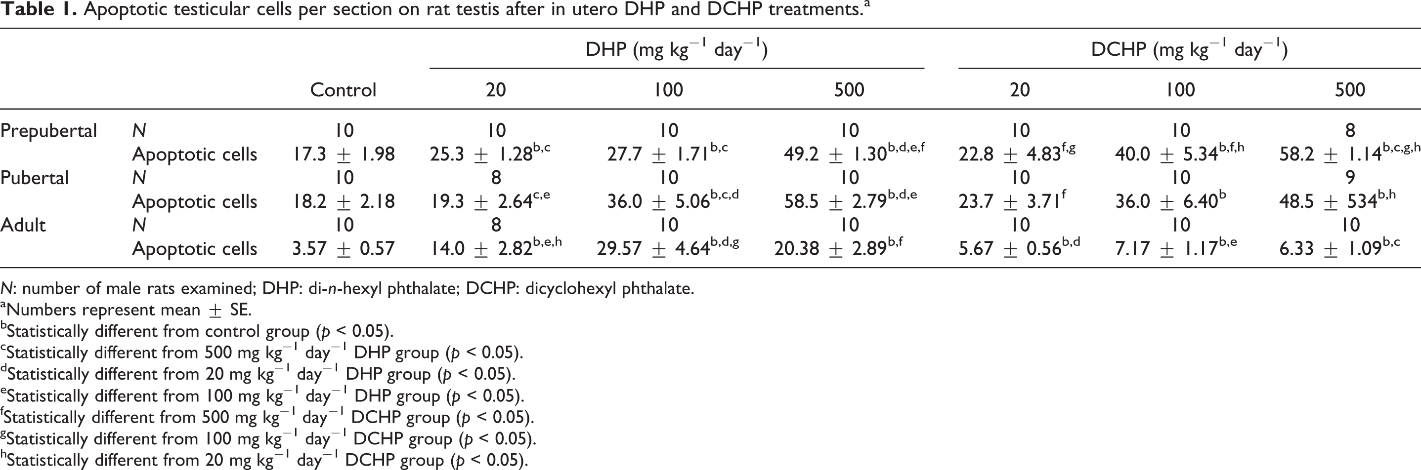
N: number of male rats examined; DHP: di-n-hexyl phthalate; DCHP: dicyclohexyl phthalate.
aNumbers represent mean ± SE.
bStatistically different from control group (p < 0.05).
cStatistically different from 500 mg kg−1 day−1 DHP group (p < 0.05).
dStatistically different from 20 mg kg−1 day−1 DHP group (p < 0.05).
eStatistically different from 100 mg kg−1 day−1 DHP group (p < 0.05).
fStatistically different from 500 mg kg−1 day−1 DCHP group (p < 0.05).
gStatistically different from 100 mg kg−1 day−1 DCHP group (p < 0.05).
hStatistically different from 20 mg kg−1 day−1 DCHP group (p < 0.05).
Comet assay
The cell viability, as tested using trypan blue dye exclusion of each treated group, was more than 80%. The amount of DNA breakage in a cell in comet assay may be estimated from the migration extent (tail length) of the genetic material in the direction of anode. 28 Furthermore, the percentage of DNA in the tail has been shown to be proportional to the frequency of DNA strand breaks. 33 OTM is a simple descriptor calculated by the computerized image analysis system considering both the migration comet length as well as the fraction of DNA migrated in the tail. 34
Table 2 presents the tail length, tail intensity, OTM, and percentage of DNA in blood and testicle samples whose mothers were administered with 20, 100, and 500 mg kg−1 day−1 DHP and DCHP from GD6 to GD19. Due to technical problems, possible genotoxicity of 100 and 500 mg kg−1 day−1 DHP could not be evaluated in blood lymphocytes.
Genotoxicity results of adult testicular and blood cells after in utero DHP and DCHP treatments.a

N: number of male rats examined; DHP: di-n-hexyl phthalate; DCHP: dicyclohexyl phthalate; NA: not analyzed.
aNumbers represent mean ± SE.
bStatistically different from 100 mg kg−1 day−1 DCHP group (p < 0.05).
cStatistically different from 500 mg kg−1 day−1 DCHP group (p < 0.05).
dStatistically different from control group (p < 0.05).
eStatistically different from 100 mg kg−1 day−1 DHP group (p < 0.05).
fStatistically different from 20 mg kg−1 day−1 DCHP group (p < 0.05).
gStatistically different from 500 mg kg−1 day−1 DHP group (p < 0.05).
hStatistically different from 20 mg kg−1 day−1 DHP group (p < 0.05).
The DNA breakage in the adult blood cells of 20 and 100 mg kg−1b.w. day−1 DCHP-treated rats increased as compared to the control values. In the testicular samples, the results indicated tail length, tail intensity, OTM, and percentage of DNA were higher when DHP content was increased. Judging from the OTM and percentage of DNA values, DHP could significantly induce DNA breakage at dose of 100 and 500 mg kg−1 day−1 compared with the control group.
Discussion
The present study is to evaluate possible genotoxic effects of prenatal DHP and DCHP exposure on testes samples at different concentrations using the comet and TUNEL assays in three different developmental stages of male rat pups. The adult blood lymphocytes of control, all DCHP, and 20 mg kg−1 b.w. day−1 DHP treatment groups were also analyzed with the comet assay.
Few published human studies have examined the effect of environmental chemicals, such as phthalates, on DNA integrity in sperm as measured by the comet assay. 9,35 The comet assay is sensitive to DNA damage and the instantaneous results may differ from whole organism response in time. Hauser et al. 9 reported that the urinary levels of phthalate metabolites were associated with modest percentage increases in study population medians of sperm DNA damage, ranging from 5% to almost 20%. In this study, the DNA damage of testicular cells was high in prenatal DHP-administrated groups. These results indicate that exposure to DHP at prenatal period leads to appear long-life effects on testicular cells. The results of high-dose DHP mostly doubled the results of control results. The effect of this chemical appeared at adulthood after in utero exposure. Recent studies indicate that DNA integrity of male gamete is one of the most discriminating parameters for fertility assessment. 36,37 The appropriate onset of gametogenesis and steroidogenesis is fundamental for the function of reproduction in the adult. Indeed, the development of germ cells, Sertoli cells, and Leydig cells during fetal life is essential for adult fertility. 38 Aberrant chromatin packing during spermatogenesis or spermiogenesis and apoptosis in the later stages of germ cell development before ejaculation result in DNA damage (strand breaks) in ejaculated sperm. 9 High levels of DNA damage contribute to an increase in the percentage of sperm DNA damage, which, in turn, may adversely affect fertility.
In this study, the TUNEL assay was performed to ascertain the mode of cell death. It is based upon the principle that TdT binds to the exposed 3-OH ends of the DNA fragments generated in response to apoptotic signals and catalyzes the addition of labeled deoxynucleotides. This assay can detect early-stage apoptosis in systems where chromatin condensation has begun and DNA strand breaks are fewer, even before the nucleus undergoes major morphological changes. 39 The DNA breaks detected in the comet assay could also stem from apoptotic cells, since the onset of apoptotic fragmentation might appear as comets. In utero exposure to DHP produced irreversible effects on reproductive development in the male offspring, similar to those reported from other developmentally toxic phthalates, like di-a-ethylhexyl phthalate (DEHP). 18 In our previous study, 40 the incidence of tubular atrophy, atrophic and damaged tubules, and germinal cell debris and apoptotic cells in testes of prepubertal, pubertal, and adult rats in all DHP and DCHP treatment groups increased as compared to the control group in histopathologic examination. In the present study, a dose-dependent increase in apoptotic cells of prepubertal and pubertal testis was observed in the groups treated in utero with DHP and DCHP as compared to the control group. The groups administered with DHP and DCHP showed a significant increase in the apoptotic cells in the adult testis as compared to the control group, but not in a dose-dependent manner.
Phthalates induce the appearance of multinucleated gonocytes in rodents. 38,41,42 In our previous study, the same treatment groups showed that the abnormal cell division that caused the formation of multinucleated gonocytes results from degeneration of progenitor cells induced by DHP and DCHP exposure. 40 In addition, we reported that there was an increase in abnormal sperm in DHP-exposed groups especially tail defects suggested that DHP affected this region of sperm seriously. In that study, 40 there was no difference in sperm head number between control and treatment groups. On the other hand, the increase in abnormal sperm percentages, sperm head, neck, and tail defect indicated that there was a decrease in the quality of the sperms. In the present study, apoptotic cells in the treatment groups increased in all treatment groups at all stages of development.
Recent epidemiological studies suggested that some phthalic acid esters, such as DEHP, increased the incidence of tumors in rodents. 43 It has been shown that di-n-butyl phthalate and di-isobutyl phthalate are genotoxic in human epithelial cells of the upper aerodigestive tract and mucosal cells and lymphocytes in in vitro studies using the alkaline comet assay (single-cell gel electrophoresis). 44,45 Also, the comet assay was used to detect DNA damage in human lymphocytes induced by in vitro exposure to DEHP and mono-ethylhexyl phthalate. On the other hand, there is no in vivo genotoxicity studies conducted on DHP and/or DCHP in literature. In this study, the DNA of lymphocytes of adult male pups is affected by prenatal exposure to low and middle dose of DCHP, not high-dose DCHP and low-dose DHP.
When comparing the DHP and DCHP treatment groups, the comet assay results of DHP treatment groups showed that DHP had more genotoxic effects on testicular cells than DCHP. According to TUNEL assay results, the genotoxicity of DHP seems to last more even during adulthood. On the other hand, the genotoxic effects of DCHP at pubertal stage were higher than DHP at this stage. One can be concluded that DCHP affects DNA of testicular cells more early and rapid than DHP. In reverse, DHP affects the DNA of testicular cells slowly but in a long-lasting manner.
In conclusion, prenatal exposure to DHP and DCHP may possess genotoxic risk to testicular cells of rats at all stages of development, even at adulthood. When compared the ability of DNA breakage of two phthalates, DHP is more effective than DCHP on testicular cells. There is a need for further studies exploring further mechanisms of genotoxic potential of phthalic esters in vivo.
Footnotes
Conflict of interest
The authors declared no conflicts of interest.
Funding
This research was partially supported by the Scientific and Technological Research Council of Turkey (TUBITAK; project number: 3106 105S073).