
Abstract
Background:
New cosmetic applications and products based on the effects of botulinum toxin (BTX) treatment have stimulated demand for this class of natural compounds. This demand generates the need for appropriate standardized protocols to test and compare the effectiveness of new BTX preparations.
Objectives:
Based on the previously described electrophysiological methods, we measured and compared the inhibitory effects of two BTX type A (BTX-A) preparations on neuromuscular transmission through split-body test.
Methods:
The effectiveness was evaluated in terms of the compound muscle action potential (CMAP) and conduction velocity after BTX-A injection. We used a split-body method to compare two different BTX-As in the rat.
Results:
Based on the changes in the CMAP, the two different BTX-As induced paralytic effect on the rat tibialis anterior muscle. However, the two different BTX-A preparations did not differ significantly in effectiveness and did not induce a delay in conduction velocity.
Conclusions:
The new BTX-A preparation used in this electrophysiological study had similar effect compared with the previously marketed BTX-A.[AQ: Please approve the edits made to the sentence “The new BTX-A preparation…”) We propose that a split-body electrophysiological protocol will be useful in establishing the comparative effectiveness of new BTX products.
Introduction
Clostridium botulinum produces neurotoxins that have been classified into seven serotypes, A, B, C, D, E, F, and G, based on their immunological characteristics. 1 Botulinum neurotoxins (BTXs) reduce muscular contractions by temporarily inhibiting acetylcholine release at the neuromuscular junction. 2 The muscle-weakening properties of botulinum neurotoxin type A (BTX-A) were first used therapeutically for strabismus. 3 Intramuscular or subcutaneous injections with BTX-A are now used widely to treat various movement disorders, mainly of focal or segmental dystonias involving the face, neck, and extremities, but also for hemifacial spasm and spasticity. 4 The list of conditions treated with the toxin has been recently expanded to include excessive involuntary movements, muscle spasms, pain, migraine, cerebral palsy, and hyperhidrosis and is increasingly used for cosmetic purposes. 5 Although other botulinum serotypes have been tested for clinical use, type A toxin is primarily used. 5
The biological activity of botulinum toxins is generally evaluated using the mouse intraperitoneal (i.p.) LD50 test. 6 This method does not assess toxin efficacy in the inhibition of neuromuscular transmission, but of toxin lethality due to respiratory muscle paralysis. Another problem with the i.p. LD50 test is that BTX is usually administered by intramuscular rather than i.p. injection. To evaluate the biological activity of BTX more appropriate techniques are needed. The in vitro enzyme-linked immunosorbent assay for BTX determines endopeptidase activity and does not use animals, but the test is less sensitive than the mouse bioassay of toxin activity, 7,8 measures only light-chain activity in many cases, and is reported to be inaccurate. The ex vivo test system using the mouse phrenic nerve–hemidiaphragm is sensitive, but it requires technical skill and results are difficult to be reproduced. 9,10 The in vivo test systems, including digit abduction scoring (DAS) and the local flaccid paralysis assay, use scores for evaluation. 11 –13 Score data, expressed as discrete quantities, have less capacity to reveal small differences than continuous data. Each testing system has both advantages and disadvantages.
To quantify the biological activity of BTX-A, Sakamoto et al. measured compound muscle action potentials (CMAPs) using electrophysiological techniques 1 as applied in the diagnosis of neurological disorders. To compare the effects of different BTXs, the authors administered the toxins to different groups of rats. However, in this study, we used a split-body protocol to closely compare two different BTX-A preparations in rats.
Materials and methods
Animals
A total of 24 male Sprague-Dawley rats were purchased from Orientbio Inc. (Seoul, Korea). These 24 male rats were randomly divided into four groups of six rats each.
BTX-A
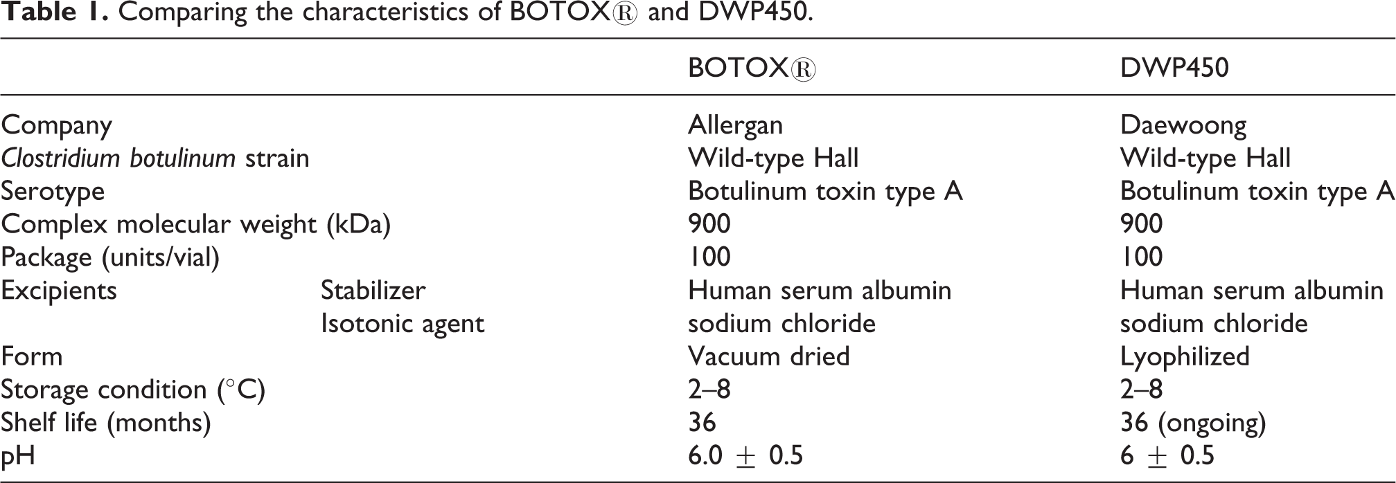
Two different BTX-A preparations, BTX-A-1 (Botox®, Allergan Inc. (Irvien, California, USA)) and BTX-A-2 (DWP450; Daewoong Pharmaceutical Co., Ltd (Seoul, Korea)), were used. The characteristics of these two preparations are almost the same (Table 1). Each preparation was diluted with 0.9% normal saline. Ketamine hydrochloride of 10 mg/kg was given by i.p. injection as anesthesia and then each toxin was injected into one of the tibialis anterior (TA) muscles of a rat (see below).
Comparing the characteristics of BOTOX® and DWP450.
In group 1, 0.08 ml of sodium chloride was injected into one TA muscle and the other TA muscle was not injected. In group 2, two units of 0.02 ml of BTX-A-1 were injected into one TA muscle and two units of 0.02 ml of BTX-A-2 were injected into the other TA muscle. In group 3, four units of 0.04 ml of BTX-A-1 were injected into one TA muscle and four units of 0.02 ml of BTX-A-2 were injected into the other TA muscle. In group 4, eight units of 0.08 ml of BTX-A-1 were injected into one TA muscle and eight units of 0.08 ml of BTX-A-2 were injected into the other TA muscle.
Assessment
CMAPs and conduction velocities were measured using CyberAmp380 and Digidata1320 (Axon Instruments.Inc. (Union City, California, USA)) to assess the inhibitory effect of BTX-A Ketamine hydrochloride of 10 mg/kg was injected into the i.p. area for anesthesia and then rats were fixed in the prone position. The electrode used was an alligator clip lead wire attached to the skin. The stimulating electrode (cathode) was placed on the skin over the popliteal area. The stimulating electrode (anode) was placed at retropubic area and greater trochanter of femur. The recording electrode was placed on the belly muscle of the tibia anterior muscle, the reference recording electrode on the left hind calcaneal tendon and the earth electrode on the sole. Electric stimulation was loaded from 1 to 5 mA. We applied two different stimulus rates, a slow stimulus rate of 2 Hz and a fast stimulus rate of 20 Hz. The paralytic effect on the TA muscle was determined by measuring peak-to-peak amplitudes of the CMAPs (dY), and the delay in conduction velocity (tC) was determined by measuring the time gap between the stimulus point and negative peak point. This analysis was performed before the BTX injection and at 3 days, 1 week, 2 weeks, 3 weeks, and 4 weeks after injection.
Statistics
The effects of BTX-A injection on CMAPs and conduction velocity were analyzed by one-way analysis of variance (ANOVA) using SAS (Version 9.2, SAS Institute Inc., Cary, North Carolina, USA). If ANOVA showed a significant result, post hoc tests were performed. The significance level was set at p < 0.05 for all comparisons.
Results
At the slow stimulus rate of 2 Hz, both BTX-A-1 and BTX-A-2 injection groups showed a paralytic effect on the TA muscle (dY) at 3 days, 1 week, 2 weeks, 3 weeks, and 4 weeks after injection (p < 0.05, Figure 1(a)). However, the effects of BTX-A-1 and BTX-A-2 did not differ significantly (Figure 1(a)). The paralytic effect on the TA muscle (dY) correlated with the dose of BTX in both BTX-A-1 and BTX-A-2 injection groups. At the fast stimulus rate of 20 Hz, both BTX-A-1 and BTX-A-2 injection groups also showed a paralytic effect on the TA muscle (dY) at 3 days, 1 week, 2 weeks, 3 weeks, and 4 weeks after injection (p < 0.05, Figure 1(b)). However, the difference between BTX-A-1 and BTX-A-2 was not significant (Figure 1(b)). The paralytic effect on the TA muscle (dY) correlated with the dose of BTX in both BTX-A-1 and BTX-A-2 injection groups.

Compound muscle action potential amplitudes induced with two different botulinum toxin-A preparations in the (a) slow-rate repetitive nerve stimulation test and (b) fast-rate repetitive stimulation test before injection and at 3 days, 1 week, 2 weeks, 3 weeks, and 4 weeks after injection (N, no injection; S, saline injection; B2, BTX-A-1, two units; D2, BTX-A-2, two units; B4, BTX-A-1, four units; D4, BTX-A-2, four units; B8, BTX-A-1, eight units; D8, BTX-A-2, eight units).
Neither the BTX-A-1 nor the BTX-A-2 injection groups showed a delayed conduction velocity (tC) at either the slow or fast stimulus rate, at 3 days, 1 week, 2 weeks, 3 weeks, or 4 weeks after injection (Figure 2).

Conduction velocities induced with two different botulinum toxin-A preparations in the (a) slow-rate repetitive nerve stimulation test and (b) fast-rate repetitive stimulation test before injection and at 3 days, 1 week, 2 weeks, 3 weeks, and 4 weeks after injection (N, no injection; S, saline injection; B2, BTX-A-1, two units; D2, BTX-A-2, two units; B4, BTX-A-1, four units; D4, BTX-A-2, four units; B8, BTX-A-1, eight units; D8, BTX-A-2, eight units).
Discussion
BTX is endocytosed at synaptic endings and cleaves a site in the protein complex that is directly involved. 14 Once inside the cell, each of the various BTX serotypes (A, B, C1, D, E, and F) cleaves one or more of the soluble N-ethylmaleimide-sensitive factor attachment protein receptor proteins required for vesicle docking and thereby blocks the neurotransmitter release. 15 This inhibition of acetylcholine release at the neuromuscular junction reduces muscular contractions. This toxicological effect has been characterized using electrophysiological methods that are widely used to study nerve and muscle physiology and neuromuscular junction diseases such as Lambert–Eaton myasthenic syndrome and myasthenia gravis.
Using similar techniques, we measured CMAP and nerve conduction velocity in rat muscle treated with BTXs. The effects on nerve conduction velocity did not differ significantly among groups injected with saline (control), Botox® and DWP450. We could not find published studies on nerve conduction velocity after BTX injection. However, in a study of intravenous drug users with wound botulism, the nerve conduction velocities were normal and this indicated that BTX did not induce acute demyelination. 16
The CMAP was significantly decreased after BTX injection in the two groups injected with either BTX preparation, but the size of effect did not differ between the groups. Sakamoto et al. measured CMAPs to quantify the biological activity of BTX-A. 1 The CMAP is generated by the contraction of muscle fibers, and the resulting microcurrent is amplified and recorded. BTX acts at nerve endings to suppress neurotransmission. Therefore, by determining the CMAP amplitude, the action of the toxin suppressing the transmission of electric stimulation to the muscle can be expressed numerically. 17 The method is very suitable for use in comparing the activity of various toxin preparations and in quality control.
The rat-CMAP test is also useful in characterizing the inhibitory effects of various BTX preparations and serotypes at the neuromuscular junction. Sakamoto et al. and Torii et al. compared the effects of different BTXs by injecting them into identical groups of rats. 1,17 However, we used a split-body method for comparing the two different BTX-As: one BTX-A was injected into the left TA muscle and the other BTX-A was injected into the right TA muscle of the same rat. By this method, we observed no difference between the neuromuscular inhibitory effects of the two BTX-As. The results of the mouse biologic assay can be affected by sex, strain, species, and route of administration. Split-body testing avoids these effects and is therefore more appropriate than group testing for comparing drug effectiveness. For this reason, split-body study already has wide usage such as evaluating the efficacy of lasers or topical agents. It can also reduce the number of animals or patients required for the study. For performing the spilt-body study, the systemic effects of BTX should be considered, because it was possible that BTX injected on one side affected the other side. Lange et al. reported systemic effects demonstrable by single-fiber electromyographic techniques at sites distant from the injection, but the effect was found only in patients who received more than 245 units of toxin. 18 Cichon et al. measured CMAP amplitude in the opposite site for investigating the systemic effect of BTX in rat. 19 Although in a five-unit injection group, transient CMAP decrease was observed, they concluded that it was not a meaningful result because only two animals remained in this group. Therefore, split-body study is appropriate to assess the effects of BTX.
Recent increase in the demand for BTX treatment is reflected in new applications in the cosmetics industry and in the development and marketing of new products containing BTX. A standardized testing protocol is therefore needed to establish the comparative effectiveness and adverse effects of new BTX preparations. The split-body study is easy and has many advantages, however, it was not performed for evaluating the efficacy of BTX. We propose that the split-body electrophysiological study may be adapted for this purpose.
Footnotes
Authors’ Note
C-SK and WSJ contributed equally to this work.
Funding
This research received no specific grant from any funding agency in the public, commercial, or not-for-profit sectors.