
Abstract
Prostatitis plays a major role in morbidity and mortality related to prostate diseases. The aim of this study was to detect whether thymoquinone (TQ) could ameliorate oxidative damage and the proliferative response induced by Escherichia coli (E. coli) in rats. A total of 42 adult male Wistar rats were used. The rats were randomly divided into seven groups (three treatment groups, three infected groups and one control group). Control group received saline and was killed 24 h after saline administration. Infected rats were killed after 24, 48 and 72 h following direct injection of E. coli into the prostate. Treatment groups were administered with 10 mg/kg dose of TQ intraperitoneally following E. coli injection and after 24 and 48 h following E. coli injection. The rats were killed at 24, 48 and 72 h after the first drug administration. Each group was compared with each other and with the control group. In addition, infected groups were compared with treatment groups. Our findings show that the treatment with TQ has a protective effect against bacterial prostatitis-induced tissue injury. Increase in malondialdehyde levels and histological damage caused by E. coli were improved markedly with TQ treatment. TQ treatment particularly increased the activity of glutathione peroxidase and decreased the activities of catalase and superoxide dismutase. These observations might be attributed, at least in part, to the antioxidant effect of TQ and suggest that it could be a clinically valuable agent in the prevention of acute prostatitis caused by E. coli.
Introduction
Prostatitis is the inflammation and/or infection of the prostate gland and has different forms like acute or chronic bacterial prostatitis, chronic abacterial prostatitis (chronic pelvic pain syndrome-CPSS) and asymptomatic inflammatory prostatitis (histological prostatitis). 1,2 Prostatic inflammation was suggested to play a vital role in the pathophysiology of benign prostatic hyperplasia as well as in the development of prostate cancer. 3
Escherichia coli (E. coli) is commonly attributable for both acute and chronic bacterial prostatitis in humans. 4,5 Bacterial products (e.g. lipopolysaccharides) stimulate the release of proinflammatory cytokines leading to the extravasation of polymorph leucocytes (PLs). 6 PLs produce toxic products, for example, free oxygen radicals causing tissue damage. 7 Inhibition of these free radicals can decrease the tissue damage to a great extent. 8
Thymoquinone (TQ), a main active constituent of the volatile oil extracted from the black seed (Nigella sativa L.), was shown to inhibit tissue inflammation and oxidative stress. 9 –12 However, there is no available information showing the protective effect of TQ in prostatic disorders. Therefore, the purpose of this study was to investigate the potential therapeutic effects of TQ to ameliorate prostatitis induced by E. coli in vivo using biochemical and histological parameters.
Materials and methods
Animals
A total of 42 adult male Wistar rats (12 weeks old), weighing from 250 to 350 g were purchased from the Laboratory Animal Production Unit of Fırat University (Elazıg, Turkey). Rats were housed as groups in plastic cages containing wood chip bedding. Rooms were under controlled temperature (22 ± 2°C) and humidity (40–60%), with food and water available ad libitum. Rats were used for experiments following 1 week of acclimation. Experiments were performed according to the Guide for Care and Use of Laboratory Animals and the rules of the Institutional Animal Ethics Committee. Experimental procedures were approved by Veterinary Faculty Ethics Committee, Mustafa Kemal University, for the use and care of laboratory animals.
Acute bacterial prostatitis (ABP) model
A strain of uropathogenic E. coli, isolated from patients with complicated urinary tract infection, was stored at −70°C and grown overnight in tryptic soy broth (Merck®, Darmstadt, Germany) at 37°C when required for inoculations. E. coli cells were centrifuged, washed three times and resuspended in sterile phosphate-buffered saline (PBS) to give 1 × 10⁸ cells/ml.
Surgical procedures and bacterial inoculation were performed following a previously described method. 13 Briefly, animals were anesthetized with intraperitoneal 1.25% sodium pentobarbital (45 mg/kg) and the lower abdomen and back were swabbed with 70% alcohol. A longitudinal incision of the lower abdomen of about 15–20 mm in length was made (medial laparotomy) to expose the prostate. Prostatitis was induced via an injection of 200 µl of E. coli diluted in sterile PBS (1 × 10⁸ CFU/ml). This bacterial solution was injected via a 30-gauge needle directly beneath the capsule of both the ventral lobes of the prostate. The peritoneum, muscle and skin were then closed with a 5-0 surgical suture. Sham injected controls received 200 µl of PBS.
Experimental groups and treatments
The rats were randomly divided into seven groups (6 rats in each group and 3 rats in each cage) as follows: the first group was used as a control group (n = 6): 200 µl PBS (pH 7.2) was administered to this group and the rats were killed after 24 h. The rats in the second group (ABP-24 h; n = 6) were killed after 24 h following E. coli injection. The third group (ABP-48 h; n = 6) was killed after 48 h following E. coli injection. The fourth group (ABP-72 h; n = 6) was killed after 72 h following E. coli injection. The fifth group (ABP + TQ-24 h; n = 6) was administered with 10 mg/kg single dose of TQ (Sigma®, St Lois, MO, USA) intraperitoneally simultaneously following E. coli injection and the rats were killed at 24 h after the drug administration. The sixth group (ABP + TQ-48 h; n = 6) was administered two doses of 10 mg/kg TQ intraperitoneally immediately after and 24 h after E. coli injection. Then rats were killed 48 h after first drug administration. Finally, the rats in the last group (ABP + TQ-72 h; n = 6) were administered three doses of 10 mg/kg TQ intraperitoneally following E. coli injection and 24 and 48 h after E. coli injection. Then they were killed at 72 h after first drug administration. Each group was compared with each another and with the control group. In addition, infected groups were compared with treatment groups.
Collection of tissue specimens and samples
The animals were killed using ketamine (50 mg/kg) and xylazine (12 mg/kg) at the end of the experiments. 13 Ventral lobes of prostate from all animals were excised for both biochemical and histological examinations.
Biochemical study
The oxidative parameters were studied in the prostate tissue. The prostate tissue samples were stored at −30°C until assayed for prostate tissue malondialdehyde (MDA) levels and activities of catalase (CAT), superoxide dismutase (SOD) and glutathione peroxidase (GSH-Px). Prostate tissues were homogenized (for 2 min at 5000 r/min) in four volumes of ice-cold Tris-HCl buffer (50 mmol, pH 7.4) using a glass Teflon homogenizer (Ultra Turrax IKA T10 Basic, Germany). Levels of MDA and protein were measured in the homogenate. The homogenate was centrifuged at 5000g for 60 min to remove debris. Supernatant fluid was collected, and CAT and GSH-Px activities as well as protein concentration were measured. The supernatant solutions were used for the assay. The supernatant solutions were mixed with an equal volume of an ethanol/chloroform mixture (5/3, volume per volume v/v). After centrifugation at 5000g for 30 min, the clear upper layer (the ethanol phase) was collected and used in the analysis of SOD activity and protein assays. All preparation procedures were carried out at +4°C.
Determination of MDA levels
The thiobarbituric acid reactive substance level was determined by a method based on the reaction with thiobarbituric acid (TBA) at 90–100°C. In the TBA test reaction, MDA or MDA-like substances (i.e. the byproduct of lipid peroxidation process of the polyunsaturated fatty acids) and TBA react together for the production of a pink pigment having an absorption maximum at 532 nm UV-1800 Spectrophotometer (SHIMADZU® Corporation Kyoto, Japan). Results were expressed as nanomoles per gram of wet tissue, by reference to a standard curve prepared from measurements made with a standard solution (1,1,3,3-tetramethoxypropane).
Determination of CAT activity
Catalase (CAT, EC 1.11.1.6) activity was measured according to the method of Aebi. 14 The principle of the assay is based on the determination of the rate constant k (dimension: s–1, k) of hydrogen peroxide (H2O2) decomposition. By measuring the absorbance changes per minute, the rate constant of the enzyme was determined. Activities were expressed as k (rate constant) per gram protein.
Determination of GSH-Px activity
GSH-Px (EC 1.6.4.2) activity was measured using the method of Paglia and Valentine. 15 The enzymatic reaction was initiated by adding H2O2 to the reaction mixture containing GSH, nicotinamide adenine dinucleotide phosphate and glutathione reductase. The change in the absorbance at 340 nm was monitored by a spectrophotometer. Activity was expressed as Units per gram of protein.
Determination of protein level
Protein measurements were performed in tissue homogenate according to the method of Lowry et al. 16
Determination of SOD activity
The principle of the total SOD (EC 1.15.1.1) activity method is based, briefly, on the inhibition of nitroblue tetrazolium (NBT) reduction by O2 − generated by xanthine/xanthine oxidase system. 17 Activity was assessed in the ethanol phase of the serum after 1 ml ethanol/chloroform mixture (5/3, v/v) was added to the same volume of the serum and centrifuged. One unit of SOD was defined as the enzyme amount causing 50% inhibition in the NBT reduction rate. The SOD activity was expressed as Units per gram of protein.
Histological study
For light microscopic examinations, prostate samples were fixed in 10% neutral buffered formalin. After routine tissue processing protocol, 5-μm-thick sections were stained with hematoxylin and eosin and evaluated with an Olympus DP20 camera attached–Olympus CX41 photomicroscope.
A histologic scoring system was used based on the extent of the lesions. Five consecutive sections were obtained for each animal. Collectively, 30 sections were examined for each group. During examination, the extent of the lesion was determined by counting the affected glandular lumens. Enlarged lumens with cellular debris and inflammatory cells were accepted as ‘affected.’ According to the extent of the lesion, sections were given points as follows: affected area less than 25%: 0 point; affected area between 25 and 50%: 1 point; affected area between 50 and 75%: 2 points and affected area more than 75%: 3 points.
Statistical analysis
The Mann-Whitney U test and the χ 2 test were used to determine differences in the distributions of scores. p < 0.05 was considered statistically significant. All experimental data were expressed as mean ± SD. Biochemical data were analyzed using a commercially available statistics software package (SPSS for Windows v. 15.0, Chicago, Illinois, USA). Distribution of the groups was analyzed with one sample Kolmogorov–Smirnov test. Groups showed normal distribution for the level of MDA and the activities of CAT, SOD and GSH-Px so that parametric statistical methods were used to analyze the data. One-way analysis of variance test was performed and post hoc multiple comparisons were made using least-squares differences. Results are presented as mean ± SEM; p < 0.05 was considered as statistically significant.
Results
Biochemical results
Effect of TQ on E. coli modulation of prostate CAT activity
When ABP groups were compared with the control group, a statistically meaningful decrease was observed in CAT activity at 72 h (p < 0.05). When ABP groups were compared with each other, CAT activity was found lower at 72 h (p < 0.05). When ABP + TQ groups were compared with the control group, a meaningful decrease was detected in ABP + TQ groups at 48 and 72 h (p < 0.01 and p < 0.05, respectively). When TQ groups were compared with each other, no meaningful difference was observed among the groups. Finally, when ABP and TQ groups were compared with each other, a significant decrease in CAT activity was observed in the ABP + TQ group at 48 h (p < 0.01; Figure 1 and Table 1).

CAT, SOD, MDA and GSH-Px levels. CAT: catalase; GSH-Px: glutathione peroxidase; MDA: malondialdehyde; SOD: superoxide dismutase.
Oxidative stress parameters in the prostate of rats (mean ± SEM, n = 6).

ABP: acute bacterial prostatitis; CAT: catalase; GSH-Px: glutathione peroxidase; MDA: malondialdehyde; PYN: pyelonephritis; SOD: superoxide dismutase; TQ: thymoquinone.
a p < 0.001 compared with control group.
b p < 0.05 compared with control group.
c p < 0.05 compared with ABP-24 h group.
d p < 0.05 compared with ABP-48 h group.
e p < 0.01 compared with ABP-48 h group.
f p < 0.01 compared with ABP-24 h group.
g p < 0.05 compared with ABP + TQ-24 h group.
h p < 0.05 compared with ABP-72 h group.
i p < 0.05 compared with ABP + TQ-48 h group.
Effect of TQ on E. coli modulation of prostate SOD activity
When ABP groups were compared with the control group in terms of SOD enzyme activities, there was a meaningful decrease in ABP groups at 24 and 48 h (p < 0.01 and p < 0.05, respectively). When ABP groups were compared with each other, there was a higher increase at 48 h compared with 24 h (p < 0.05) and at 72 h compared with 48 and 24 h (p < 0.01). When ABP + TQ groups were compared with the control group, a meaningful decrease in SOD enzyme activities was detected in all ABP + TQ groups (p < 0.05). When ABP + TQ-48 h and ABP + TQ-72 h groups were compared with ABP-48 h and ABP-72 h groups, TQ groups had lower values (p < 0.05). When ABP + TQ groups were compared with each other, there was a meaningful increase in the ABP + TQ-72 h group compared with the 24 h group (p < 0.05; Figure 1 and Table 1).
Effect of TQ on E. coli modulation of prostate lipid peroxidation
When ABP groups were compared with the control group in terms of MDA levels, a statistically significant increase was observed in all ABP groups (p < 0.01). When ABP groups were compared with each other, there was a meaningful increase especially in ABP-72 h group (p < 0.05). When ABP + TQ groups were compared with control group, a meaningful increase was found in all of the groups (p < 0.01 and p < 0.05, respectively). When ABP + TQ groups were compared with each other, MDA level was low in ABP + TQ-48 h and 72 h (p < 0.05). When ABP groups were compared with ABP + TQ groups in terms of MDA levels, a meaningful decrease was observed in ABP + TQ groups at 48 and 72 h (p < 0.05) (Figure 1, Table 1).
Effect of TQ on E. coli modulation of prostate GSH-Px activity
A statistically meaningful increase in GSH-Px enzyme activities was detected in all the ABP groups compared with control groups (p < 0.01). When ABP groups were compared with each other, this parameter was found to be significantly lower at 48 and 72 h (p < 0.05). When ABP + TQ groups were compared with the control group, a meaningful increase was found in all ABP + TQ groups (p < 0.01). When ABP + TQ groups were compared with each other, a meaningful increase was detected especially in ABP + TQ-72 h (p < 0.05). When ABP groups were compared with ABP + TQ groups, GSH-Px enzyme activity was found to be significantly higher in ABP + TQ-72 h (p < 0.05; Figure 1 and Table 1).
Histological data of the prostate
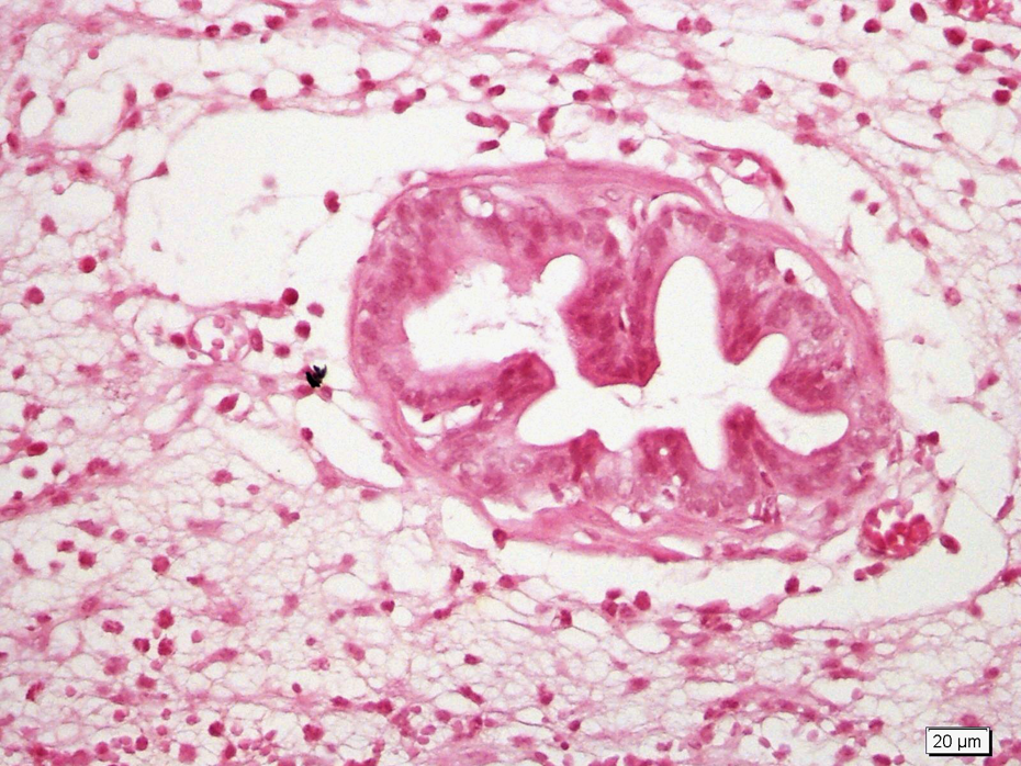
Statistical analysis of histological scores of main findings is shown in Table 2.Microscopic examination revealed normal prostate tissue in the control group. In group 2 (24 h E. coli group), nearly half of the glandular lumens in the section were filled with cellular debris and leucocytic infiltrate. Degeneration was significant compared with the control group (p < 0.05). Degeneration was more severe and significant in 48 h E. coli group (group 3) compared with controls (p < 0.05). More than half of the glands in the section were degenerated and filled with leucocytic infiltrate. In group 4 (72 h E. coli group), all glandular lumens were distended with leucocytic infiltrate and cellular debris (Figure 2). Degeneration was significant compared with the control group (p < 0.05).

Glandular lumens were distended with leucocytic infiltrate and cellular debris (HEX40).
Grading levels of inflammation in ABP- and TQ-treated groups as determined by histopathological examination.a
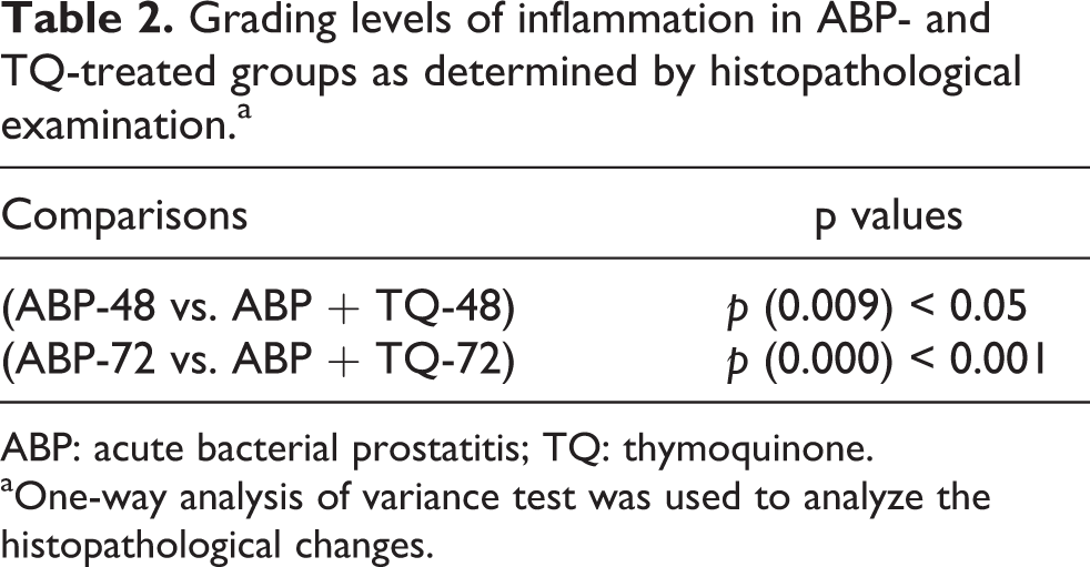
ABP: acute bacterial prostatitis; TQ: thymoquinone.
aOne-way analysis of variance test was used to analyze the histopathological changes.
TQ administration improved the general structure of prostate in group 5 (TQ + 24 h E. coli group). However, improvement was not statistically significant (p > 0.05).
In group 6 (TQ + 48 h E. coli group), improvement was statistically significant (p < 0.05) when compared with group 3. Remaining glands (more than half of the glands in the section) were close to normal. In group 7 (TQ + 72 h E. coli group), there was an intact area without any debris in glandular lumens (Figure 3). When compared with group 4, changes were significant (p < 0.05) and it was noticed that glandular lumens were not totally obliterated with cellular debris and infiltrate and were protected to some extent in a localized area.
Partially protected glands (HEX40).
Discussion
SOD, GSH-Px and CAT show antioxidant activities involving the detoxification process of superoxide anions and, in subsequent reactions, of H2O2. 18,19 Kim et al. 20 found that SOD and GSH-Px activities in prostate tissue decreased in the rats with estradiol-induced chronic prostatitis. Lou et al. 21 also declared that CAT and SOD activities decreased in the patients with chronic bacterial prostatitis. At the same time, Zhou et al. 22 emphasized that the erythrocyte SOD, CAT and GSH-Px activities in patients with chronic bacterial prostatitis were significantly decreased. Similarly in our study, CAT and SOD activities decreased in E. coli-infected groups when compared with control rats. We also found that the activities of GSH-Px significantly increased in all E. coli groups when compared with the control group.
The antioxidant activities of TQ have been previously reported. Kanter et al. 23 reported that intraperitoneal TQ decreased CAT, GSH-Px and SOD enzyme activities in a study of oxidative stress induced in rats by cadmium. Mansour et al. 9 reported that TQ given orally decreased CAT, GSH-Px and SOD enzyme activities in liver tissues in mice, but it decreased only SOD enzyme activities in cardiac tissue and did not affect CAT and GSH-Px enzyme activities. In TQ administered rats, we detected that the activities of CAT and SOD decreased, whereas the enzyme activity of GSH-Px increased. These findings were similar to those of Kanter et al. 23 and Mansour et al. 9 Orally administered TQ increased the activities of CAT and GSH-Px enzymes in studies carried out by Nagi et al. 10 and Fouda et al. 24 They also reported an increase in GSH-Px enzyme activity. In contrast to our study, there was an increase in CAT enzyme activity. These differences were possibly due to the differences in tissues studied, administration periods and administration routes of agents. Furthermore, TQ is expected to have an influence on GSH-Px activation rather than SOD and CAT activation in the antioxidant process in the prostate tissue.
Reactive oxygen species (ROS) cause membrane injury by producing lipid peroxidation. 25 Kim et al. 20 Lou et al., 21 Zhou et al. 22 and finally Orsilles and Depiante-Depaoli 26 found increased MDA levels in their clinical and experimental prostatitis studies. In our study, it was shown that MDA levels were significantly increased in the prostate tissues of all E. coli-infected rats when compared with controls. Quintar et al. 13 reported that prostatitis reached the peak level at 72 h in the rat model of E. coli-induced ABP. In our study, MDA levels significantly increased in E. coli-infected rats especially at 72 h when compared with the control rats.
TQ could have a protective effect on lipid peroxidation through its reported antioxidant defense against the generation of ROS in different tissues. 27,28 Fouda et al., 24 Alenzi et al. 11 and Helal 12 found that the administration of TQ led to decreased MDA levels in different toxicity studies. We similarly showed in our study that MDA levels were significantly decreased with TQ administration when compared with the E. coli-infected rats. Fouda et al. 24 reported that the maximal protection offered by TQ treatment was particularly noticeable at 48 and 72 h after the administration of the toxic agent. In our study, similar results indicated that MDA levels significantly decreased especially in the ABP + TQ-72 h group when compared with E. coli groups.
In this study, we also histologically showed the antibacterial effect of TQ in prostate tissue. TQ improved the general histologic structure in prostatic tissue and downgraded the degree of inflammation as time passed. Fouda et al. 24 reported that the maximal histologic improvement offered by TQ treatment was particularly noticeable at 48and 72 h after the administration of the toxic agent. We noted similar histologic results. Quintar et al. 13 reported that histologic injury became evident at 48 and 72 h post infection. We found similar findings in our acute infection groups.
The antibacterial effects or antiproliferative activity of TQ against prostate cancer have been previously reported. 29,30 However, our study is the first in the literature reporting the protective role of TQ against ABP.
Conclusion
This study demonstrated for the first time that ABP induced by E. coli could regress following TQ administration as shown with histological and biochemical findings. Further studies are needed to show its antibacterial effect against other agents with more detailed morphological and biochemical analysis.
Footnotes
Funding
This research received no specific grant from any funding agency in the public, commercial, or not-for-profit sectors.
Acknowledgement
The authors of this article greatly acknowledge Murat M. Rifaioglu for his contribution to the study in terms of statistical analysis.