
Abstract
Dioxin (2,3,7,8-tetrachlorodibenzo-p-dioxin; TCDD) is one of the most powerful environmental toxins and causes a variety of toxic effects in humans. Since it makes first contact with bronchial epithelial cells as an atmospheric contaminant, we identified differentially expressed genes (DEGs) in TCDD-treated human bronchial epithelial cells (HBE4-E6/E7) using an annealing control primer (ACP) system. Six genes, five upregulated and one downregulated, were isolated and their expression patterns were confirmed by reverse dot blot analysis. Their genomic sequences were used for identification, and the upregulated proteins were found to be acyl-coenzyme A dehydrogenase (VLCAD), S100 calcium binding protein A6 (S100A6), nuclear receptor co-repressor 2 (NCOR2), ribosomal protein, large, P1 (RPLP1), and tubulin α 1c, and the downregulated protein was shown to be tubulin β2. Among them, the expression of the S100A6 was further analysed by northern hybridization because of its relationship with TCDD. These results suggest that this new method was simple and convenient to identify DEGs regulated by a specific agent. Moreover, these isolated genes may provide important information to better understand the mechanisms of TCDD toxicity in human bronchial epithelial cells.
Introduction
The 2,3,7,8-tetrachlorodibenzo-p-dioxin (TCDD or simply called as dioxin) is one of the most toxic environmental chemicals made by humans and induces a variety of biological disorders. 1 Dioxin, known as a potent tumor promoter, activates the aryl hydrocarbon receptor (AhR), a ligand-activated transcription factor belonging to the basic helix-loop-helix-PAS family, to enhance tumorigenesis via unknown mechanisms. 2 However, the exact mechanisms of the TCDD carcinogenesis are still largely unknown as well as the biological consequence of the dioxin toxicity in the respiratory system. Results from animal studies have shown that a high dose of TCDD exposure can cause cancer in the lungs.3–5 Lung cancer is generally subdivided into two large groups: small cell lung cancer (SCLC), which accounts for about 20–25% of bronchogenic carcinomas, and nonsmall cell lung cancer (NSCLC), which accounts for the remainder. The NSCLC can be further divided into three major subtypes: large cell lung carcinoma (LCLC), adenocarcinoma, and squamous cell carcinoma. The LCLC may represent highly undifferentiated squamous carcinomas and adenocarcinomas. Adenocarcinoma can be either bronchial derived or bronchioalveolar in origin. In contrast, squamous cell carcinoma results from malignant transformation of the epithelial cell lining of the bronchi. 6 Tissue and cell specificity is a crucial factor in the biochemical and toxic effects of TCDD. The lung and bronchial epithelium are a known target organ for TCDD and other polyhalogenated aromatic hydrocarbons (PHAHs) in humans. Identifying signaling pathways that are altered by TCDD and its carcinogenic mechanisms in pulmonary tissue are important. Thus, the objective of the present study was to identify novel genes that are regulated by TCDD in a normal human bronchial epithelial cell line. To accomplish this goal, differentially expressed gene (DEG) profiling using a new reverse transcription–polymerase chain reaction (RT-PCR) that involves annealing control primers (ACPs) was applied to TCDD-treated human bronchial epithelial cells.
Methods
Cell culture and TCDD exposure
HBE4-E6/E7, a human bronchial epithelial cell line was obtained from the American type culture collection (Rockville, MD, USA) and maintained using the supplier’s instruction. TCDD was purchased from the AccuStandard Inc. (New Haven, CT, USA) and dissolved in dimethyl sulfoxide (DMSO; Sigma, St. Louis, MO, USA). The cultured cells were treated with either TCDD (10, 100 nM) or the vehicle (DMSO) for 24 h for RT-PCR using ACPs.
RNA preparation and ACP RT-PCR
Total RNA was isolated using the Tri-reagent (Sigma) according to the manufacturer’s instruction. First-strand complementary DNA (cDNA) was synthesized using the deoxythymidine (dT)-ACP1 primer (Gene-Fishing™ DEG kits, Seegene, Seoul, Korea) by RT and then the first-stage PCR for second-strand cDNA synthesis was conducted using arbitrary ACPs in only one cycle. The second-stage PCR for amplifying the second-strand cDNA was performed using dT and one of 20 arbitrary ACPs at the 3′- and 5′-end, respectively, during 35-40 cycles. Only authentic PCR products were amplified at that time. After all PCR processes, DEGs were detected on an agarose gel and separated for further experiments.
Reverse dot blot hybridization
The amplified products were ligated into the pGEM-T Easy vector (Promega Co., Madison, WI, USA) and the recombinant vector was transferred into JM109 Escherichia coli. The amplified insert DNA in plasmids were extracted and subjected to reverse dot blot hybridization. Isolated DEGs were blotted on a nylon membrane using the Bio-Dot Microfiltration System (Bio-Rad, Hercules, CA, USA). The 32 P-labeled first-strand cDNA probes, originating from the TCDD-treated or non-treated RNA, were synthesized using a Random Primer DNA Labeling Kit (Takara Korea Biomedical Inc., Seoul, Korea) and purified with Quick Spin Columns (Sephadex G-50, Roche Applied Science, Mannheim, Germany). The DEG-spotted reverse dot blot was hybridized with the labeled cDNA probes, exposed to a phosphoimager screen (Packard Bioscience, Boston, MA, USA) for 1 h, and imaged using a Phosphoimager (Typhoon scanner, GE Healthcare, Little Chalfont, UK). The band intensity was quantified using image analysis software (ImageQuant Version 5.2, Molecular Dynamics).
Sequencing
Purified plasmids were subjected to dideoxy chain termination sequencing. The identity of each product was confirmed by sequence homology analysis using the Basic Local Alignment Search Tool (BLAST).
Northern blot hybridization
For analysis of the dose dependency of selected genes, the cultured cells were treated with either TCDD (0.1, 1, 10, 100 nM) or the vehicle (DMSO) for 24 h. Total RNA was extracted using the Tri-reagent (Sigma) and subjected to northern blot analysis with selected DEG.
Results and discussion
In this study, a new ACP-differential display RT-PCR technique was used to identify genes that were altered in HBE4-E6/E7 cells after treatment with 10 and 100 nM of TCDD. This method involved an ACP that had a unique tripartite structure in that its distinct 3′- and 5′-end portions were separated by a regulator.7,8 Using this technique, six DEGs were identified in TCDD-treated cells at doses of 0, 10, and 100 nM. As shown in Figure 1 , five of the DEGs were upregulated and one was downregulated. They were isolated and subjected to reverse dot blot analysis to confirm their expression patterns. The five upregulated DEGs coincided with the ACP RT-PCR results, while the downregulated DEG did not show any change in the reverse dot blot analysis (Figure 2 ). Among the upregulated genes, DEGs 2 and 5 were dramatically increased when exposed to TCDD. Thus, genomic sequences were analysed to identify their origins using the dideoxy chain termination method. By using this approach, the DEGs were determined to be acyl-coenzyme A dehydrogenase (VLCAD), S100 calcium-binding protein A6 (S100A6), nuclear receptor co-repressor 2 (NCOR2), tubulin-β2, ribosomal protein, large, P1 (RPLP1), and tubulin α 1c (Table 1 ).

Photographs of the agarose gel containing the amplified complementary DNA (cDNA) products obtained from TCDD-treated HBE4-E6/E7 cells. The samples were obtained from cells treated with 0, 10, and 100 nM TCDD. Arrows indicate DEGs induced by TCDD intoxication. DEG: differentially expressed gene, 1~20: arbitrary annealing control primers (ACPs), M: Marker, 0, 10, 100 nM TCDD.

Reverse dot blot analysis of the six DEGs induced by TCDD. Salmon sperm DNA (200 ng) and human β-actin (2.5, 5, 10, 20, 40 ng) were used as negative and positive controls, respectively. N/C: negative control, P/C: positive control.
Sequencing results of the differentially expressed genes (DEGs)

The VLCAD is one of the four acyl-CoA dehydrogenases that have various chain-length specificities. The VLCAD catalyses the initial step of mitochondrial fatty acid oxidation and plays a critical role in energy metabolism. 9 Exposure to di-(2-ethylhexyl) phthalate (DEHP), a common environmental toxin, was previously shown to result in VLCAD induction in the HepG2 cell line and rat liver.10,11 Kurts et al. reported that acetyl-CoA, a product of VLCAD, provides a majority of the energy required in the liver for ketogenesis under stressful conditions, such as fasting, strenuous exercise, and sepsis. 12 In another study, upregulated fatty acid metabolism in colorectal cancer (CRC) was found to be associated with CRC proliferation. 13 However, at this point, further studies would be needed to determine why this enzyme was induced after exposure to TCDD.
S100A6, also named as calcyclin, was sharply upregulated in TCDD-intoxicated human bronchial epithelial cells, which was confirmed by reverse dot blot analysis and northern hybridization due to a novel gene induced by TCDD (Figures 2 and 3). The S100 is a family of nonubiquitous Ca2+-modulated proteins and is found in a wide range of cells. These proteins are thought to be involved in diverse intracellular and extracellular activities, such as metastasis, cell cycle regulation, protein degradation, transcription factor regulation, modulation of enzyme activities, cell growth and differentiation, and Ca2+ homeostasis.14,15 Emberley et al. reported that S100 proteins may promote cancer progression due to its role in cell survival and apoptosis pathways. 16 Among the S100 proteins, S100A6 induction prevented TNF-α-induced cardiac myocyte apoptosis, which is suggested to play a homeostatic role in programmed cell death. 17 Overexpressed S100A6 was also proposed to be associated with decreased metastasis and inhibition of cell migration in human osteosarcoma. 18 In addition, oxidants stimulated S100A6 gene expression due to an increase in nuclear factor-E2-related factor 2 (Nrf2)-binding affinity to antioxidant response element (ARE) sequences. 19 In a recent study related to TCDD, S100A6 was found to be associated with thymocyte differentiation–stimulated antigen, which was remarkably inhibited by TCDD exposure. 20 Although the precise role of this protein is poorly understood, highly upregulated S100A6 (Figures 1–3) may play a defensive role against TCDD toxicity in human bronchial epithelial cells.
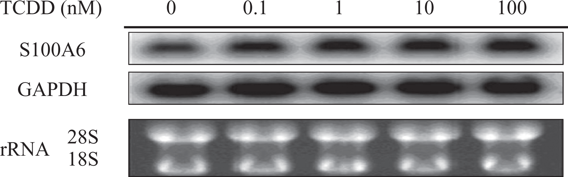
Northern blot analysis of S100 calcium-binding protein A6 (S100A6; calcyclin) in TCDD-treated HBE4-E6/E7 cells. Lane 1 is dimethyl sulfoxide (DMSO) only and 2 to 5 are HBE4-E6/E7 treated with TCDD (0.1, 1, 10, and 100 nM).
NCOR2, DEG3, is a silencing mediator of retinoic acid and thyroid hormone receptors (SMRT). 21 This protein plays a crucial role as a transcription co-repressor molecule of inflammatory transcription factors, such as activator protein (AP)-1 and nuclear factor (NF)-κB. 22 Interleukin (IL)-6 production in kupffer cell by NF-κB activation contributes tumor cell survival, growth, and carcinogenesis promotion. 23 In addition, IL-8 expression using the same pathway also results in angiogenesis and metastasis in tumor cells. 24 Both cytokines 25 may be overexpressed through the inhibition of NCOR2. Derepression of NCOR2 via phosphorylation and nuclear export is a prerequisite for NF-κB transcription and cell survival, suggesting that overexpressed NCOR2 may lead to cytotoxicity though disrupting the survival pathway. 26 In an animal study, 4-week exposure to phenobarbital, which was used to induce tumorigenesis, resulted in hypermethylation of the NCOR2 sequence and expression was decreased in both C57BL/6 and tumor-prone B6C3F1 mice.27,28 Therefore, NCOR2 induction may play a role in inhibiting IL-6 and IL-8; thus upregulation of this protein can inhibit cytokine-related cell death, which may result in TCDD-issued tumorigenesis in normal human bronchial epithelial cells as well as cytotoxicity.
The β-tubulin family consists of 15–20 dispersed genes in humans. The tubulin β2, identified as DEG4, is abundantly expressed in the testis and at lower levels in many other tissues. 29 In this study, tubulin β2 was decreased by TCDD intoxication in ACP RT-PCR; however, this trend was not observed in the reverse dot blot analysis.
Ribosomes are essential factors to various cellular responses. Among them, the 40S and 60S ribosomes are core components for amino acid elongation; however, over 80 ribosomal proteins have been shown to be involved in this process. 30 Therefore, the exact mechanism of biogenesis and function of all ribosomesare still poorly understood. Highly upregulated RPLP1 was identified as DEG5, which is a member of the P group of ribosomal proteins. The RPLP1 overexpression resulted in senescence and immortalization of primary cultured mouse embryonic fibroblasts (MEFs). In addition, upregulation of this ribosome was identified in tumors with mutant p53, which disrupts wild-type p53 function and promotes tumorigenesis. Indeed, elevated RPLP1 mRNA was found in 16 of 26 human colon cancers, and overexpression of mutant p53 was observed in 6 of the 16 upregulated specimens. 31 Therefore, induction of this ribosome by TCDD may be relevant to human cancer development.
Homo sapiens tubulin α 1c was identified as a DEG6. α-tubulin, one of the cytoskeleton proteins, is found in high concentrations in many kinds of cells and is related with movement, reproduction, and death of the cell. The regulation of tubulin α was examined in neuronal cells, and microtubule trafficking was negatively impacted by organophosphate (OP) pesticide exposure. 32 In addition, upregulation of tubulin α in medaka fish exposed to diazinon might compensate for impaired microtubule function caused by OP pesticide diazinon. 33 Tubulin α induction in this study was suggested to play a similar compensatory role against environmental toxins in neuronal cells.
Together, the DEGs could be related with their roles on TCDD-induced cytotoxicity and carcinogenic process as follow. Enhanced metabolism of fatty acids via VLCAD, mechanisms of defensive role and enhancing cell survival via S100A6, and possible roles of NCOR2 on decreasing cell survival and immunosuppressive effect might be involved in both TCDD toxicity and/or carcinogenesis. The RPLP1 and tubulin α 1c may be relevant to human cancer development and compensatory role against environmental toxins impairing microtubule function in neuronal cells, respectively.
DEGs by TCDD, which included five upregulated genes and one downregulated gene, were identified in normal human bronchial epithelial cells. Among them, S100A6 and RPLP1 showed obvious overexpression in the presence of TCDD. Although their functions are still not known, the ACP RT-PCR technique is a simple and convenient method of identifying DEG against a specific agent. In future studies, the exact role and mechanisms of the altered genes related to TCDD intoxication will be studied.
Footnotes
This work was supported by the Inje Research and Scholarship Foundation in 2010.