
Abstract
The aim of this study is to evaluate the antibacterial activity of dental material containing nanoparticles of the extract of S. persica against Streptococcus pneumonia, Streptococcus mutans and Haemophilus influenza, and to examine their triboligical and mechanical properties. The specific properties of flowable dental material containing different percentage of the extract are investigated using the below different kinds of tests: - Antibacterial activity (including agar diffusion test), - Tribological behavior (including a pin-on-disk tribometer, surface profilometer and scanning electron microscopy (SEM)), - Mechanical behavior (including compressive strength, bending strength and bending modulus). The agar diffusion test reveals significant difference between the samples, whereas the antibacterial one demonstrates that the bacterial growth is diminished by increasing the nanoparticle content. The bending strength and the bending modulus remain unchanged by a 10% incorporation of the nanoparticles while the compressive strength increases significantly. The extract containing resins show higher wear resistance. The production of dental material with antibacterial activity, with suitable wear resistance, and particularly without any significant sacrificing effect on the mechanical properties is practically desirable in dental material science.
Keywords
Introduction
Denture teeth materials are frequently used to replace missing teeth. The material compositions have witnessed not only regular improvements but also revolutionary developments. However, the dental prosthesis is not without intricate challenges as the chewing forces and the presence of the acidic biofilms notably affect the prosthesis.
As a matter of fact, direct restorations have presented an annual failure, rating up to 7.9% with the main reasons of secondary caries and bulk fracture.1–3 It was reported that the 5-year failure rate of fixed dental prostheses was more than 10%, due to the common complications of caries and endodontic diseases.4,5 Although the implant survival has ranged from 92.8 to 97.1% reached over a follow-up period of 10 years, the peri-implantitis cannot be ignored. It is mainly originated by the biofilm accumulation.6,7 Dental materials are subjected to an aggressive attack by bacteria. The oral cavity seems of a complex ground for a noticeably high humidity, a moderate temperature, and an abundance of nutrients. It actually motivates the development of differentiated microorganisms as well as microbial biofilms.8,9
Accordingly, it is critically important to develop a denture teeth material with the capacity of minimizing the cropping up occurrence of recurrent caries and the presence of acidic biofilms. Research has focused on imparting antimicrobial properties to biomaterials using silver, zinc oxide, and quaternary ammonium compounds,8,10 which exhibit strong antibacterial effects but may pose risks such as cytotoxicity. In contrast, medicinal plant particles offer a natural and biocompatible alternative, rich in diverse bioactive compounds that reduce the risk of resistance and provide antioxidant benefits. Specifically, particles from S. persica particles have been widely used in medical and pharmaceutical engineering owing to their strong bactericidal effect against a broad spectrum of oral bacteria, as compared to other fillers. The powder of S. persica was previously incorporated into PMMA dental resin. It achieved successful antibacterial results. 11 However, the improvement of the antibacterial activity of this composite is needed for further limitation of the differentiated bacteria and microbial biofilms.
Moreover, there has been much discussion about the antibacterial activity of the extract of S. persica for pulp tissue regeneration therapy. It has been reported that the extract of S. persica possesses various biological properties against organisms. It is considered important that we should develop the dental plaque as well as the periodontitis. 12
The extraction of the S. persica and its incorporation into denture teeth material can improve its antibacterial activity. In this vein, the objectives of this study are as follows: firstly to develop new formulations of dental material with antibacterial properties; and secondly, to investigate the effects of incorporating the extract of S. persica on the tribological properties and the mechanical performances, which are directly associated with the durability of the dental prosthesis. The tested hypothesis is mainly to develop a new bioactive dental material with an antibacterial activity by incorporating the extract of S. persica into the PMMA dental resin. It is expected to achieve acceptable tribological and mechanical properties within the international organization of the ISO standardization, as a recommended range.
Materials and methods
Materials
A denture teeth material has been manufactured as a composite material by reinforcing resin with filler, classifying it within the composite material category. It is based on PMMA, which is used as an organic matrix. At the same time, different percentage of S. persica extract is used as filler. The resin (Nic Tone, MDC dental, USA) is used as the polymer/monomer ratio of 2:1.
Firstly, S. persica sticks (Miswak, Sewak alBaraka, Pakistan) are dried at room temperature. Later, the dried sticks are blended in a food-processing blender (coffee grinder, Moulinex, China). The alcoholic extract is done by a cold maceration of 20 g of S. persica fine powder in 200 mL of ethanol. The mixture is maintained at rest for 48 h. The resulting extract is filtered; meanwhile the solvent is completely removed using a rotary evaporator. The extract stored at +4°C until further use. 13 The extract contains bioactive compounds, including benzylisothiocyanate (BIT), limonene, α-pinene, flavonoids, alkaloids, saponins, and tannins, which contribute to its antimicrobial, antioxidant, and anti-inflammatory properties.11–13 The extract is expected to demonstrate significant antibacterial activity but exhibits lower stiffness compared to PMMA. The filler particles display isotropic behavior due to their spherical shape.
Measurement of filler particle size
Extract particle size distributions have been measured on Malvern’s Dynamic Light Scattering (DLS) instruments, the Zetasizer Nano S. This Zetasizer is a device, which measures the sized particles, having particle size of particles having a diameter, which ranges from 0.4 nm to 10 µm.
Composites processing
In order to improve the filler dispersion in the resin, the mixture of PMMA and the extract of S. persica are mechanically stirred at a high shear rate. A great majority of authors working at laboratory scale with nanocomposites use the same manufacturing process, as a direct way. It mainly consists of mechanical stirring. Indeed, this process is described by the company Ashland. 14 The polymerization of the composites is affected in a pressure device, known as Mini Major 2000, Major, Italy, at 60°C during 10 min under 0.5 MPa pressure.
Antibacterial activity tests
The following oral cavity pathogen strains are selected to conduct antimicrobial activity tests, notably Streptococcus pneumonia (CIP 49619), Streptococcus mutans (CECT 479), and Haemophilus influenza (ATCC 10211). The antimicrobial testing was performed on Mueller-Hinton agar supplemented with 5% defibrinated horse blood + β-NAD (MHF, Biomerieux) using the agar diffusion method. S. mutans and S. pneumoniae are grown on blood agar, while H. influenzae is grown on chocolate agar. A suspension of 0.5 McFarland Standard was prepared and spread on MHF agar plates according to the EUCAST recommendations. The sample of composite is immersed in 400 mL of sterile water for 1 day so that in order to allow the composite can release the antibacterial substances. The required number of wells, each 6 mm in diameter, were cut out of the agar using a sterile glass capillary. Then, wells were filled with 100 µL of S. persica extract or composite extraction solution. After incubating the plates at 37°C with a 5% of the CO2 for 24 h, the diameters of the inhibition zone around the samples are measured. The experiments are performed in triplicate.
Tribological tests
The experiments are conducted using a pin-on-disk tribometer (Figure 1). The dimensions of the cylindrical samples are of Ø25 mm × 4 mm per disk. The applied counter-face is an Alumina ball with a diameter of 6 mm per pin. The two loads of 5 and 10N are applied during the wear tests. The sliding velocity is 300 rpm for 2 hours meanwhile the diameter of the wear track is 6 mm. Actually, the sliding distance is equal to 0,678 Km. The set of tests are conducted under dry conditions at room temperature. For a more stimulation of the real wear conditions of the human teeth, the choice of these parameters is based on both the clinical experience and the literature itself. During the chewing process of human beings, the magnitude of the masticatory force in the oral cavity ranges from 3 to 36 N.
15
Furthermore, the dental restorative materials and teeth are subjected to 0.6 km per year sliding distance.
16
Pin-on-disk tribometer.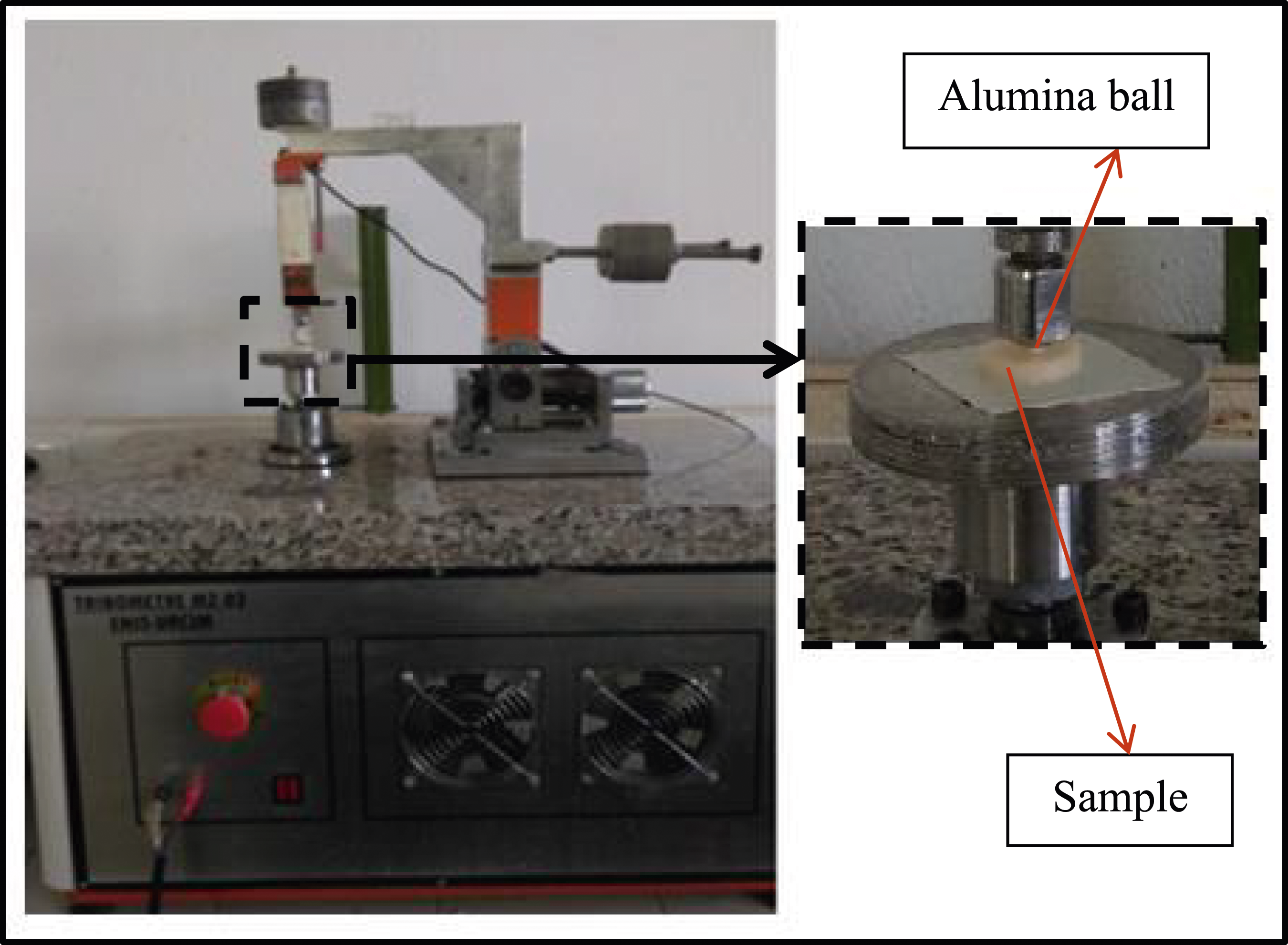
The worn surfaces of the specimens were scanned using a surface profilometer to obtain the wear volume (Figure 2). For each sample, ten passes were made, and the average was multiplied by the perimeter of the wear track to calculate the wear volume. Equally, in order to determine the wear mechanisms, the wear tracks are examined using a scanning electron microscope (ZEISS 1450VPSE). Profilometer investigation of typical wear track.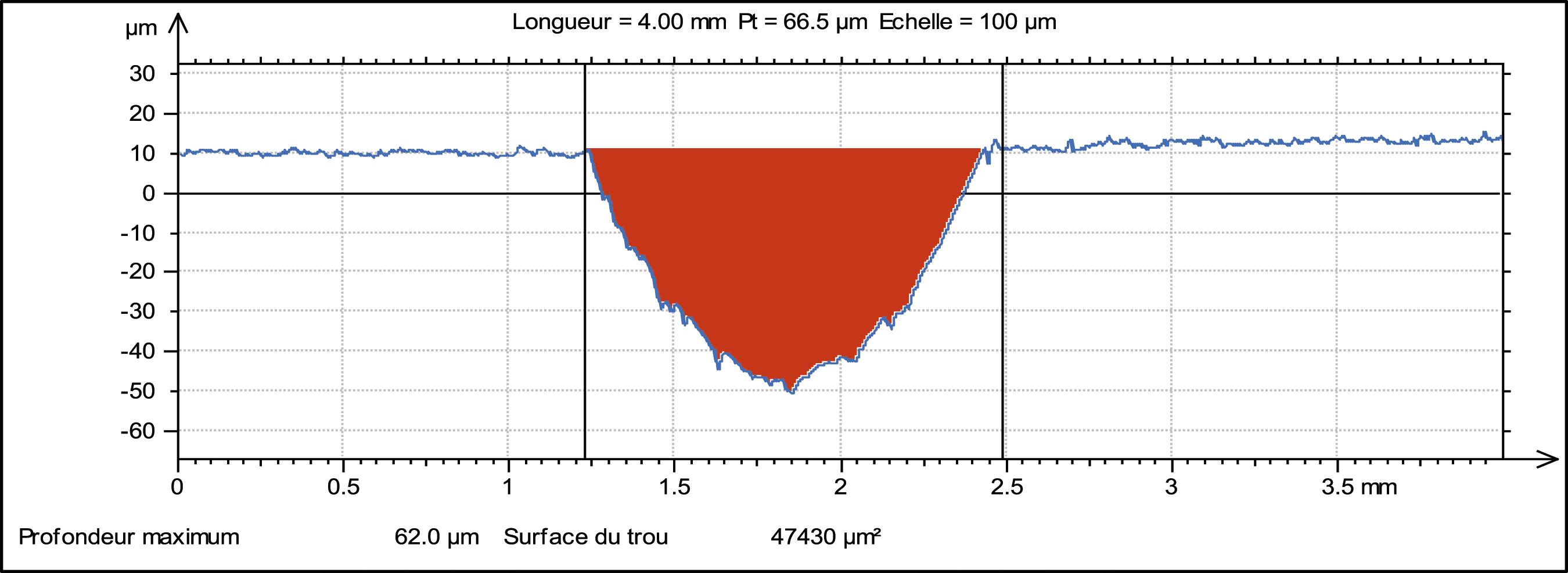
Mechanical tests
The bending strength, the bending modulus and the compressive strength of the elaborated composites are measured using a universal mechanical testing machine, known as TIME WDW-50E. The rectangular bar specimens with the dimensions of 64 mm × 10 mm × 3.3 mm are prepared for the three-point bending test, notably the 50 mm span and the 5 mm/min crosshead speed, However, the dental prosthesis is not without intricate challenges, notably the chewing forces and the presence of the acidic biofilms, which affect the prosthesis.
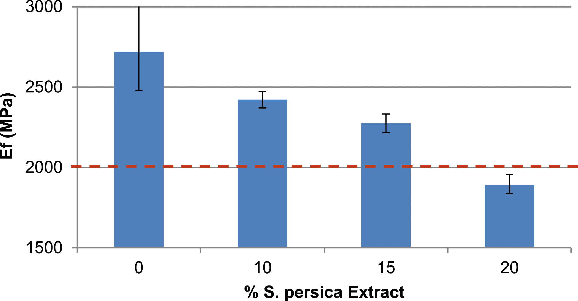
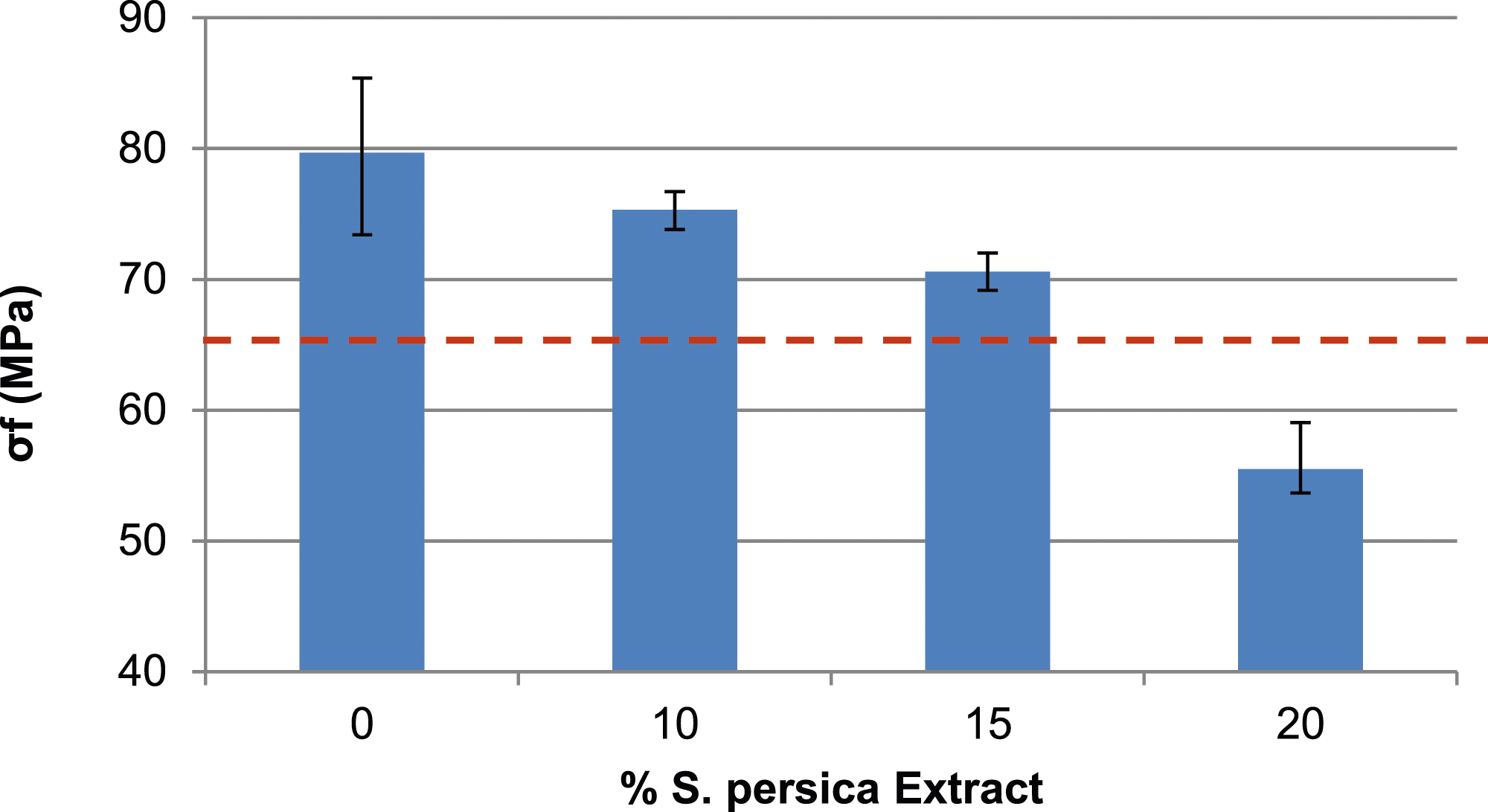
Complied with the requirements of the 20795-1:2013 ISO norm. The standard norm sets a limit of at least 65 MPa for bending strength and 2000 MPa for bending modulus to qualify composites as dental materials.
For the compressive test, the cylinder specimens dimensionally identified as Ø13 mm × 26 mm are prepared in accordance with ASTM D695 to measure compressive strength, using a loading rate of 1.3 mm/min.
Results and discussion
Extract of the particles size
The S. persica extract particle size distribution is embodied in Figure 3, which displays that the 70 nm is the value of the mean particle size. Particle size distribution of S. persica extract.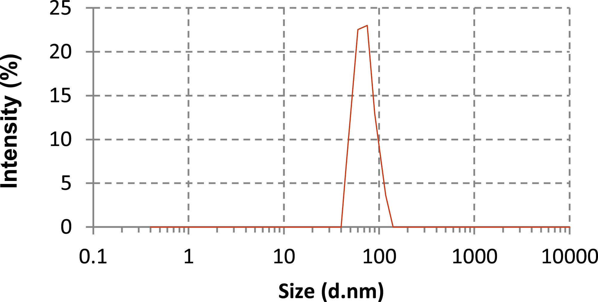
According to Damien et al, a nanocomposite is a multiphase solid material where one of the phases is nanofiller. Generally, a nanofiller is a filler, which has three distinct dimensions of less than 100 nm. 14 Accordingly, the S. persica extract is subsumed under the family of the nanofillers. In this respect, the composite, which contains this extract, will be recognized as a nanocomposite.
Antibacterial activity
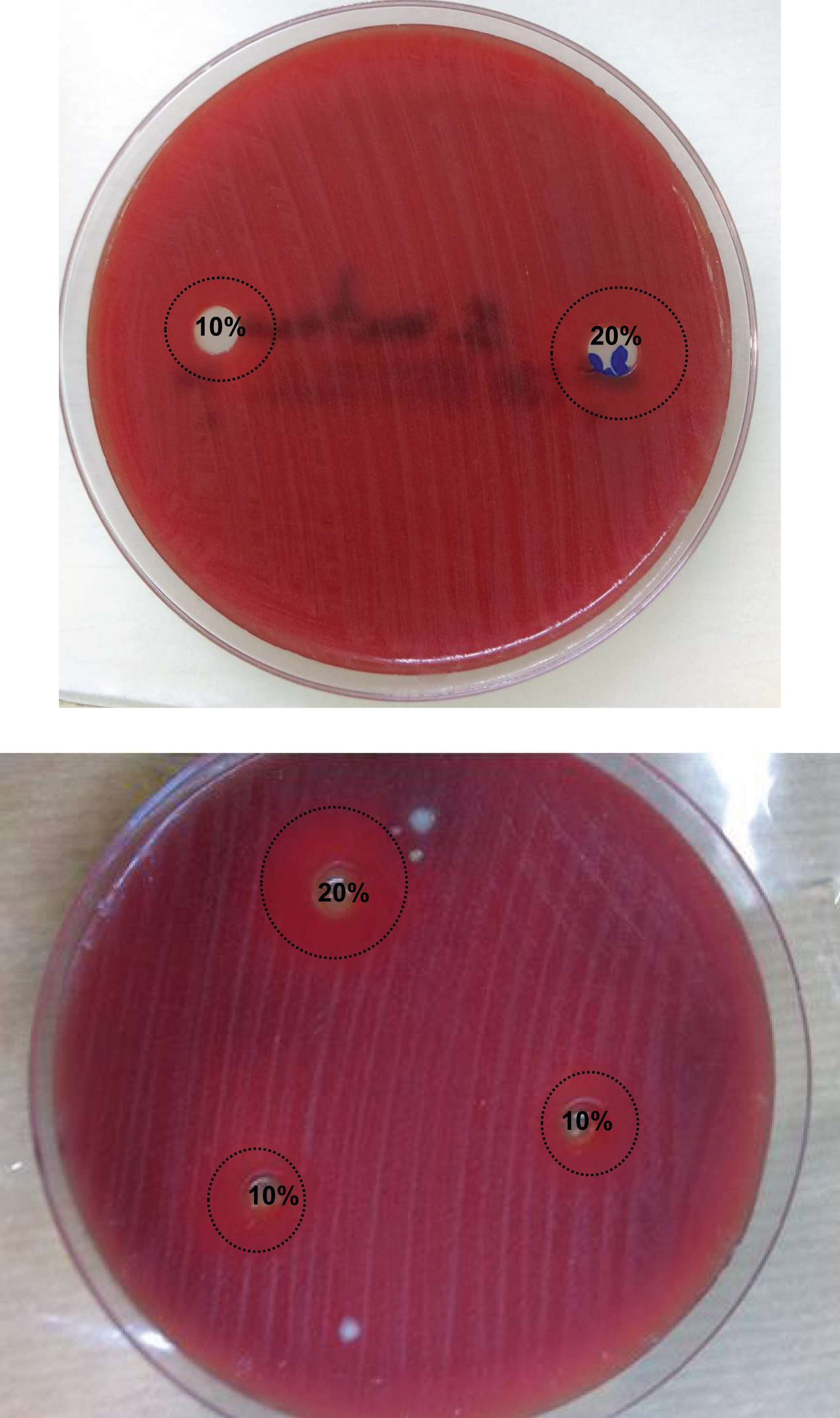
The antibacterial activity of the S. persica extract and its containing composite against the S. pneumonia, the S. mutans, and the H. influenza are shown in Figures 4 and 5. The results show a varying degree of the antibacterial activity. In fact, the extract is predominantly effective on all the strains, ranging from 16 to 18 mm with respect to the literature, one of the study indicates that the S. persica extract reveals a strong antibacterial activity.17,18 In addition, the 10% and 20% extract of the composites have less inhibitory effects on the growth of bacteria than the S. persica extract. However, they remain important for the 8 and 13 mm. An investigation carried out by Chaaben et al. on the antibacterial effect of the S. persica and the composite containing 30% of its powder on the same oral bacteria, confirms that the diameter of the inhibition zone of the composite ranges from 8 to 10.5 mm.
11
In fact, the composite, containing 30% of the S. persica powder, has the same antibacterial activity as the one with 10% of the extract of such a plant. Consequently, the composite with the S. persica, has a powerful antibacterial activity. Antibacterial activity of the extract of S. persica and the composites. Inhibition zone of composites containing 10% and 20% of the extract of S.persica against S.mutans bacteria.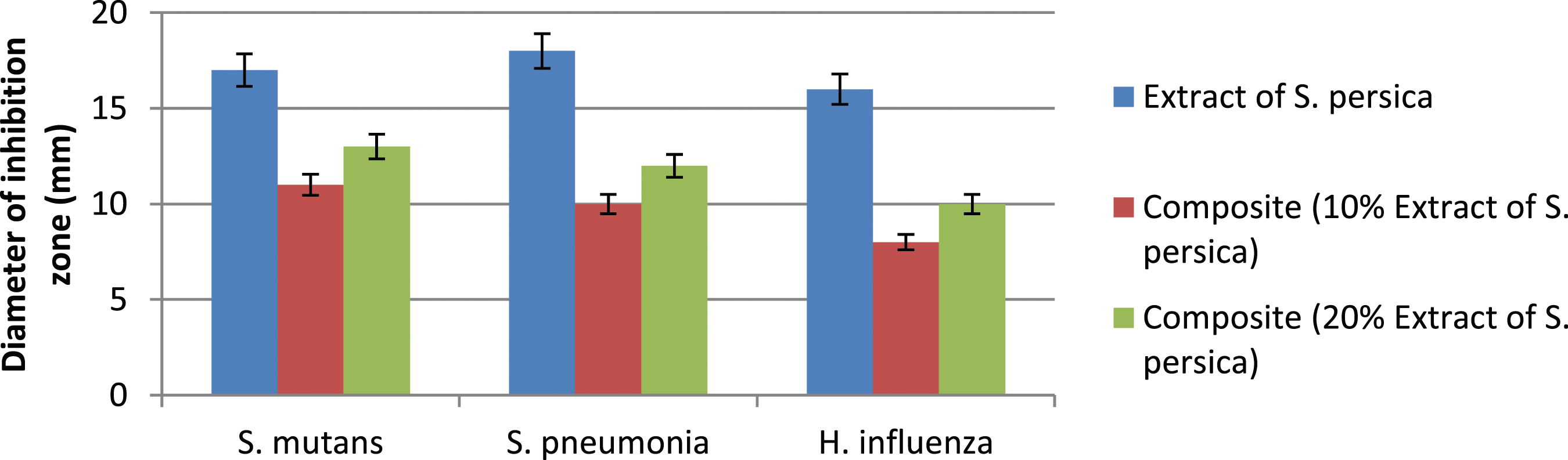
Once the antibacterial activity is confirmed, a check of the mechanical behavior of the material is necessary for dental use. Hence, the tribological and the bending characterization will be done. The findings are presented in the following sections.
Tribological response
Two important properties are investigated, which consist of the friction and the wear of the biocomposite with different rate of S. persica nanoparticles.
Friction coefficient
The evolution of the friction coefficient for the composites containing different percentage of the extract, against different applied normal load, is displayed in Figure 6. It is clearly noted that the friction coefficient increases with the applied load. This finding is mainly due to the plastic deformation of the asperities in the contact. Specifically, the friction coefficient goes minimally, matching a transitional course from the elastic contact to the plastic one. It is worth noting that the load is likely to change the temperature of the visco-elastic transitions in polymers and accordingly the friction mechanism itself.
19
Friction coefficient of composites containing different percentage of the extract of S. persica.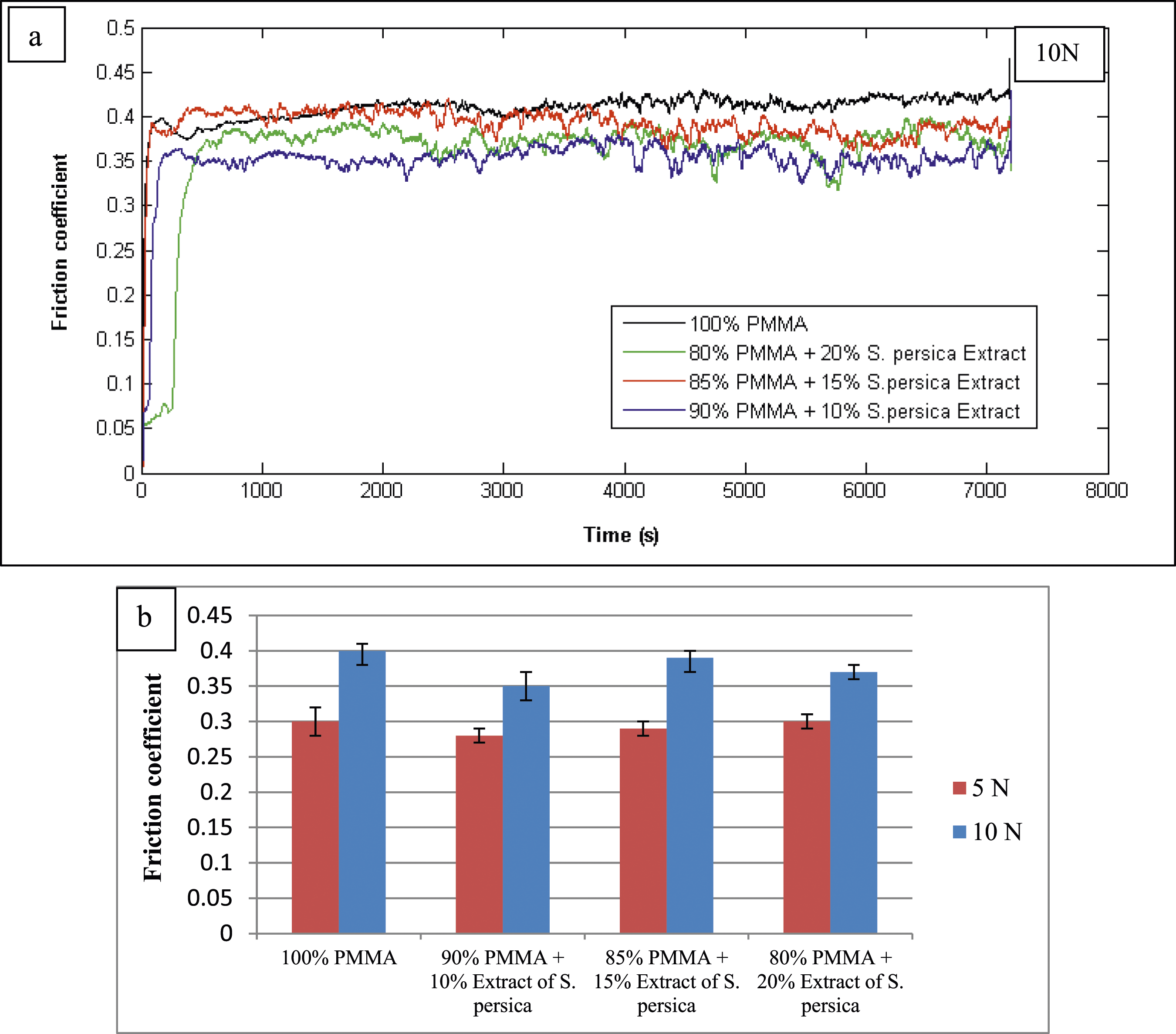
More importantly, the findings reveal that the evolution of the friction coefficients of composites/alumina is not affected by the filler’s addition (Figure 6(b)), with the exception of the incorporation of 10% of the filler causing the decrease of the friction coefficient. Aguilera-Camacho et al. have found similar results with the incorporation of the calcium oxide (CaO) nanoparticles into the PMMA. 20 The incorporation of a small percentage of CaO shows a lower mean friction coefficient value, as an average for all the loads, compared to the PMMA. 20
Wear volume
Figure 7 shows the evolution of the wear volume in accordance with the percentage of the S. persica extract with two applied loads. The wear volume which increases with the applied load becomes greater and greater. In this respect, it is influenced by the incorporation of the extract. The wear resistance is enhanced by the addition of 10% of the extract with all the applied loads. Previous research has demonstrated that the incorporation of nanofillers in the resin have the potential of enhancing the wear resistance of the composites.20,21 However, beyond 10% of the extract, the wear resistance is reduced particularly when the 10N is applied. This is possibly caused either by a lack of a coupling agent between the fillers and the polymer matrix phase or by a bad dispersion of nanoparticles, while forming particle agglomerates. Karthikeyan et al.
22
note that the tribological performance of a material depends on various factors occurring with the interfacial adhesion matrix/filler and its type (chemical or mechanical). Equally, they mention that the agglomeration of the nanoparticles increases the stress concentration at specific sites. It is actually attributed to the decrease in strength as well as the wear resistance performance.
22
Wear volume of different composites under 5 and 10N normal loads.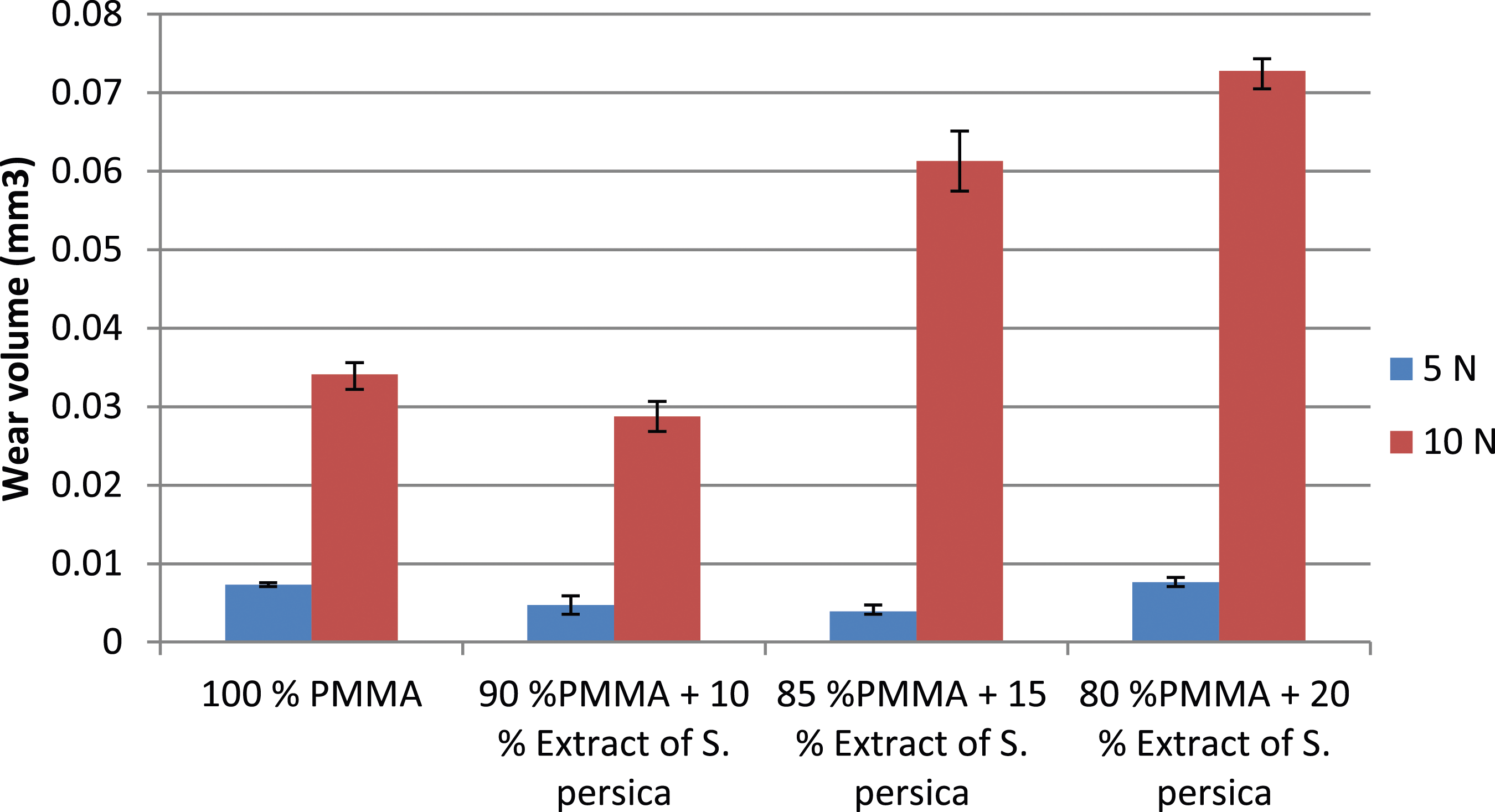
As a conclusion, the optimal fraction of the extract, which gives higher wear resistance, is 10% with both applied loads.
Wear mechanism
The SEM micrographs, which are tested through 5 N and 10 N normal loads show wear tracks of PMMA.
From Figure 8(a) and (b), plastic deformations are found. Indeed, a crack network proves that the wear mechanism of PMMA at the lower normal load is both an adhesive and a fatigue wear. In fact, the adhesive wear is evidenced by the presence of the galling forms at the contact. A general principle of the feature of adhesive wear is the transfer of material from one surface to another due to localized bonding between the contacting solid surfaces.20,23 Several studies have reported similar adhesive wear for PMMA sliding against different counter-face.20,24,25 In addition, some micro-cracks are noticed due to the micro-fatigue phenomenon and the stress concentration on the surface of PMMA. These micro-cracks facilitate material removal, in the form of debris, during alumina ball sliding. Indeed, PMMA is a fragile material, demonstrating low fracture toughness KIC.
26
Consequently, the considerable ability of PMMA to create debris is related to its low fracture toughness. SEM of PMMA wear track under different normal load 5N (a and b) and 10N (c and d).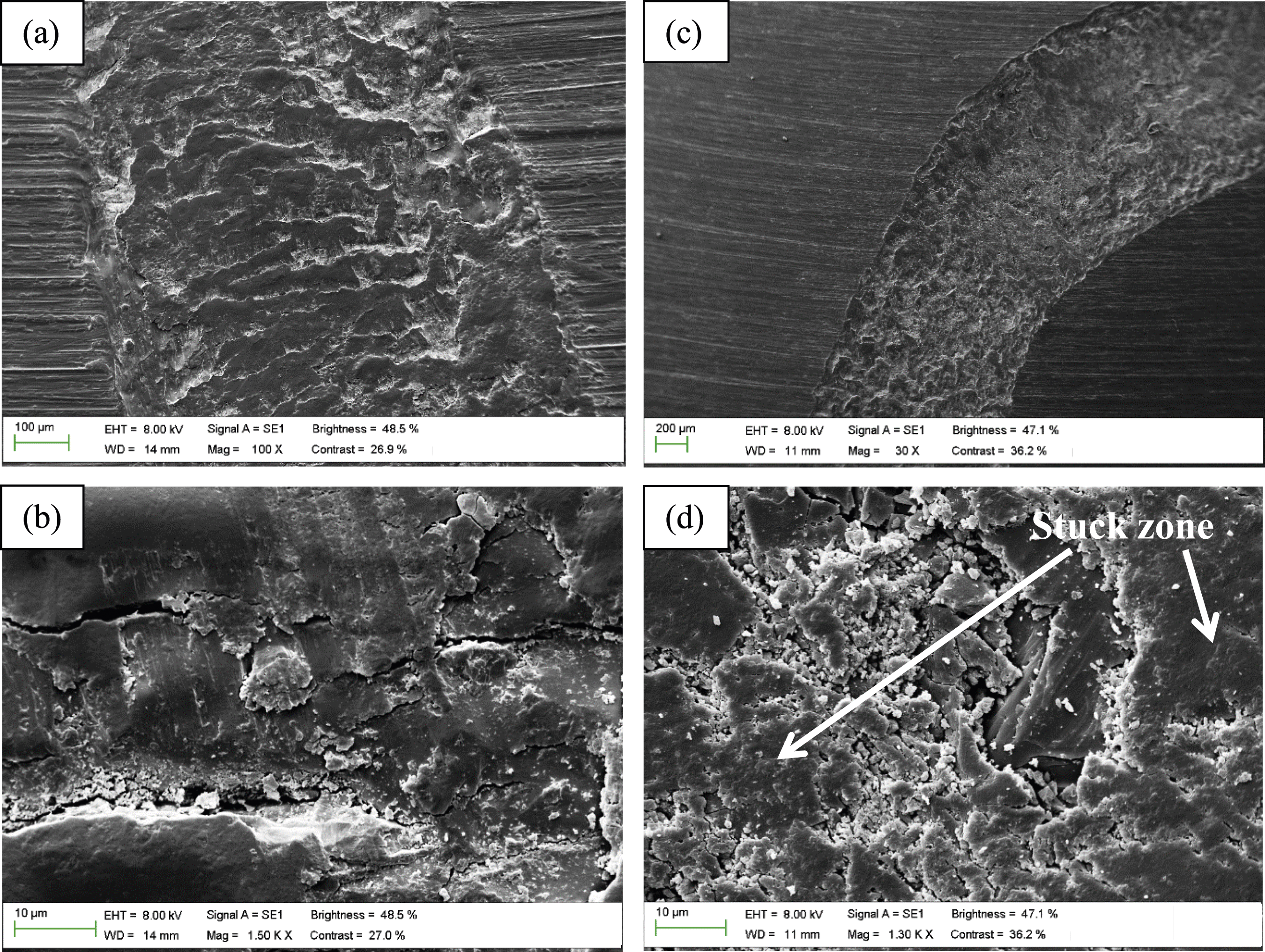
As a result, the more the normal load increases to 10N (Figure 8(c) and (d)), the more complicated the wear mechanism becomes. It is referred to the high deformation of the plastic. During the running-in period, the mechanism still remains a combination of adhesive and fatigue wear, leading to slight damage on the contact. With the evolution of cycles, the fatigue wear mechanism becomes much more severe and the generation of debris becomes more and more important. Consequently, the debris produced from the highly loaded surface contacts by fracture are trapped between the two sliding surfaces. They are crushed into finer particles. Consecutively, they become compacted into the wear track to form a stuck zone and smooth area at the contact surface. Just after the stuck zones are submitted to plastic, deformations and fractures appear, which again allow the detachment of the material and their evacuation outside the wear track. This mechanism is cyclically repeated.
After the incorporation of the filler, the SEM micrographs for ‘PMMA/10% S. persica Extract’ worn surface exhibit changes in the surface topography following the wear measurements (Figure 9). It is noticed that the wear track width significantly decreased (Figure 9(a) and (c)). Indeed, the fatigue wear discloses an evident change due to the extract nanoparticles incorporation. Cracks would have been less likely to develop. Therefore, less wide and less deep cracks are observed at higher magnification (Figure 9(b) and (d)). Consequently, lower production of debris is perceived in the case of the composite. In this respect, the wear volume decrease is observed. This composite is accounted for the softened fatigue wear. SEM observation of PMMA (a and b) and composite (10% Extract of S. persica) (c and d) wear track (case of 5N normal load).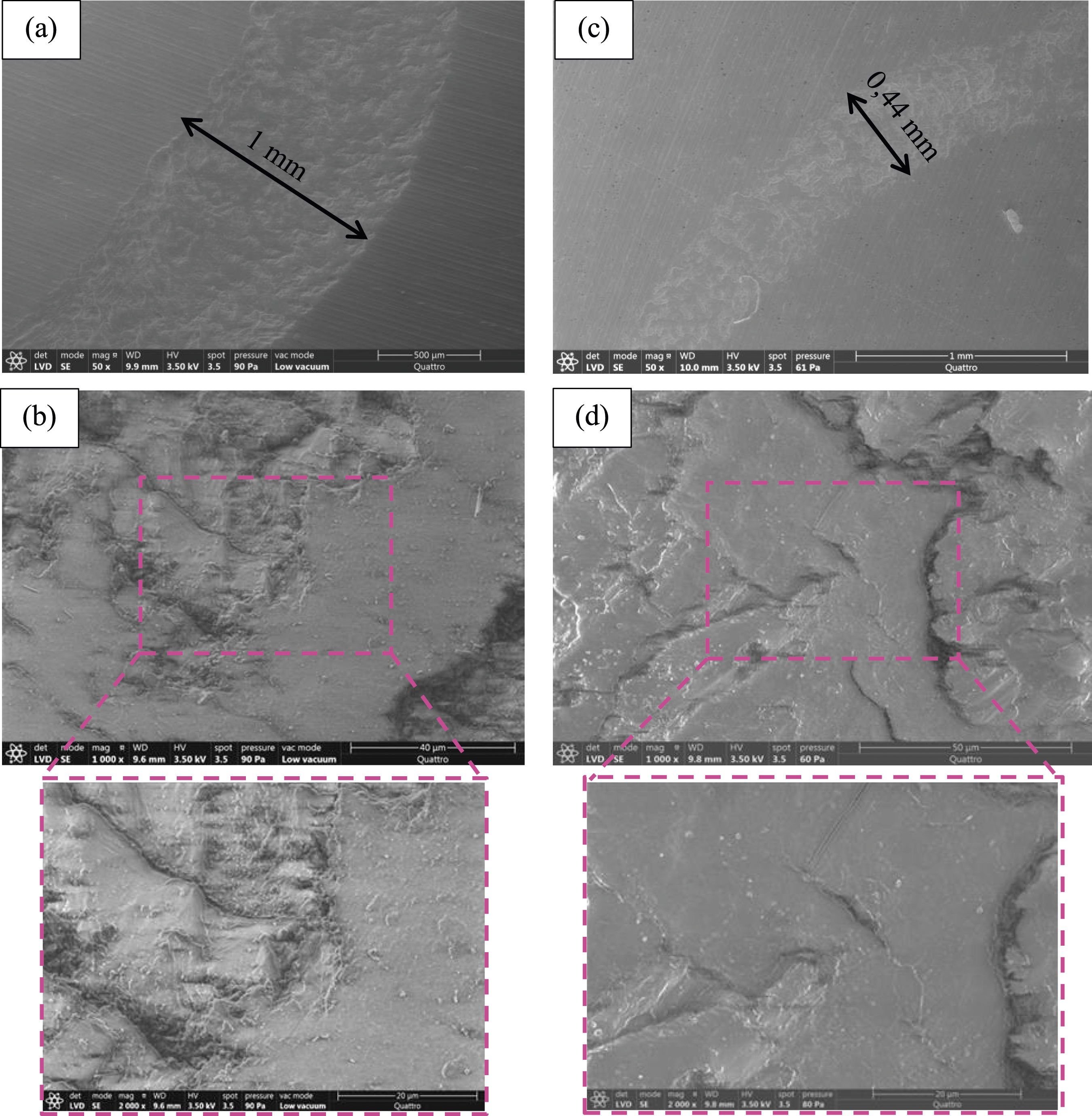
Mechanical properties (bending properties and compressive strength)
The bending strength (σf), the bending modulus (Ef) and the compressive strength (σc) of dental composites are presented in Figures 10–12 respectively. These figures show an obvious change with regard to the extract nanoparticles incorporation. Comparing to PMMA, the incorporation of the 10% extract has an enhancing effect on the final composite compressive strength. Nevertheless, the addition of the 15% and 20% extract lead to a reduction in the compressive strength. The bending strength (σf) and the bending modulus (Ef) are decreased by the increase of the filler’s percentage. However, they remained above the indicated limits of the standard norm by adding either 10% or 15% of the filler. Compressive strength (σc) of composites reinforced with different percentage of S. persica extract. Bending modulus (Ef) of composites reinforced with different percentage of S. persica extract. Bending strength (σf) of composites reinforced with different percentage of S. persica extract.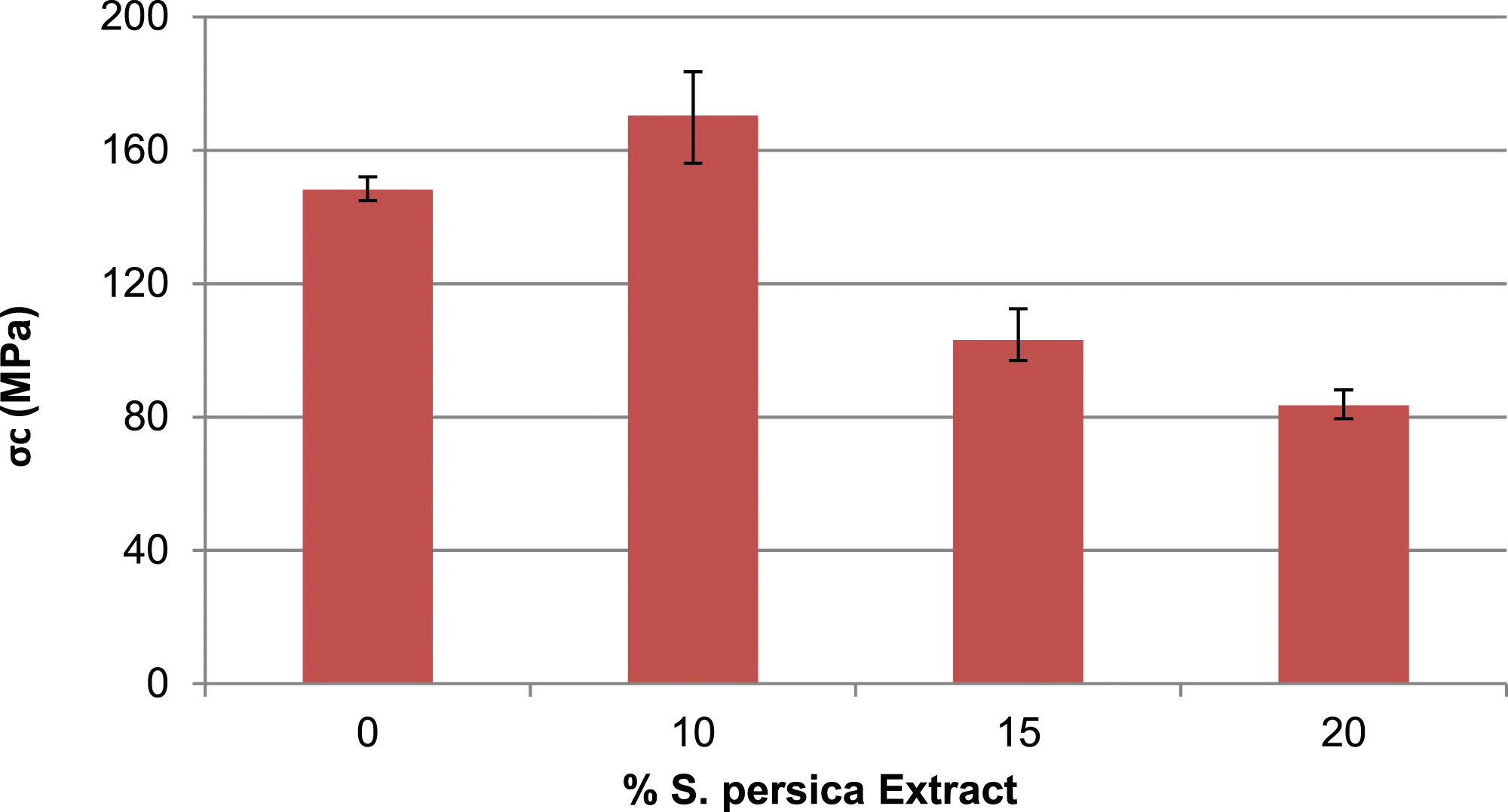
These findings are possibly associated with the size, the particles dispersion quality and the volume percentage of the nanofiller.14,27 Over the past few decades, research on dental composites has primarily focused on reducing the size of filler particles, transitioning from several microns (>10 µm) in traditional composites to sizes below 1 µm in modern composites. Smaller particles offer a more favorable surface area-to-volume ratio, leading to increased interaction with the organic matrix.14,28 It is expected that the mechanical properties of composites filled with nanoscopic extract can be enhanced by achieving a more homogeneous dispersion of nanoparticles in the organic matrix. The findings for the nanocomposite containing 10% extract demonstrate that the improvement in polymer compressive strength is associated with well-dispersed nanoparticles of the extract. However, if the percentage of incorporated nanoparticles increases, they tend to form mesoporous agglomerates.23,28 This agglomeration negatively affects the adhesion between the filler and the polymer, consequently impacting the mechanical properties.
Moreover, one of the key parameters governing the properties of nanocomposites is the interaction between the filler and the matrix. This interaction is largely influenced by the application of chemical treatments to the surface of the filler,14,29 which is feasible for most types of nanofillers. However, in the present case, the antibacterial activity of the used extract is significantly impacted, potentially leading to its degradation when the surface structure is altered.
Conclusions
A new denture teeth material incorporating S. persica extract as a bioactive agent has been developed, demonstrating strong antibacterial activity against oral bacteria. This innovative composite material exhibits excellent wear resistance without compromising its mechanical properties, largely depending on the composition of the mixture. The inclusion of an optimal amount of S. persica nanoparticles (10%) has optimized the properties of the composite.
Footnotes
Declaration of conflicting interests
The author(s) declared no potential conflicts of interest with respect to the research, authorship, and/or publication of this article.
Funding
The author(s) received no financial support for the research, authorship, and/or publication of this article.