
Abstract
Polylactic acid (PLA) has been attributed as one of the most significant biodegradable polymers that hold great potential to replace petroleum-based polymers in the near future. This has motivated the chemists, surgeons, industrial engineers as well as research scholars to consider PLA and its copolymers for various biomedical applications including scaffolds, sutures in tissue engineering, and drug carrier vessel for delivery. However, the intrinsic limitations of the PLA in terms of surface integrity, cell adhesion, and degradation are restricting the practical implication at par with the required capabilities. Therefore, in the present review article, an attempt has been made to categorize the various posttreatment processes which can benefit the users to eliminate the existing barriers. Along with, the impacts of the various classes of posttreatment technologies, for PLA and its composites, have been summarized on the basis of outcomes. Overall, the review article will provide technical pathways for the betterment of PLA with practical insights.
Introduction
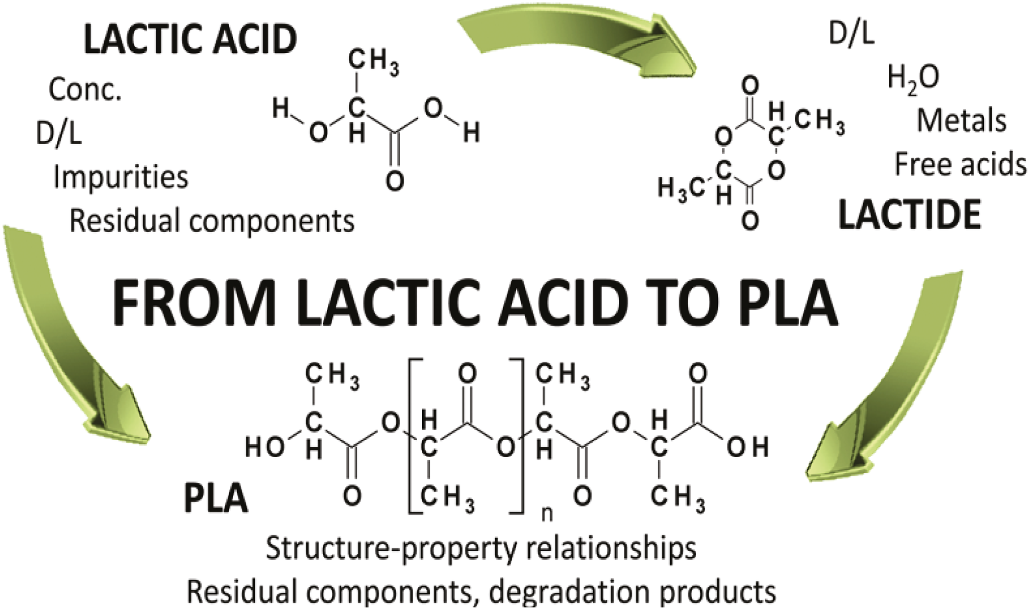
Second half of the 20th century gave rise to a range of materials for restoration and replacement of some tissues inside and outside the human body. 1 Usually, metals (such as stainless steel and titanium alloys), ceramics (hydroxyapatite), and natural polymers (chitosan) are utilized for orthopedic or medical applications. Due to their existing limitations, these materials are being replaced by new and versatile polymeric systems. Keeping these facts in mind, large considerations are being given to natural polymers in biomedical field due to which the biodegradable materials consumption is increasing day-by-day. 2 Polylactic acid (PLA) and its copolymers (such as poly(lactic-co-glycolic acid) (PLGA)) are biodegradable polymer that is extremely versatile and derived from 100% renewable resources. Therefore, due to agricultural origin and biodegradability, PLA is one of the most promising materials for various applications. It is generally prepared by the ring-opening polymerization of lactide, as shown in Figure 1. PLA provides outstanding results, while being cost-effective, owing to its desirable molecular weight. For instance, it plays a significant role for repairing internal fixation of damaged bones and joints. Table 1 shows the selected physical and chemical properties of various types of biopolymers.
PLA production method. 3
Some selected physical and chemical properties of PLA-based polymers. 4
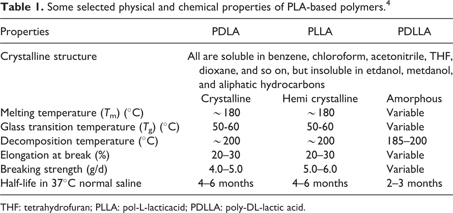
THF: tetrahydrofuran; PLLA: pol-L-lacticacid; PDLLA: poly-DL-lactic acid.
Further, the elastic modulus of pure PLA is quite low (2–7 GPa) in comparison to natural cortical bone (3–30 GPa); hence, the combinations of PLA are used in distinctive applications. 5 –9 Drug delivery systems, vascular prosthesis, orthopedic surgery, and so on, are some of the major areas of its clinical applications of PLA. The applications of different types of biopolymers (PLA, its copolymers, and others) for vaccine development such as delivery traditional drugs, proteins, and nucleic acids for gene delivery are summarized in Table 2.
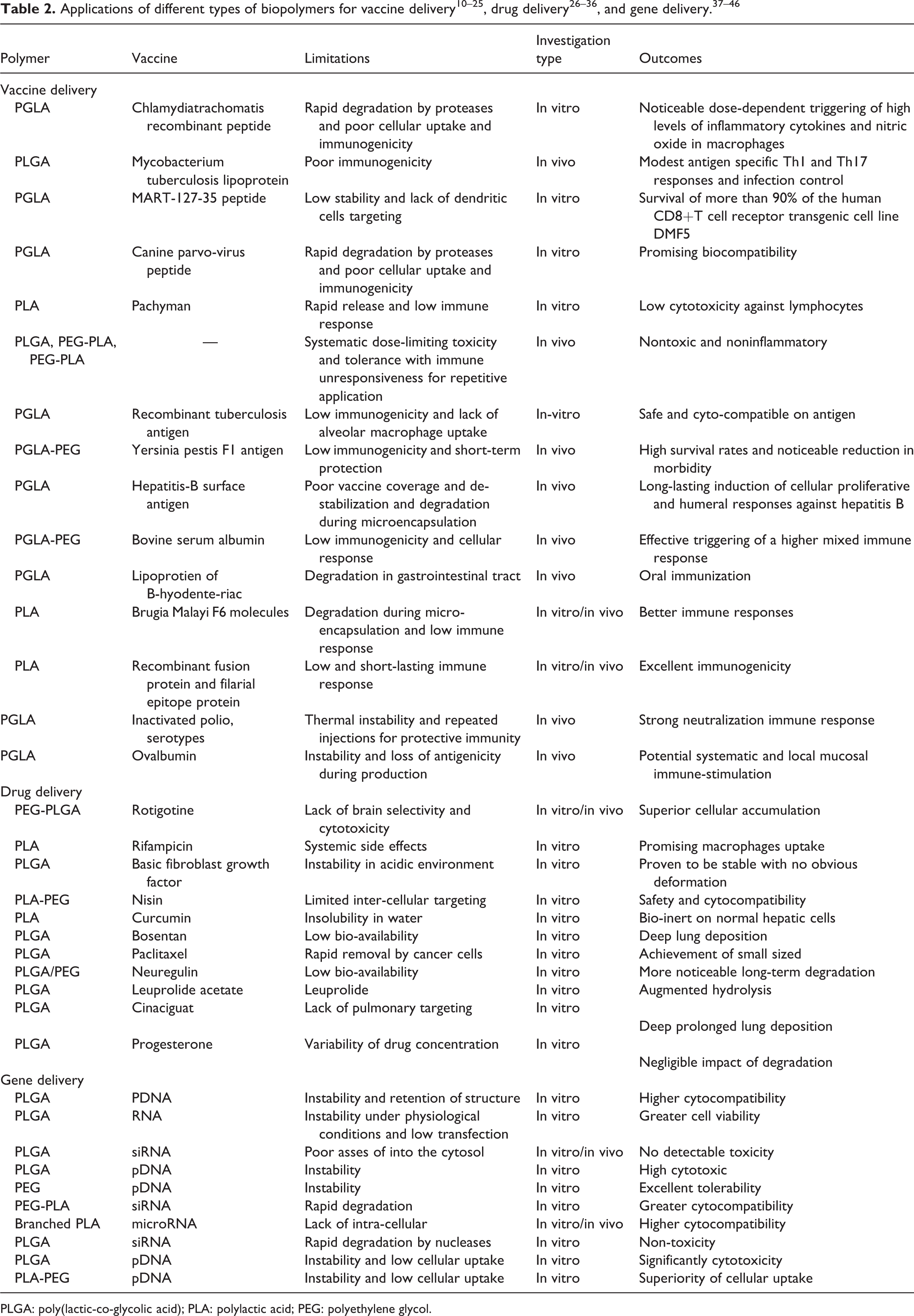
PLGA: poly(lactic-co-glycolic acid); PLA: polylactic acid; PEG: polyethylene glycol.
Moreover, PLA implants are highly biocompatible and do not have any carcinogenic or toxic effects on the tissues. As these polymer implants remain in the body for a short time and then disappear upon degradation, therefore, it is not essential to remove. 47 Thus, they are approved to be in direct contact with biological fluids as per the Food and Drug Administration (FDA). PLA implants are much easier to process when compared to polyethylene glycol (PEG) or materials such as polycaprolactone (PCL). These are being manufactured using film extrusion, injection molding, thermoforming, and fiber spinning. 48 Moreover, the production processes for PLA are much more efficient than those used for petroleum-based polymers because they consume 25–55% less energy and are thus relatively cheaper. 49 Indeed, PLA can be used successfully for biomedical applications if its surface properties such as hydrophobicity, reactive functionalities, and roughness can be controlled for specific applications. PLA has some limitations as well, having poor toughness, lack of reactive side-chain groups, slow degradations, and hydrophobicity when compared with the other competent biopolymers. However, the only disadvantage related with PLA is its high brittleness, which restricts it for some practical applications. However, the flexibility of pol-L-lacticacid (PLLA) has been enhanced by a number of modifications. For the time being, the use of PLA is limited for plastics which need deformation at high-stress levels. 48 –50 Despite all, the degradation, molecular weight, and molecular weight distribution of PLA can be controlled through its crystallinity, which usually lend it a longer in vivo lifetime. 51 –54 Being hydrophobic, PLA has low cell affinity and can cause inflammation in living host tissue when in direct contact with biological fluids. 55,56 Further, toughness and degradation rate of PLA can be improved using bulk modification and its hydrophilicity and surface integrity can be controlled using surface modification as required in biomedical engineering. Indeed, both surface and bulk properties are essential for fabricating a successful biomedical device. The mechanical properties of the polymeric products majorly depend on the process conditions of the adopted methodology. Therefore, negligible scope for the further enhancement exists. However, the surface characteristics could be significantly affected through the posttreatment methods and hold a high importance in the successful candidature presented by the resulting products. Thus, the present review article is focused on discussing different types of posttreatments available for obtaining an efficient biomedical device. 57 –60
Surface modification of PLA
To determine the applications of the materials, their surface properties play a crucial role, especially biocompatibility. PLGA has received the maximum attention among various other polymers because of its favorable degradation characteristics and biocompatibility. 61 Moreover, its physicochemical properties can be customized by controlling their synthesis metabolic pathways. The surface interactions of any material play a vital role in deciding its various properties under different conditions. The treatment of reinforcement for polymer composites for biomedical applications has emerged as an important domain to be explored. Therefore, it very essential to understand the behavior of PLA while working for biomedical applications. 62 Various synthetic and natural polymers are used these days for customizing the properties of PLA substrates by using various processing techniques as shown in Table 3. Therefore, a lot of research has been carried out to modify the surface of PLA to make it appropriate for specific biomedical applications. The modification of PLA and its copolymers is performed to improve its surface chemistry and bulk properties. 53 Therefore, many methods have been investigated to modify the surfaces of biopolymers, to make these suitable for human tissues, through capitalizing on quantum modification or copolymerization with other lactone-type monomers. Moreover, in these types of materials, monomers with functional play vital role in effecting the degradation rates and surface properties of PLA-type polymers thus, can be considerably enhanced. Surface modifications of the polymers via coating and plasma treatment can further enhance affinities of PLA polymers. This peculiar competency to enhance the properties of PLA-type polymers empowers them with exceptional biocompatibilities, biodegradability, and cell affinities. 53 The “ideal” polymer particles must be decomposable, with toxicologically safe in vivo degradation products, which can be easily eliminated possess promising future for tissue engineering, other human health and patient care as pure PLA can cause minor inflammatory reaction upon implantation. Therefore, much research is going on this field for enhancing the properties of PLA-based devices. This portion of the review highlights the various strategies required for sophisticated applications.
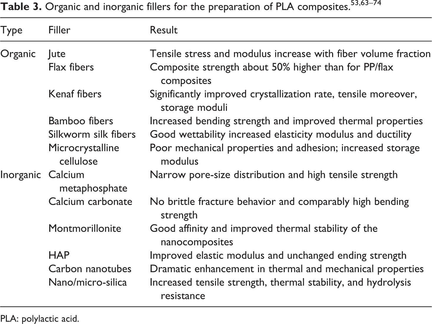
PLA: polylactic acid.
Nonpermanent surface modification methods
Coating
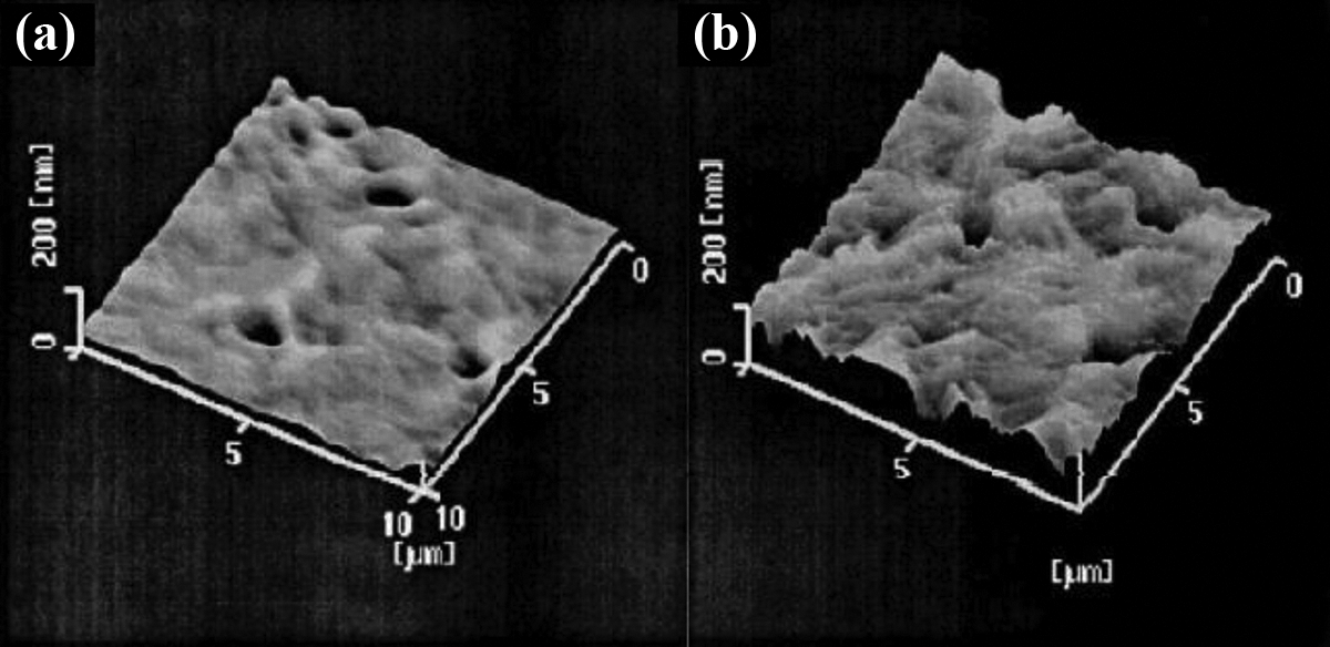
The interface between artificial materials and living tissues is a highly critical research area mainly with respect to the use of scaffolding materials in tissue engineering and reformative medicine. Numerous aspects are considered in the design of scaffolding materials for specific applications such as the release of toxic degradation products or biomechanical interactions between cells and scaffolds. The surface modification of PLA can be done through the coating, by depositing a layer of modifying material on the surface. Bio-mimetic apatite and extracellular matrices (ECMs) proteins (such as fibronectin, collagen, and vitronectin) are used for controlling the cell interactions of PLA. Chen et al. 75 investigated the biomimetic coating of PLA/PLLA by using a phase separation technique, while surface cell morphologies of PLLA scaffolds were studied using scanning electron microscopy (SEM) as depicted in Figure 2. The results revealed that the biomimetic processes can enhance interfaces amid osteoblasts and the polymeric scaffoldings. In another similar study, investigations were made on the surface hydrolysis of polyester scaffolds by Atthoff and Hilborn. 76 The examination of the influence of hydrolysis on protein adsorption carried on three materials, such as PLA, polyethylene terephthalate (PET), and polyglycolic acid (PGA), with the utilization of X-ray photoelectron spectroscopy (XPS) technique and SEM showed an increment in adsorption rate with no in vivo degradation of proteins on polymer. Further, post-hydrolysis treatment using acetic acid instead of counter-ion enhanced the protein attachment to the surface. The weight loss results for PLA and PGA are shown in Table 4. Specifically, all the measurements were taken for the contact angle to check the hydrophilicity. Many more significant researches were performed for studying the effect of coatings, for instance, Cronin et al. 77 compared the efficacy comparison of coated and noncoated PLLA fibers for refurbishing impaired skeletal muscle tissues. The fibers upon coating with extracellular matrix proteins can provide a scaffold for the progress of skeletal muscle tissue for engineering and cell transplantation applications. The micrographical difference can be clearly shown in Figure 3.
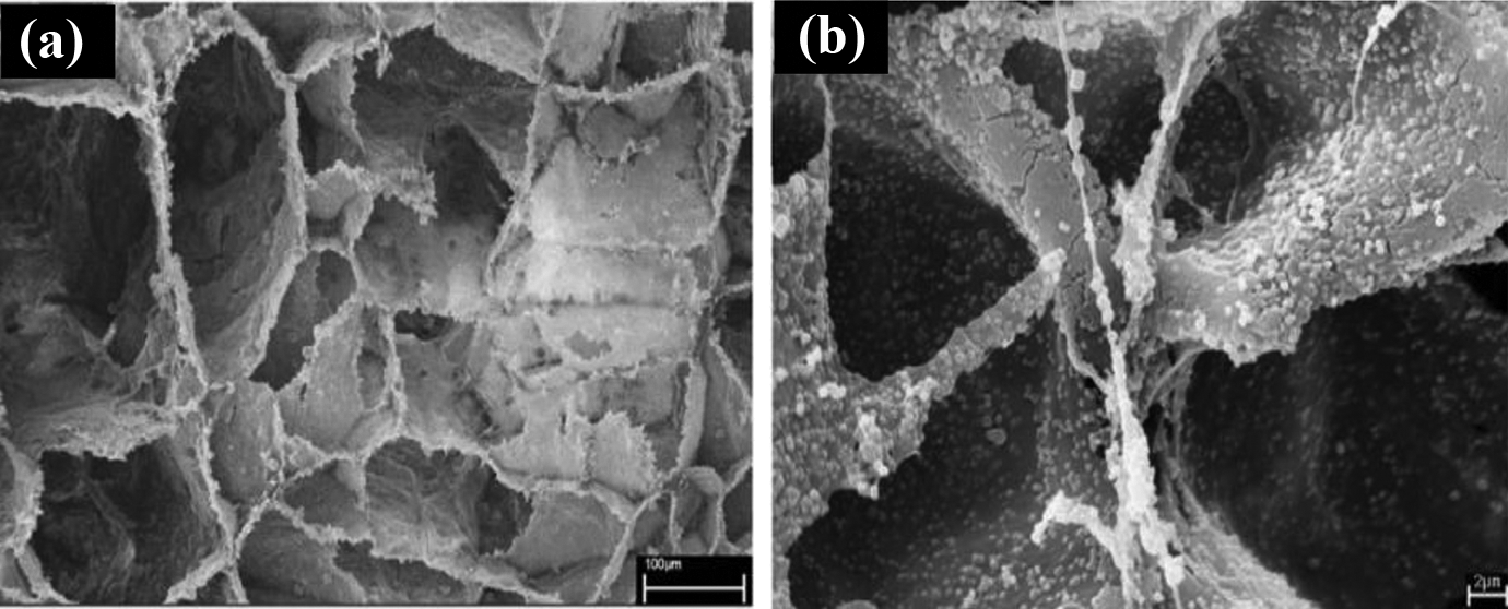
SEM images (a) with apatite coating and (b) apatite/collagen coating. 75
Effect on weight and contact angle of PLA and PGA. 76
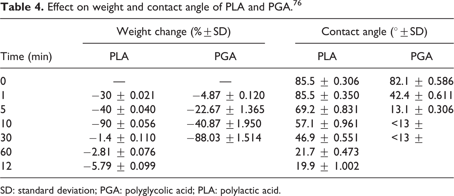
SD: standard deviation; PGA: polyglycolic acid; PLA: polylactic acid.
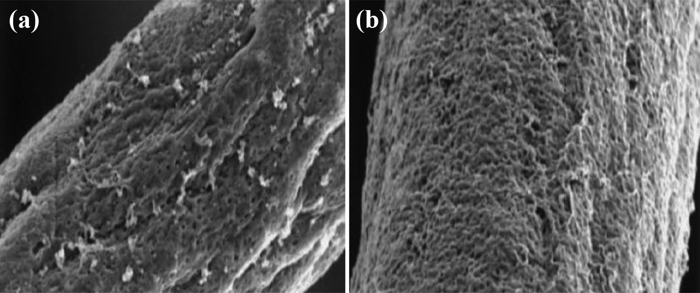
SEM images of coating (a) PPLA fibers before and (b) PLLA after ECM gel coating. 77
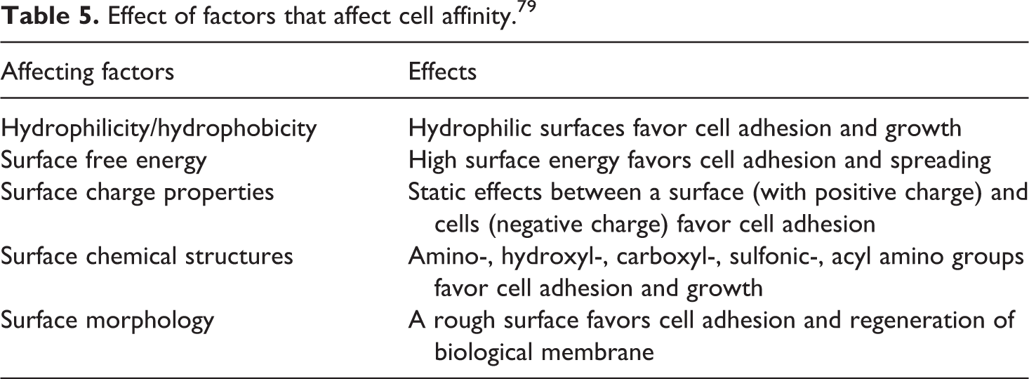
Micropatterning technique for pattering proteins and cells on PLA and PLGA substrates that are used as scaffolds in engineering tissues was presented by Lin et al. 78 It can be used for the positioning of the cells specifically on the substrate. Although pattering technique is very much used for cell patterning using scanning electron microscopy (SEMs) on metal substrates, but it is very limited for biomaterials. Therefore, in this study, poly-(oligo-ethylene-glycol-methacrylate) (poly-OEGMA) or poly-(oligo-ethylene-glycol methacrylate-co-methacrylic-acid) (poly-(OEGMA-co-MA)) was micro-contact printed onto substrates to create cell resilient areas. Proteins adsorbed onto the unprinted regions, whereas the polymer-printed regions successfully repel nonspecific protein adsorption. This process for controlling the spatial morphology and distribution of cells on synthetic biomaterials could play an important role in tissue engineering (Table 5). Eid et al. 79 studied the surface modification of PLGA through the use of synthetic peptides as a coating medium. The assessment was carried out on unicortical tibial osseous rat wounds (5 mm diameter). The results indicated that the new osteo-compatibility stages were enhanced in-growth by arginine-glycine-aspartic acid (RGD) coating. It reveals that the coatings of bio-ceramics improved the mechanical, biological, and microscopical efficiencies of the produced polymeric products, significantly. However, being delicate and fragile, there exist numerous barriers for developing a uniform layer of coatings on PLA. The stability of micropatterned NIH3T3 fibroblasts on PLGA films is shown in Figure 4. Additionally, fibronectin (FN) immobilization on PLA can improve cell adhesiveness, which may further enhance cell attachment for tissue regeneration. 80 Further, the laminin-coated scaffolds and their applications have been witnessed widely. It assists in the development of skeletal muscle tissue substitutes and may also be used for in vitro 3D models to study the development of muscle as well as for drug detection. 81 In another study, polyelectrolytes (PEs) were coated on low-molecular weight PLA by using a layer-by-layer technique for the production of nano-particulate drug delivery systems with better biocompatibility and continuous/targeted release of drug substances. 82 Coating with the help of phase-locked loop highlighted an appropriate approach for functionalizing the polymer surface and improving adhesion, propagation, and mineralization of jaw periosteal cell. 83,84 Moreover, the bio-inspired coating synthetic PLA polymer can be used as a simple technique to render the surfaces of synthetic scaffolds active, thus allowing them to direct the specific responses of human adipose-derived stem cells. 85 In this research, the preparation of polymer blend films between PLA and poly-(butylene-succinate) coated with various chitosan concentration was performed to introduce antibacterial activity to the films. Corona treatment with different electricity input was used to modify film surfaces before chitosan coating to increase ability of films to adhere with chitosan. 86 In a study, it has been found that the initial cell attachment of human gingival fibroblasts can be enhanced by immobilizing fibronectin onto PLA through condensation reaction by the use of water-soluble carbodiimide. The resulting products can be used as biomedical adherent for the skin, capping materials for dental pulp, or as lesion-dressing material. 87 Similarly, researchers have loaded the lapatinib onto PLA microspheres and used the same for delivering drugs and tissue engineering. 88
Effect of factors that affect cell affinity. 79
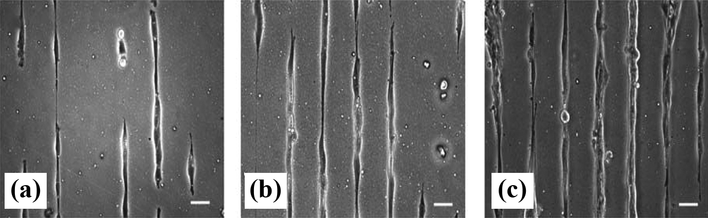
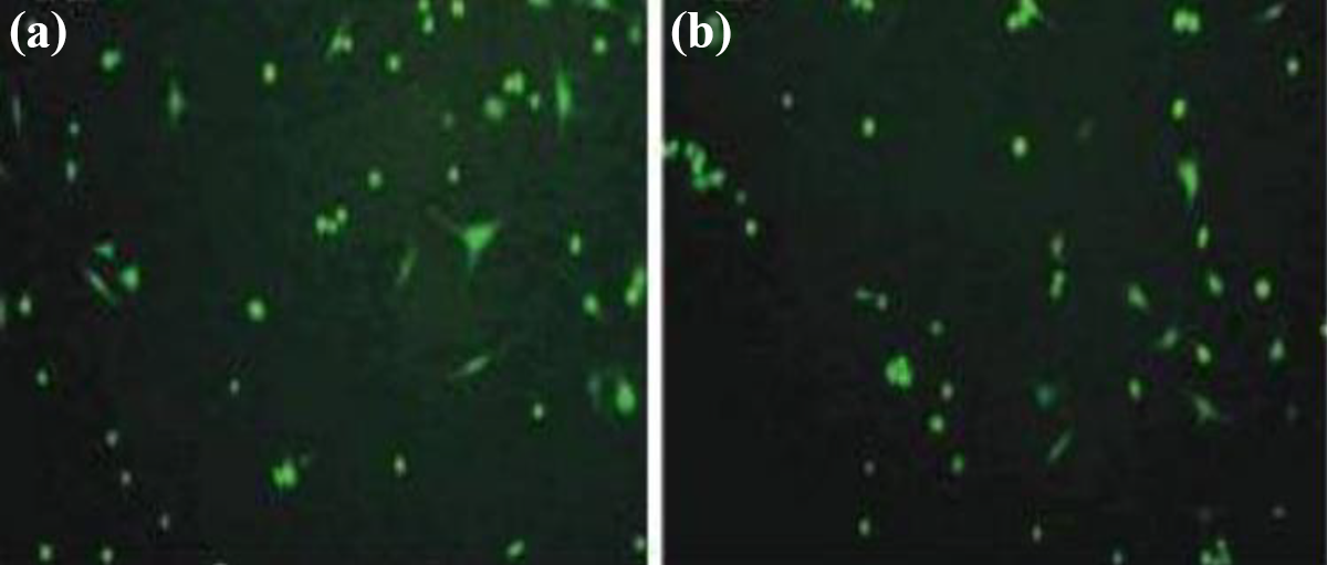
NIH3T3 fibroblasts on PLA films 78 (reproduced after permission).
Entrapment
Entrapment process is a surface modification method utilizing surface physical interpenetration network (SPIN). This is done by performing modifications on the polymer surface by incorporating altering species into the polymer surface region using the reversible swelling property in a categorical solvent/nonsolvent substrate. The advantage of this process is in terms of its capability of rapid entrapment for highly dense feedstock materials. Moreover, it is a simple method that does not require any further synthesis of incipient polymer structures as depicted in Figure 5. Zhu et al.
89
studied the modification of poly-
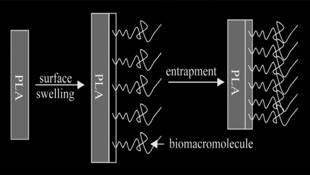
Schematic diagram of the entrapment process 89
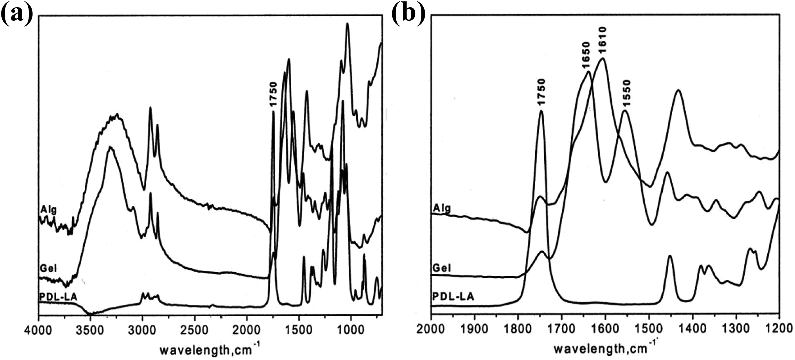
ATR-FT-IR spectra of PDL-LA films modified with biomacromolecules (a) and PDL-LA virgin films gelatin-modified, and virgin films (b).
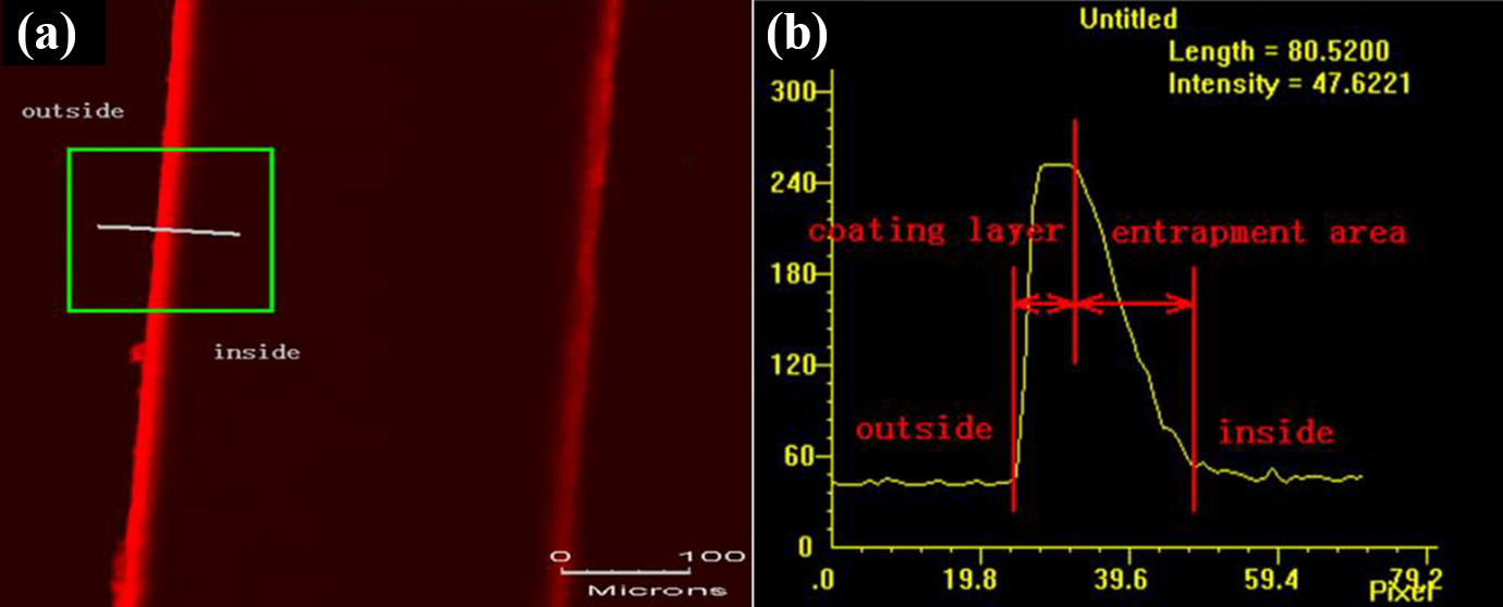
(a) Gelatin modified PDLA film (b) gelatin layer from outside to inside. 89
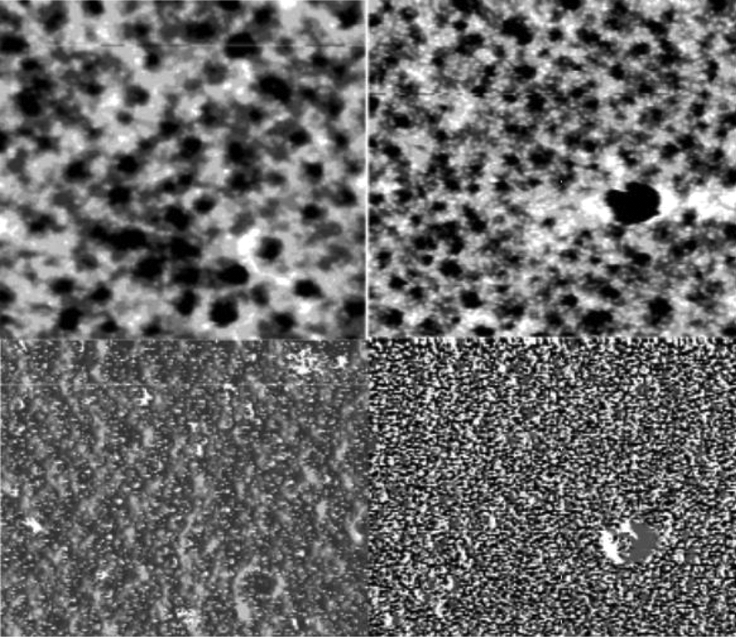
On similar lines, Zhang et al. 90 studied the surface modification of PLA materials using entrapment method for controlling protein adsorption in greater density values of PEG. The change in physical properties was observed using scanning thermal microscopy (SThM). The results showed that the addition of PEG caused a reduction in the glass transition temperature (Tg) of altered PLA, significantly. Additionally, there was no such change was observed for PEG-modified PLA region. Further results showed that the entrapped PEG mixed with PLA were consistently dissolved. The results indicated that the observed thermal dependence exhibited a divergence from the temperature depending on the thermal conductivity of the parent PLA. Lin et al. 78 investigated and maintained the lung epithelium using ECM proteins, covered on tissue culture plastic and poly-DL-lactic acid (PDLLA). The results showed that that selection of the coating protein could significantly disturb the differentiation of embryonic stem cells (ESCs). Moreover, ECM-degradable scaffold combination can be used for lung tissue constructs. Quirk et al. 91 altered the PEG-modified PLA by entrapment technique (the reversible gelation of the PLA surface followed by a solvent/nonsolvent mixture exposure). The diffusion of the PEG in the swollen surface region was carried before it collapsed after the addition of excessive nonsolvents that caused the localized physical entrapment of the diffused material. The readings have also shown that entrapment may be used to restrain numerous changing species, thus generating a material that can avoid undesirable cell/protein communications yet aggravate anticipated reactions with correct cell types through the surface exhibition of specific adhesion receptor ligands.
Fluorescence microscopy images of BAE cell attachment to various PLA surfaces at several stages are shown in Figure 8. Rasal et al. 92 modified the surface properties of PLA and polyhydroxyalkanoate (PHA) using photografting method. The benzophenone photographed on the film surface in the first step. This is followed by the next step where the hydrophilic monomers acrylamide and acrylic acid were photopolymerized from the film surfaces. The results revealed that there was monomer diffusion on the surface of films. Lu et al. 93 investigated surface modification of PDLLA membrane surface using entrapment method. The characterization of the material carried with using XPS and observed that cytocompatibility of the modified surface was enhanced using the entrapment process. The similar study has been conducted to investigate the surface modification of PDLLA films by depositing chitosan and gelatin by using the entrapment method. 89
(a) Untreated PLA (b) TFE-treated PLA (10%v/v TFE, 5 min). 92
Surface modification using migratory additives
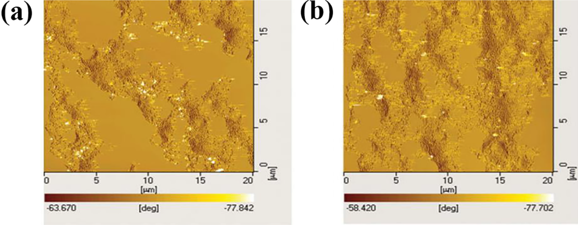
Some additives with specific functional groups are used to blend PLA to customize the biological properties. Irvine et al. 94 studied the surface modification of scaffolds for using in biomedical applications where the surface segregation of peptides was blended with PLA. The surface modifications of PLA and PLGA were carried with Pluronic block copolymers using the solvent casting technique and the molecular weight of monomer was also calculated for the same. The AFM topography and phase images of PLA are shown in Figure 9.
AFM images of PLA blends. 94 (reproduced after permission).
However, many significant works were performed in the field of surface modifications. One major work was conducted by Kiss et al., 95 who studied the surface properties of PLA for using it in polymeric drug delivery systems. Poly-(ethylene oxide) (PEO) was applied for reducing the hydrophobicity of the PLA surfaces, and solvent casting method was used to obtain the films of polymeric blends. Improvement of wettability was found to be proportional to the absorption of Pluronics in the surface layer derived from the XPS measurements. XPS measurements also showed a significant growth of Pluronics on the surface layer of the blend films. As an example, carbon and oxygen signals of PLA+PE6800 blend film are displayed with their components in Figure 10.
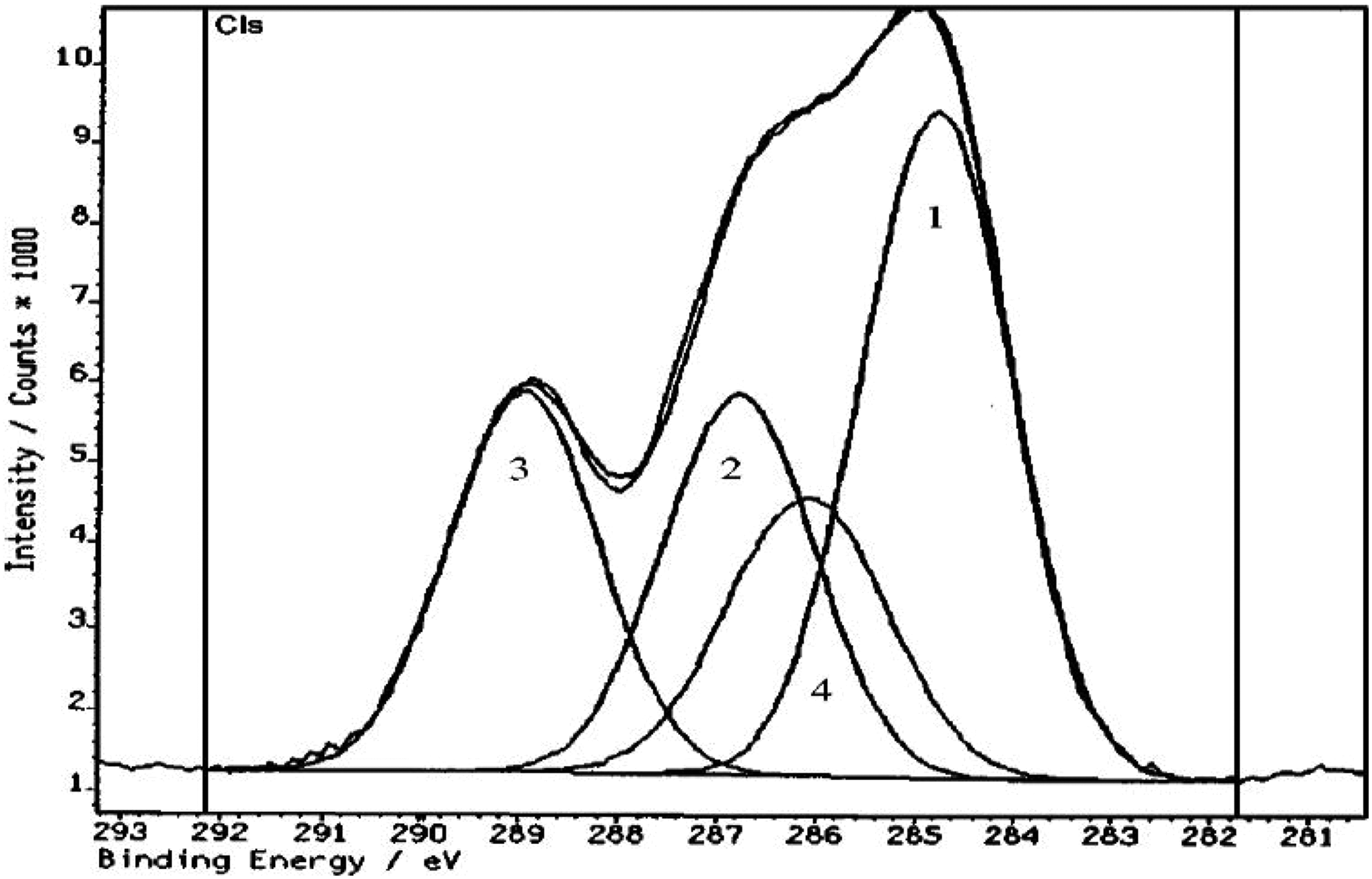
XPS signals of a PLA film.
Yu et al. 96 studied the surface modification of PLA for promoting chondrocyte attachment. The fabrication was achieved through a coupling reaction between PLA and PEG with the help of methylene diphenyl disocyanate. The diblock copolymer chain was water insoluble and also eroded in the biological environment due to its biodegradable properties. The chondrocyte test results revealed that there was a significant improvement for the chondrocyte compatibility which has useful applications for tissue engineering. The atomic force microscopy (AFM) phase images of PLA with different compositions of the PLE64 copolymer are presented in Figure 11.
(a) PLA modified with 5% of PLE64 (b) PLA modified with 10% of PLE64. 96
Plasma treatment
There are many applications of perfluorinated polymers due to their excellent chemical stability. However, they lack their effectiveness in applications where adhesive bonding is required with another surface as their hydrophobic nature prevents some good interfacial bonding in some applications. Therefore, some chemical groups are introduced onto the fluoro-polymer surface before bonding of these materials in which hydroxyl and amine groups play an important role. The plasma modification (Figure 12) is of great interest, nowadays. The surface modification of fluorinated ethylene propylene (FEP) and poly-tetra-fluoro-ethylene (PTFE) mainly carried using this technique. Xie et al. 98 investigated the surface of FEP and PTFE with plasma treatment setup, arranged in water as well as with ammonia gas. Both treatments were compared, and it was observed that contact angles were considerably low when the material was exposed to NH3 plasmas. Moreover, there was an improvement in the hydrophilic properties of the material. Figure 13 reproduces C-1 s XPS spectra of ammonia plasma-modified FEP took at the emission angles of 0° and 75°, and surface alterations of PTFE and FEP by plasmas of water vapor or ammonia gas were effective in providing more hydrophilic surfaces with short exposure times. Air/water contact angles were significantly lower on ammonia plasma treatment than on water vapor plasma treatment.
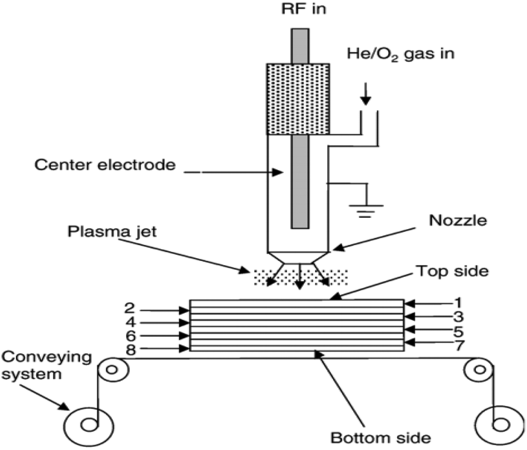
Atmospheric pressure plasma treatment system. 97
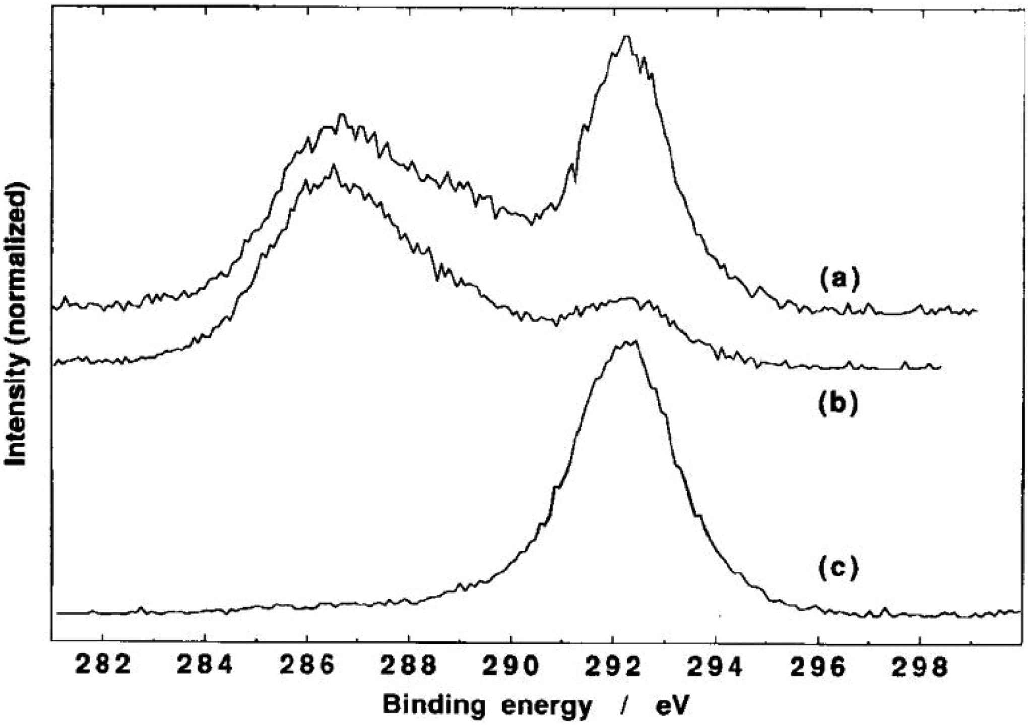
A XPS C I spectra of ammonia plasma-treated FEP.
Wang et al. 97 studied the surface treatment of polyester fabrics using various types of plasmas to understand the penetration of plasma on the surface. The fabrics to be modified were exposed to atmospheric pressure plasma jet and AFM was used to understand the surface morphology of the fabrics. The results revealed that plasmas penetrated more into the fabrics with larger average pore sizes. The comparison of the mechanical strength of adhesive-bonded specimens of various polyesters treated with activated helium and with activated oxygen studied by Hall et al. 99 The results revealed that the oxygen and helium were both effective with certain PE. Low-pressure radio-frequency plasma treatment for achieving a sufficient thickness of high-porous poly PDLLA scaffolds was developed by Safinia et al. 100 using NH3 plasma treatments. High porosity and the void fraction observed in and bubble point measurements and SEM (Figure 14) highlighted the interconnectivity of macrospores with microspores as the porous matrix. Moreover, the exposure of the material to this treatment showed improved wetting behavior. Yang et al. 101 investigated the surface modification of PLA and PLGA scaffolds for tissue engineering in different pore structures. The cell affinity and hydrophilicity of scaffolds were improved by using NH3 plasma treatment. Hirotsu et al. 102 fabricated the PLA sheets with a melt extrusion method with oxygen (O2), He, and ammonia (N2) gases as plasma treatment for improving the wettability. It was observed that plasma treatment do not have much effect on biodegration rate in soil.
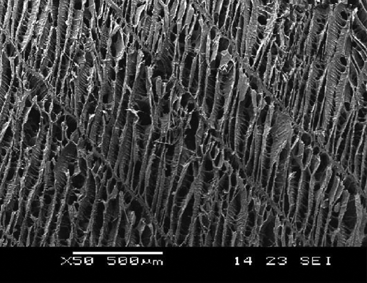
SEM image of PDLLA scaffold. 100
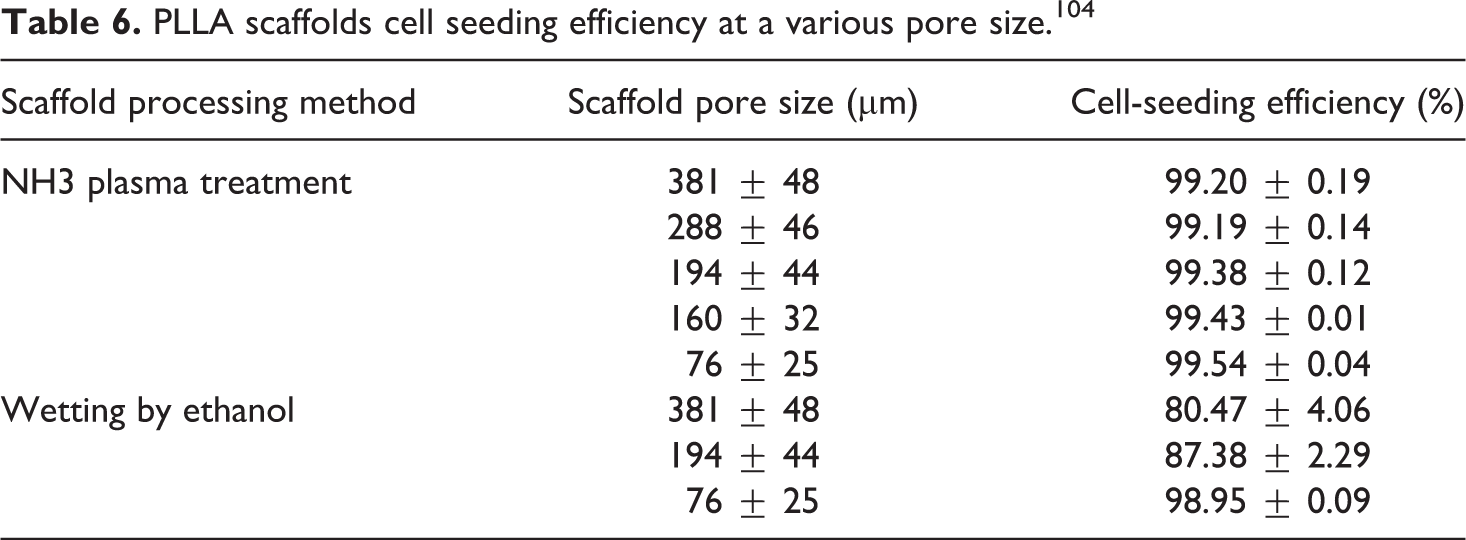
Yang et al. 103 studied the surface treatment of PDLLA using a combination of plasma treatment and collagen modification. The plasma treatment was carried using O2 and anhydrous N2 gas. The assessment of surface contact angles to water between the untreated samples and plasma-treated PDLLA samples was done and the surface morphology of the modified material was observed using SEM. The cell affinity evaluated mouse 3T3 fibroblasts model cells and results showed that the hydrophilicity was enhanced by using plasma treatment. Effect of the pore size on cell seeding efficiency is shown in Table 6. Khorasani et al. 104 studied the O2 plasma treatment for modification of PLLA and PLGA for biomedical applications. The evaluation of cell affinity of plasma-treated film was observed using B65 cell culture. It was observed that the hydrophobicity properties of the material enhanced after the O2 plasma treatment.
PLLA scaffolds cell seeding efficiency at a various pore size. 104
Wan et al. 105 investigated the plasma treatment of PLA for hydrophobicity of scaffolds for tissue engineering applications, as this technique is useful for enhancing the hydrophobicity of the scaffolds. There was an increase in modifying the depth of scaffolds after the treatment, but the degradation of the scaffolds improved with increasing treatment time and plasma power. The weight loss of the plasma-modified scaffolds (height 1 mm) was shown in Figure 15.
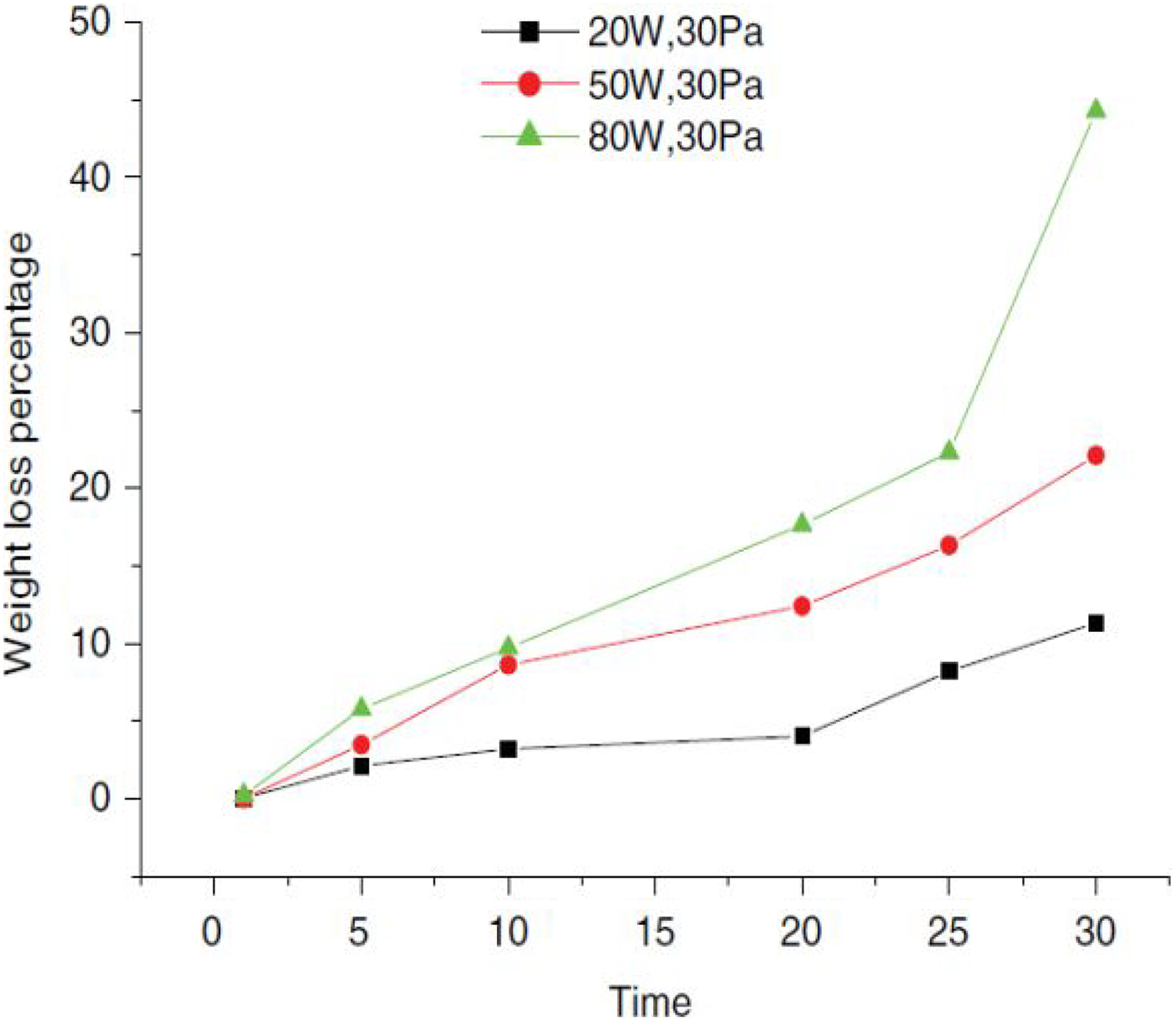
Effect of NH3 plasma treatment condition on weight loss of PLLA scaffold.
Permanent surface modification methods
Modification with conjugation using wet chemistry
Wet chemistry is a form of methodical chemistry that uses classical methods such as observation to analyze materials. It is called wet chemistry because most of investigating is done in the liquid phase. Endowing material surface with cell-adhesive properties is a common approach in biomaterial research and tissue engineering. Therefore, most crucial challenge while working with the surface modification of PLA is to control its dissolution in organic solvents such as benzene and chloroform.
105
Cai et al.
106
investigated the modification of ethylene-co-acrylic acid (EAA) copolymer film by surface grafting of amino acid intermediates covalently. The process was carried out in four steps at room temperature. The observations of the modified surface using ATR-FTIR spectroscopy showed that the hydrophilicity and surface roughness properties of the material were enhanced. Yang et al.
107
studied the modification of PLLA cell scaffold for improvement of cell affinity by using a mixture of aqueous 0.26 M NaOH/ethanol. Ren et al.
108
highlighted the hydrophobic properties of poly(
Zang et al. 109 investigated the reaction between thionyl-chloride and polyethylene-co-acrylic acid films for the surface modification of PEAA film. The results revealed that the improved film can be beneficial for material for tethering biomolecules. Zhu et al. 110 studied the modification of PLLA by reacting with 1,6-hexanediamine using glutaraldehyde as a coupling agent and it surface morphology is shown in Figure 16. It has been observed that the NH2 density was enhanced using this method.
PLLA membrane (a) and PLLA membrane analyzed at 50°C (b). 110
Photographing
The chemistry of the surface profoundly influences the behavior of any solid substrate in many applications, for example, in printing and adhesion. Although there were various methods used for the modification, the photoinduced grafting process is playing an utmost important role to produce homo-polymer and cross-linked polymers. Ma et al. 111 investigated the photografting technique for grafting monomers with abstractable hydrogen’s to support polymer. It was observed from the kinetic studies that the maximum reaction rate depends upon the BP concentration. Bae et al. 112 studied the surface modification of hydrophobic PLA fabrics using ultraviolet (UV) photografting with silica particles and possessing with vinyl functional groups. The results showed that the hydrophobic properties of the material were enhanced significantly using this method. The modification of poly(methyl-methacrylate) films was investigated by Rahane et al. 113 by using the photoiniferter-mediated photo polymerization (SI-PMP). For studying termination reactions, kinetic models were developed with PMP reactions. It was observed that there was a significant improvement in the surface properties of the material. Rasal et al. 53 investigated the effect of photo-induced grafting reaction on the bulk properties PLLA (PHBHHx) blend films. The dynamic mechanical analysis of films showed that the blend appeared to be noncompatible. Janorkar et al. 56 investigated the surface modifications of PLA films using UV irradiations. The results showed that there was a loss of molecular weight of PLA.
Liquid phase photografting
It is the covalent integration of functional additives to a polymer matrix or polymer surface using a light-induced mechanism. The “grafting from” method has been used more extensively than the “grafting to” approach for modifications of PLA surface. The activation of PLA surface is done by using plasma treatment which is followed by followed by photo polymerization of vinyl or acrylic monomers from the surface. Zhu et al. 114 investigated the surface treatment of PLA using chitosan with a photosensitive crosslinking reagent, 4-aminobenzoic acid. It has been observed that the reaction between an acid group of the cross-linking reagent and a free amino group of chitosan supported the bonding. The cross-linking of chitosan on PLA for coating and modifications was carried out by irradiating with UV light and introducing it to photolyze azide groups. It was observed that the modified surface offered an excellent platelet adhesion. The covalent binding of proteins and peptides PLLA grafted with polyacrylic acid was investigated by Steffens et al. 115 Amino groups of the protein/peptide were used for achieving the covalent attachment with carboxyl groups of the graft polymer using water-soluble carbodiimide and n-hydroxysuccinimide. Ma et al. 116 studied the surface modification of PLLA membrane surfaces using induced grafting copolymerization method and studied the cell behavior with chondrocyte culture. It was observed that cytocompatibility of the modified surface was enhanced after processing using this technique.
Vapor phase photografting
A common approach for the chemical alteration of polymeric substrates is the surface activation by using preparative high-energy radiation. Edlund et al. 117 studied nondestructive method for chemical surface modification of PLA using vapor phase of vinyl monomers, by benzophenone photo initiation under solvent-free conditions. The process improved wettability and revealed that this method could be useful to attain the desired properties of graft-chain pendant groups. Källrot et al. 118 investigated the solvent-free and nondestructive photographing method for covalently grafting n-vinylpyrrolidone onto the surfaces of degradable polymer, for instance, PLLA. The characterization of the modified surface was carried out using XPS, ATR-FTIR, and SEM. The results revealed that the wettability was tremendously enhanced. Källrot et al. 119 studied the modification of PLLA films using the vapor phase nondestructive photographing method. Grafting was carried out for 25 min with monomer acrylamide incubation in vitro in a phosphate-buffered saline solution at 38°C for 155 days. The results indicated that the grafted polymers possessed improved degradation rates owing to the produced covalent bonds. Edlund et al. 120 modified PLLA and PCL surface by grafting as shown in with heparin, covalently, with acrylamide with the nondestructive method. The biocompatibility measures showed that there was much improvement in the attachment mesenchymal stem cells (MSCs).
Summary
In the present work, various research methodologies aimed at the improvement of PLA-based bio-medical devices and tools were reviewed, critically. Based on the observations recorded, the following conclusions have been drawn: It is observed that various characteristics, including renewability, biocompatibility, thermo-plasticity, biodegradability, and environmentally friendliness of PLA, make it a potential biomaterial. The virgin PLA itself does not possess the essential features, which act as critical barriers for the broad spread utility of the same. However, the posttreatment of PLA via coating, entrapment, migratory additives, plasma treatment, modification with conjugation using wet chemistry, photographing, liquid phase photografting, and vapor phase photografting techniques enhanced the bio-characteristics of PLA, resulted in better performance standards. Most of the experiments were carried out in in vitro conditions; therefore, the generalized outputs of the review generalize before their in-depth in vivo validations. Moreover, the empirical and analytical relationships between functionalities and compositions of various PLA combinations, for instance, cell adhesion and degradability, should be developed. Also, the focus of the future research efforts should be on the realistic design for a novel carrier for biomedical uses as it allows for the development of good alternative in drug development as for consumer and biomedical applications, where PLA can be a prospective candidate. The degradation behavior of some proteins, such as fibronectin, should also be investigated which will help to explore the applications of coated PLA and its polymers in biomedical and tissue engineering. Further on the basis of exhaustive literature survey, it can be concluded that PLA, PLGA, and other biopolymers possess capabilities to replace most of the traditional materials in the biomedical fields. These materials have been utilized for vaccine development, encapsulating conventional drugs as well as in gene delivery.
121
Nanoparticles can be utilized to modify the properties of PLA and other copolymers. Looking at the success rate of nanoparticles with conventional plastics, it would not be unexpected to see more future work on PLA-nano composites. The other exciting area that needs attention is shape memory properties of PLA as an implantable biomaterial.