
Abstract
Background
Although the peripheral and central sensitizations of trigeminal nervous system may be one of the important factors of migraine, the precise mechanism is not fully understood. In this study, we examined the influence of the sensitization of the second division of the trigeminal nerve (V2) by chronic constriction injury (CCI) of the infraorbital nerve (ION) on migraine headache, using the capsaicin-induced migraine model.
Methods
Male Sprague-Dawley rats were assigned to four groups: (a) sham surgery and topical-dural vehicle application (Sham + Vehicle) group, (b) CCI-ION and topical-dural vehicle application (CCI-ION + Vehicle) group, (c) sham surgery and topical-dural capsaicin application (Sham + Capsaicin) group, (d) CCI-ION and topical-dural capsaicin application (CCI-ION + Capsaicin) group. Behavioral testing and immunohistochemical staining were performed.
Results
In the behavioral test, the Sham + Capsaicin group showed significantly longer duration of immobilization and shorter duration of exploration compared with the Sham + Vehicle group, which is similar to clinical features of migraine patients. Moreover, CCI-ION enhanced these effects in the CCI-ION + Capsaicin group. Immunohistochemical staining for phospho-extracellular signal-related kinase (pERK) in the trigeminal ganglion (TG) containing first and second divisions of the trigeminal nerve and the trigeminocervical complex (TCC) revealed that pERK expression was significantly increased in the CCI-ION + Capsaicin group compared with the other groups. However, comparing between effects of the peripheral and central sensitizations (in the TG and TCC), from our results, peripheral sensitization would play a much less or not significant role.
Conclusions
These data demonstrate that the sensitization of V2 could influence the activation and the sensitization of the first division of the trigeminal nerve in the TCC, subsequently exacerbating pain sensation and pain-related behaviors. We have shown for the first time that the existence of the central sensitization of V2 can be an exacerbating factor for migraine related nociceptive thresholds/activation.
Keywords
Introduction
Many groups have tried to elucidate the mechanism of migraine pathophysiology using experimental animal models, by stimulation of the dura mater with capsaicin, mustard oil or inflammatory soup, to directly activate the trigeminovascular system or by cortical spreading depression induced by mechanical, chemical or electrical stimulation (1–5). In addition, several gene mutations associated with migraine have recently been identified, and genetically modified mice have been generated and used for animal studies (6).
Cutaneous allodynia is the perception of pain or discomfort resulting from non-noxious thermal and mechanical stimuli applied to normal skin (7). Burstein et al. reported that cutaneous allodynia was observed in cephalic and extracephalic skin of patients with migraine and animal experimental models during migraine attacks. They hypothesized that cutaneous allodynia is induced by the sensitization of second-order trigeminal neurons in the spinal trigeminal nucleus and third-order neurons in the thalamus (7,8). Recently, it has been reported that cutaneous allodynia is not only a clinical marker for the central sensitization in the pathological condition of migraine, but also a predictor of migraine chronification (9,10). It can be hypothesized from these reports that the repetitive onset of migraine attacks occurs by activation of the first division of the trigeminal nerve (V1) and the central nervous system, which gradually sensitizes neurons innervating to cephalic (V2, maxillary nerve and V3, mandibular nerve) and extracephalic regions and induces cutaneous allodynia in these regions. The activation of these sensitized neurons then lowers the nociceptive threshold of V1, resulting in the aggravation and chronification of migraine.
Some migraine patients experience atypical pain in the V2 and V3 innervating regions, which present as non-odontogenic toothache or lower-half facial migraine instead of headache during the migraine attack period (11,12). These clinical conditions suggest that the activation of the trigeminovascular system via V1, which is associated with the migraine pathophysiology, can cause pain in any of three divisions of the trigeminal nerve (TN), indicating the interaction among the three divisions of TN.
Chronic constriction injury (CCI) has been known to induce peripheral and central sensitizations along the nociceptive pathway, manifesting as chronic neuropathic pain such as allodynia or hyperalgesia in regions innervated by the injured nerve. Several CCI animal models have already been established and widely used for experiments using the sciatic nerve or TN (13–15).
The purpose of this study was to determine if the sensitization of a division other than V1 enhances the responses in a migraine model by behavioral assessment, using capsaicin-induced migraine and CCI models. We also tried to demonstrate the behavioral results using immunohistochemical techniques.
Methods
Animals
A total of 90 male Sprague-Dawley rats (250–300 g, Charles River Laboratories Japan, Yokohama, Japan) were used in this study. Rats were housed under 12 h/12 h dark/light cycle conditions and had free access to food and water. All experiments were conducted in accordance with the National Institutes of Health Guidelines for the Care and Use of Laboratory Animals. The study protocol was reviewed and approved by the Institute of Experimental Animal Sciences at Osaka University Graduate School of Dentistry, Japan.
CCI-ION model
Rats were anesthetized with 50 mg/kg sodium pentobarbital (Kyoritsu Seiyaku, Tokyo, Japan) intraperitoneally, then placed on a thermostatically-controlled heating pad (ATB-1100: Nihon Kohden, Tokyo, Japan). A small incision was made at the edge of the right whisker pad, and the right ION was exposed. Two nylon sutures (4-0 thread) were placed 2 mm apart around the ION, and the wound was sutured after ligation of the ION. These ligations reduced the diameter of the nerve noticeably, but did not occlude it completely (16). The left ION was intact. For the sham surgery, the right ION was exposed but did not undergo nerve ligation.
Rats were placed on an acrylic box (25 × 25 × 25 cm) and allowed to acclimatize to this environment for 20 minutes before any stimulation. Then mechanical stimulations (0.6, 1, 2, 4, 8, 15 and 26 g) were applied to the center of the whisker pad using von Frey filaments (Muromachi Kikai, Tokyo, Japan). The head-withdrawal threshold of the mechanical stimulation on the whisker pad skin was defined as the minimum pressure needed to evoke at least three reactions by five stimuli. The cutoff to prevent tissue damage was determined as 26 g. Before CCI-ION surgery, stimulation with the 26 g filament did not induce any positive nociceptive behaviors (withdrawal of the head, or an attack or escape reaction). The stimuli were applied before CCI-ION surgery or the sham surgery, and 7, 14, and 21 days after surgery to confirm the sensitization of V2, which manifested as the development of tactile allodynia.
Preparation of capsaicin solution
Capsaicin (3.1 mg) was dissolved in 100% ethanol (65.6 μl) (6.5%), tween 80 (65.6 μl) (6.5%) (Kanto Chemical Co., Inc, Tokyo, Japan) and saline (871.8 μl) (87%). The vehicle solution contained the same concentration of 100% ethanol, tween 80 and saline as the capsaicin solution, but without the capsaicin.
Experimental groups
Rats were randomly assigned to one of four groups as follows: (a) the Sham + Vehicle group, which underwent sham surgery on the right infraorbital nerve (ION), then vehicle application on the right dura mater; (b) the CCI-ION + Vehicle group, which underwent CCI of the right ION, then vehicle application on the right dura mater; (c) the Sham + Capsaicin group, which underwent sham surgery on the right ION, then capsaicin application on the right dura mater; and (d) the CCI-ION + Capsaicin group, which underwent CCI of the right ION, then capsaicin application on the right dura mater.
Behavioral tests
Cannula setting on the dura mater for the capsaicin/vehicle application
At day 14 after CCI-ION, rats were anesthetized with sodium pentobarbital (50 mg/kg) intraperitoneally, and fixed to a brain stereotaxic apparatus. A medial incision was made on the scalp to expose the skull. A burr hole (1 mm diameter) was made in the right skull to expose the dura mater on the right transverse sinus, and the cannula was fixed using dental resin to the burr hole by simply touching the surface of the dura mater.
Behavioral recording of freely-moving rats
At day 21 after CCI-ION (seven days after setting the cannula), rats were placed in a transparent acrylic box (25 × 25 × 25 cm) with a mirror on the back surface of the interior for video recording, and 20 µl of either 10 mM capsaicin or vehicle was infused using a micro-syringe through the cannula. Experiments were conducted between 09:00 and 16:00. We recorded spontaneous behaviors for 30 minutes after infusion, and three major types of behavior were analyzed, which were immobilization (freezing or resting), exploration, and face grooming. These three behaviors analyzed in our study were defined according to the previous publication by Melo-Carrillo et al. (17).
Double-labeling immunofluorescent examinations of the trigeminal ganglion, spinal trigeminal nucleus and upper cervical cord for phospho-extracellular signal-related kinase and NeuN
At day 21 after CCI-ION, rats were anesthetized with sodium pentobarbital (50 mg/kg) intraperitoneally, and fixed to a brain stereotaxic apparatus. The skull was then exposed and a 3 mm diameter burr hole was made to expose the dura mater on the right transverse sinus. Two hours later, a 2 mm diameter cotton ball, soaked in either 10 mM capsaicin or vehicle, was applied to the dura mater. Four minutes later, rats were perfused with 0.01 M phosphate buffer saline (PBS), followed by 4% paraformaldehyde (PFA) in 0.01 M PBS. The right trigeminal ganglion (TG), spinal trigeminal nucleus (Vc) and upper cervical cord (C1–2) were harvested and post-fixed in 4% PFA for three days at 4℃. Tissues were then transferred and stored in 30% sucrose in PBS at 4℃ for cryoprotection.
Ten micrometer thick tissue sections of the TG were cut using a cryostat (Leica, Welzlar, Germany) and mounted on Superfrost Plus glass slides (Matsunami Glass Ind., Osaka, Japan). Tissue sections were rinsed in 10% normal goat serum (NGS) (Vector Laboratories, Burlingame, CA, USA) and then incubated in rabbit anti-phospho-p44/42 MAPK (phospho-extracellular signal-related kinase: pERK) antibody (1:1000, Cell Signaling Technology, Danvers, MA, USA) and mouse anti-NeuN (neuronal marker) antibody (1:1000, Merck Millipore, Billerica, MA, USA) for 24 hours at room temperature. After washing in PBS, tissue sections were incubated in Alexa Fluor 488 anti-rabbit IgG and Alexa Fluor 568 anti-mouse IgG (1:400, Invitrogen, Carlsbad, CA, USA) for 2 hours. After that, slides were coverslipped with Fluoromount (Diagnostic BioSystems, Pleasanton, CA, USA).
Forty micrometer thick tissue sections were cut from Vc and C1–2 using a freezing microtome (Yamato Kohki, Saitama, Japan). Every third section was collected in PBS. The following double-labeling immunofluorescent procedures were similar to those performed on the TG, as described above. Tissue sections were mounted on gelatin-coated slides, and slides were coverslipped with Fluoromount.
Following double-immunofluorescent staining, the TG, Vc and C1–2 were examined under a confocal laser-scanning microscope (Zeiss, Jena, Germany) or BZ-X700 fluorescence microscope (Keyence, Osaka, Japan).
Immunohistochemical examinations of the trigeminal ganglion, spinal trigeminal nucleus and upper cervical cord
The right TG, Vc and C1–C2 were prepared in the same way as described in the previous section.
Ten micrometer thick tissue sections of the TG were incubated in 1% NGS in PBS for 30 minutes at room temperature and then incubated in rabbit anti-phospho-p44/42 MAPK (pERK) antibody (1:1000, Cell Signaling Technology) for 24 hours at room temperature. Then, the sections were incubated in biotinylated goat anti-rabbit IgG (1:200, Vector Laboratories) for 1 hour. After washing, sections were incubated in peroxidase-conjugated avidin-biotin complex (Vector Laboratories) for two hours at room temperature, followed by reaction with 0.05% diaminobenzidine tetrahydrochloride, 0.1% ammonium nickel sulfate and 0.01% hydrogen peroxide in 0.05 M Tris-HCl buffer (pH 7.2) (Vector Laboratories). Slides were then rinsed in 0.01 M PBS, dehydrated in a series of alcohol and coverslipped with Permount (Fisher Scientific, Pittsburgh, PA, USA).
Forty micrometer thick tissue sections were cut from Vc and C1–2 using a freezing microtome. Every third section was collected in PBS. The following phospho-p44/42 MAPK (pERK) immunohistochemical procedures were similar to those performed on the TG, as described above. Tissue sections were mounted on gelatin-coated slides and underwent alcohol dehydration before being coverslipped with Permount. We also performed immunohistochemical staining without the primary antibody for phospho-p44/42 MAPK (pERK) as a control, in which no immune products could be observed (data not shown).
In the TG containing V1 and V2, we analyzed every 10 sections. Three images were taken from each section and pERK-immunoreactive (IR) cells and all the cells in each image were counted. Then the percentage of pERK-IR cells in each animal was calculated by the following formula: 100 × total number of pERK-IR cells in images/total number of cells in images (18). In Vc and C1–2, the number of pERK-IR cells was counted in every sixth section from the obex (the upper margin of the area postrema) to 6480 µm caudally (19–21).
Statistical analysis
Data were expressed as mean ± standard deviation (SD). The power and sample size calculations were performed by a priori and post hoc analyses. In anticipation of potential technical failures, extra rats were added a priori to the capsaicin administration groups in behavioral tests. The time-dependent change of head-withdrawal thresholds for the mechanical stimulation (von Frey test) in each group was analyzed using repeated measures one-way analysis of variance (ANOVA), followed by post hoc Tukey–Kramer test. A two-tailed Student’s t-test was performed for differences in head-withdrawal thresholds for the mechanical stimulation between two groups for each day. One-way or two-way ANOVA followed by post hoc Tukey–Kramer test were performed for the behavioral tests, and analysis of the percentage of pERK-IR cells in the TG and the number of pERK-IR cells in Vc and C1–2. The analysis of behavior recordings, counting the cells for immunohistochemical experiments, and all statistical analysis were performed blindly. A value of p < 0.05 was considered statistically significant.
Results
Development of tactile allodynia after CCI-ION
The head-withdrawal threshold for the mechanical stimulation to the whisker pad of the ipsilateral side was significantly decreased at days 7, 14 and 21 after CCI-ION compared with that before CCI-ION (day 0) (p < 0.001 at all time points, 95% confidence interval of difference (95% CI of difference): 14.6–20.5, 14.9–20.8, 18.2–24.1 at each time point) (Figure 1) . The head-withdrawal threshold of CCI-ION rats (n = 42) was significantly lower than sham-operated rats (n = 48) at days 7, 14 and 21 after CCI-ION (p < 0.001 at all time points) (Figure 1).
Time course of the development of tactile allodynia after chronic constriction injury (CCI) of infraorbital nerve (ION). Data are expressed as mean ± standard deviation (SD). To prevent tissue damage, 26 g was determined as a cutoff. Asterisks indicate the head-withdrawal thresholds that are significantly different from day 0 (before the surgery) (repeated measures one-way analysis of variance followed by post hoc Tukey–Kramer test, p < 0.001 at all time points). Hashes indicate the head-withdrawal thresholds that are significantly different from the sham surgery group on each day (a two-tailed Student’s t-test, p < 0.001 at all time points). CCI-ION (n = 42) gradually decreased the threshold, the effect lasting at least three weeks, whereas the sham surgery group (n = 48) did not show any change to the threshold.
Behavioral changes induced by dural application of capsaicin
The total duration of immobilization in the Sham + Capsaicin group (n = 14, 661 ± 354 seconds) was significantly longer than in the Sham + Vehicle group (n = 8, 179 ± 185 seconds, p = 0.028, 95% CI of difference: 41–922) (Figure 2(a)). The total duration of immobilization in the CCI-ION + Capsaicin group (n = 13, 1069 ± 422 seconds) was longest compared with the Sham + Vehicle (p < 0.001, 95% CI of difference: 443–1336), CCI-ION + Vehicle (n = 5, 312 ± 334 seconds, p = 0.002, 95% CI of difference: 233–1279) and Sham + Capsaicin groups (p = 0.033, 95% CI of difference: 25–791) (Figure 2(a)).
Changes in behaviors correlated to migraine-related nociceptive activation. Data are expressed as mean ± standard deviation (SD). Asterisks indicate the significant difference between each group (one-way analysis of variance followed by post hoc Tukey–Kramer test, p < 0.05). (a) Total duration of immobilization after vehicle or capsaicin application. The CCI-ION + Capsaicin group (n = 13) showed significantly longer duration than any other group (p < 0.05); (b) total duration of exploration after vehicle or capsaicin application. The CCI-ION + Capsaicin group had decreased duration of exploration compared to the Sham + Capsaicin group (n = 14) (p = 0.052), and significantly shorter duration than the Sham + Vehicle (n = 8) and CCI-ION + Vehicle (n = 5) groups (p < 0.05); (c) total duration of face grooming after vehicle or capsaicin application. Although the CCI-ION + Vehicle group showed slightly longer duration than any other group, there was no significant difference between all groups.
The total duration of exploration in the Sham + Capsaicin group (n = 14, 993 ± 336 seconds) was significantly shorter than in the Sham + Vehicle group (n = 8, 1414 ± 309 seconds, p = 0.045, 95% CI of difference: 8–834) (Figure 2(b)). The total duration of exploration in the CCI-ION + Capsaicin group (n = 13, 637 ± 348 seconds) was shortest compared with the Sham + Vehicle group (p < 0.001, 95% CI of difference: 358–1196), CCI-ION + Vehicle group (n = 5, 1252 ± 283 seconds, p = 0.009, 95% CI of difference: 125–1106) and Sham + Capsaicin group (p = 0.052, 95% CI of difference: −3–716) (Figure 2(b)).
Although there were no statistically significant differences among the four groups, the CCI-ION + Vehicle group (n = 13) showed a tendency for the longest duration (151 ± 73 seconds) of face grooming behavior compared with the Sham + Vehicle group (n = 8, 96 ± 71 seconds, p = 0.49, 95% CI of difference: −49–159), Sham + Capsaicin group (n = 14, 77 ± 42 seconds, p = 0.17, 95% CI of difference: −21–169) and CCI-ION + Capsaicin group (n = 13, 70 ± 75 seconds, p = 0.097, 95% CI of difference: −11–183), which was consistent with a previous report (14) (Figure 2(c)). Capsaicin application alone, or the combination of CCI-ION and capsaicin application, did not have any effect on face grooming behavior, while CCI-ION itself did have an effect (Figure 2(c)).
Phosphorylation/activation of ERK in TG neurons
To confirm if pERK-IR cells were neurons in the TG, we performed double-labeling immunohistochemistry using an anti-NeuN antibody for neuronal labeling and anti-pERK antibody. Since all pERK-IR cells were also NeuN-positive, these cells were neuronal-specific (Figure 3(a–c)).
Double labeling for phospho-extracellular signal-related kinase (pERK) (a) and NeuN (b) immunoreactivity (IR) in the trigeminal ganglion (TG) containing first and second divisions of the trigeminal nerve after chronic constriction injury of the infraorbital nerve and capsaicin application to the dura mater. (a) Open arrows indicate pERK-IR cells; (b) hatched arrows indicate NeuN-IR cells; (c) merged image of (a) and (b). Arrowheads indicate pERK-IR cells co-localized with NeuN, showing that all of the cells expressing pERK are neurons. Scale bar = 50 µm.
The percentage of pERK-IR cells in the TG containing V1 and V2 in the CCI-ION + Capsaicin group was significantly higher (n = 5, 9.9 ± 2.2%) compared with the Sham + Vehicle group (n = 5, 3.0 ± 0.4%, p < 0.001, 95% CI of difference: 4.0–9.7), CCI-ION + Vehicle group (n = 5, 4.7 ± 1.6%, p = 0.001, 95% CI of difference: 2.3–8.0) and Sham + Capsaicin group (n = 5, 5.9 ± 0.5%, p = 0.005, 95% CI of difference: 1.2–6.9), however, there was not a significant interaction between the surgery effect and the dural-treatment effect (two-way ANOVA, p = 0.12) (Figure 4(a–e))
Immunohistochemical analysis of phospho-extracellular signal-related kinase (pERK)-immunoreactive (IR) cells in the trigeminal ganglion (TG) containing first (V1) and second (V2) divisions of trigeminal nerve. (a–d) Representative photomicrographs in the TG containing V1 and V2 after sham surgery and vehicle application (a), or CCI-ION and vehicle application (b), or sham surgery and capsaicin application (c), or CCI-ION and capsaicin application (d). Arrowheads indicate pERK-IR cells. Scale bar = 100 µm; (e) histogram summarizing the quantitative data for pERK-IR cells in the TG containing V1 and V2. Data are expressed as mean ± standard deviation (SD). The asterisk indicates significant difference compared to the Sham + Vehicle (n = 5), CCI-ION + Vehicle (n = 5) and Sham + Capsaicin (n = 5) groups (one-way ANOVA followed by post hoc Tukey–Kramer test, p < 0.05). The CCI-ION + Capsaicin group (n = 5) was the only group where there was a significant increase in pERK-IR cells in the TG containing V1 and V2.
Phosphorylation/activation of ERK in the spinal trigeminal nucleus and upper cervical cord
To confirm if pERK-IR cells were neurons in Vc and C1–2, we performed double-labeling immunohistochemistry using an anti-NeuN antibody and anti-pERK antibody. Since all pERK-IR cells were also NeuN-positive, these cells were neuronal-specific (Figure 5(a–c)).
Double-labeling for phospho-extracellular signal-related kinase (pERK) (a) and NeuN (b) immunoreactivity (IR) at 4320 µm caudal to the obex in the upper cervical cord after chronic constriction injury of the infraorbital nerve and capsaicin application to the dura mater. (a) Open arrows indicate pERK-IR cells; (b) hatched arrows indicate NeuN-IR cells; (c) merged image of (a) and (b). Arrowheads indicate pERK-IR cells co-localized with NeuN, showing that all of the cells expressing pERK are neurons. Scale bar = 50 µm.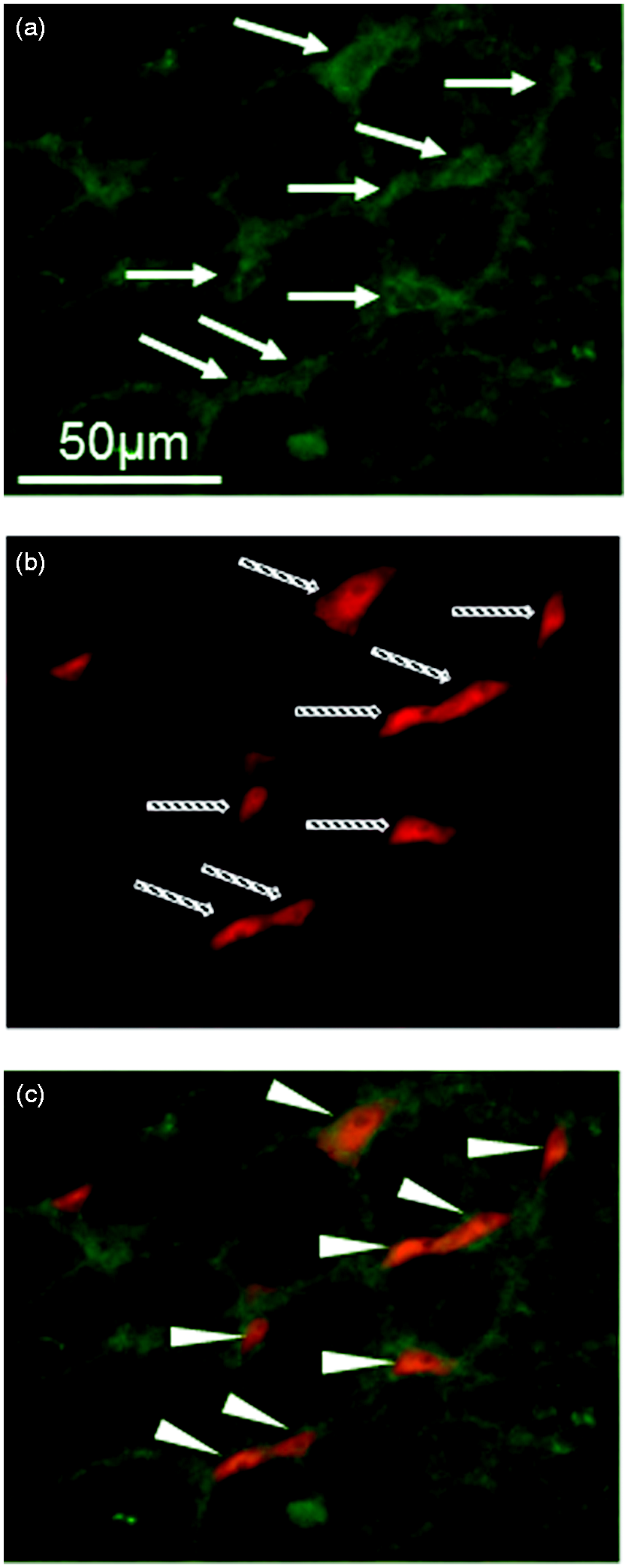
A large number of pERK-IR cells were observed in the superficial laminae (laminae I and II) of Vc and C1–C2 (Figure 6(a–d)).
Immunohistochemical analysis of phospho-extracellular signal-related kinase (pERK)-immunoreactive (IR) cells in the spinal trigeminal nucleus (Vc) and upper cervical cord (C1–C2).
At 3600 µm caudal to the obex, the mean number of pERK-IR cells was significantly higher in the Sham + Capsaicin group (n = 6) compared with the Sham + Vehicle group (n = 6, p = 0.032, 95% CI of difference: 1.0–29.0) (Figure 6(e)). In addition, at 4320 µm caudal to the obex, the mean number of pERK-IR cells in the Sham + Capsaicin group had a strong tendency to be increased compared with the Sham + Vehicle group (p = 0.079, 95% CI of difference: −1.3–30.6) (Figure 6(e)). No significant difference was observed in other regions between the Sham + Vehicle and Sham + Capsaicin groups (Figure 6(e)). Comparing the effect of CCI-ION on capsaicin-induced pERK expression, the mean number of pERK-IR cells at 3600 µm and 4320 µm caudal to the obex were significantly higher in the CCI-ION + Capsaicin group (n = 8) compared with the Sham + Capsaicin group (p = 0.003, 95% CI of difference: 5.8–31.9 at 3600 µm caudal and p = 0.006, 95% CI of difference: 5.2–35.1 at 4320 µm caudal to the obex), which were the same regions in which the capsaicin effect on pERK-IR cells was observed when comparing the Sham + Vehicle and Sham + Capsaicin groups (Figure 6(e)).
Discussion
Although some preclinical and clinical data have revealed the role of the sensitization of the trigeminal nervous system to migraine headache, few reports have determined the effect of the sensitization and the interaction among three divisions of TN (V1, V2 and V3) in the TG, and also V1 and C1–2 (known as the trigeminocervical complex (TCC)) on migraine-related nociceptive thresholds/activation. Thus, we used the chronic constriction injury method to sensitize one of the TN other than V1, and then tested the effect of this sensitization on migraine related nociceptive thresholds/activation using the capsaicin-induced migraine model.
To establish the sensitization model of V2, we performed CCI surgery to ION. CCI models using the sciatic nerve, V2 or V3 have already been well established (13,14) and used for chronic neuropathic pain research. It is known that after CCI, nociceptive neurons are gradually sensitized peripherally at peripheral terminals of primary sensory neurons and centrally at the dorsal horn of the spinal cord (22–24). Furthermore, a few weeks after CCI surgery, animals exhibit changes in non-evoked and evoked behaviors due to hyperalgesia or allodynia (25–29), and increased c-fos and pERK expressions in the dorsal horn (16,30). In this study, we confirmed the establishment of the CCI-ION model (sensitization of V2) by observing tactile allodynia, which was analyzed by von Frey filaments.
To establish the animal models of migraine, we applied capsaicin to the dura mater on just the right transverse sinus. The most widely used migraine experimental models are electrical stimulation of the TG or chemical stimulation of the dura mater on the transverse sinus or the superior sagittal sinus using capsaicin, mustard oil or inflammatory soup (1–4). In this study, we chose the capsaicin model. Shimizu et al showed the data that TRPV1 was expressed on the dura mater (31). And pERK expression induced by the capsaicin application on the dura mater or cortical spreading depression was suppressed by TRPV1 antagonist (2). In addition, Zhang et al recently reported that botulinum neurotoxin type A, which reduces the number of days of chronic migraine, inhibited the intracranial meningeal nociceptors’ responses to stimulation of TRPV1 (32). These results suggest that TRPV1 may play a key role in migraine pathophysiology. Although there is still something inconclusive (33), TRPV1 agonist capsaicin application on the dura mater can be one of the migraine models.
It has been reported that migraine animal models using inflammatory soup or cortical spreading depression showed increased immobilization behavior and decreased exploratory behavior (17,34), which resemble the clinical features observed in migraine patients during migraine attacks (35). According to these reports of migraine-related behaviors, we observed the unstimulated behaviors, immobilization, exploration and face grooming, immediately after applying capsaicin or vehicle on the dura mater of freely-moving rats. Capsaicin increased the immobilization behavior compared with vehicle, whereas it decreased the exploratory behavior. Interestingly, CCI-ION enhanced the effects of capsaicin on immobilization and exploration. Our results suggest that the sensitization of V2 by CCI may lower the migraine-related nociceptive threshold. The behavioral test of face grooming is rather for assessing the effect of CCI-ION on the sensitization of V2, not V1. So, in our data, CCI-ION itself showed a tendency to increase the duration of face grooming compared to other groups, which was consistent with previous papers reporting the effect of CCI-ION on face grooming behavior (14,36). On the other hand, CCI-ION did not increase the duration of face grooming when comparing the Sham + Capsaicin and CCI-ION + Capsaicin groups, because the capsaicin administration increased immobilization, which may counteract movements including face grooming.
ERK is a member of MAPKs, and is activated by upstream MAPK/ERK kinase (MEK) (37). It is well known that ERK is activated (phosphorylated) in primary afferent neurons or spinal dorsal horn neurons by peripheral noxious stimulation and inflammation (38,39). In addition, Iwashita et al. recently reported that capsaicin application to the dura mater or cortical spreading depression induced the expression of pERK in the TG (2). Furthermore, inhibition of ERK activation using MEK inhibitors reduced inflammatory or neuropathic pain (23,38). Therefore, pERK is now widely investigated in pain studies as a better marker for peripheral and central activations or sensitizations in pain transduction pathways than c-Fos, which has been used as a marker for more than 20 years (40).
In this study, we observed that pERK expression in the TG containing V1 and V2 was strongly increased by capsaicin application to the dura mater alone (p = 0.054) and significantly increased in the CCI-ION + Capsaicin group compared with any other group. Shinoda et al. reported that inflammation in the lower lip increased nerve growth factor (NGF) in first-order neurons of V3 in the TG, and that NGF influenced first-order neurons of V2 in the TG, resulting in the sensitization of V2 and ectopic heat hyperalgesia at the whisker pad (41). Takeda et al. also reported that temporomandibular joint inflammation increased the content of substance P (SP) in TG neurons of V3, and SP was released via a paracrine mechanism resulting in up-regulation of neurokinin 1 receptors in TG neurons innervating the facial skin (V2), causing mechanical allodynia around the facial skin (42). As we didn’t distinguish V1 neurons from V2 neurons in TG in this study, and also didn’t see a significant interaction between the surgery effect and the dural-treatment effect, it’s hard to conclude from our results at the moment that peripheral sensitization via possible mechanisms reported by previous studies is responsible for affecting migraine-related behaviors. With regard to this point, further investigations would be needed.
In the TCC, pERK-IR cells were expressed ventrolaterally at the lamina I and II, following capsaicin application to the dura mater. According to the somatotopic organization of Vc reported previously, our observation reflects the activation of V1 by capsaicin to the dura mater (43). Regarding rostrocaudal distribution in the TCC, pERK-IR cells were significantly increased at −3600 µm (p = 0.032) and had a strong tendency to be increased at −4320 µm (p = 0.079) by capsaicin application to the dura mater compared with the Sham + Vehicle group. Only in these regions, the combination of CCI-ION and capsaicin application (CCI-ION + Capsaicin group) significantly increased pERK expression compared with the Sham + Capsaicin group. Strassmann et al. reported that c-Fos expression showed a bimodal distribution, around the obex and between caudal Vc and C2, by stimulating the dura mater over the transverse sinus using mechanical stimulation (44). Conversely, pERK was expressed in rostral Vc when the whisker pads of CCI-ION animals were mechanically stimulated immediately before perfusion (16). It has been reported that the injury of the cervical nerve or V3 induces the central sensitization, manifested by c-Fos or pERK expression in neurons or astroglial cell activation in the TCC, while also causing hypersensitivity to mechanical and heat stimulations in extraterritorial regions (21,45). According to these reports, regions in which pERK expression was significantly increased by the combination of CCI-ION and capsaicin application in this study were V1-input, not V2, suggesting that the sensitization of V2 neurons facilitated the activation and the sensitization of V1 neurons in the TCC as the central sensitization.
Taken together, our results suggest that CCI-ION continuously activated and sensitized not only V2, but also V1 in the TCC, resulting in exacerbation of migraine pain and migraine-related behaviors. In terms of clinical implications, the sensitization of V2 due to nerve injury could exacerbate migraine pain. In addition, our data can contribute to elucidating the correlation between the sensitization of the trigeminal nervous system and allodynia as the associated neurological symptom of migraine, and the mechanism for the development of chronic migraine.
Article highlights
Sensitization of the infraorbital nerve affects migraine-related behaviors. This sensitization enhances the pERK expression in the trigeminocervical complex of the migraine rat model. Although CCI-ION and capsaicin application significantly increased the pERK expression in the trigeminal ganglion of the migraine rat model, it was not clear that the peripheral sensitization had an important role in affecting migraine-related behaviors. Sensitization of the second division of the trigeminal nerve may worsen the migraine headache via central sensitization.
Footnotes
Declaration of conflicting interests
The authors declared no potential conflicts of interest with respect to the research, authorship, and/or publication of this article.
Funding
The authors disclosed receipt of the following financial support for the research, authorship, and/or publication of this article: This study was supported by JSPS KAKENHI Grant Number 22592257.