
Abstract
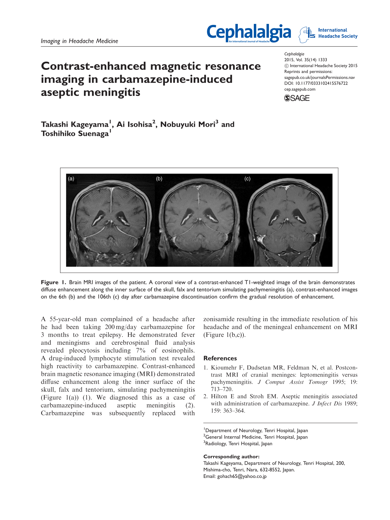
A 55-year-old man complained of a headache after he had been taking 200 mg/day carbamazepine for 3 months to treat epilepsy. He demonstrated fever and meningisms and cerebrospinal fluid analysis revealed pleocytosis including 7% of eosinophils. A drug-induced lymphocyte stimulation test revealed high reactivity to carbamazepine. Contrast-enhanced brain magnetic resonance imaging (MRI) demonstrated diffuse enhancement along the inner surface of the skull, falx and tentorium, simulating pachymeningitis (Figure 1(a)) (1). We diagnosed this as a case of carbamazepine-induced aseptic meningitis (2). Carbamazepine was subsequently replaced with zonisamide resulting in the immediate resolution of his headache and of the meningeal enhancement on MRI (Figure 1(b,c)).
Brain MRI images of the patient. A coronal view of a contrast-enhanced T1-weighted image of the brain demonstrates diffuse enhancement along the inner surface of the skull, falx and tentorium simulating pachymeningitis (a), contrast-enhanced images on the 6th (b) and the 106th (c) day after carbamazepine discontinuation confirm the gradual resolution of enhancement.