
Abstract
Objectives:
Studies have reported an association between migraine and white matter hyperintensities on T2-weighted brain magnetic resonance imaging (MRI) in adults. The aim of the present study was to evaluate white matter MRI brain findings in pediatric patients with migraine.
Methods:
The medical files and imaging scans of all 194 patients who underwent brain MRI at the headache clinic of a tertiary medical center in 2008–2011 were reviewed.
Results:
Mean age was 10.9 ± 3.5 years. Migraine was diagnosed in 131 patients and other disorders in 63. In the migraine group, findings on physical and laboratory examinations were within normal range. White matter lesions were identified on MRI scan in 14 children with migraine (10.6%) and none of the children with other disorders (p = 0.006). In 13/14 patients, the lesions were focal with a variable distribution; in the remaining patient, confluent periventricular hyperintensities were documented.
Conclusions:
In a headache clinic of a tertiary pediatric medical center, white matter lesions are found in about 10% of pediatric patients with migraine.
Introduction
Headache is a very common symptom in adults and children, accounting for 18 million outpatient visits in the United States each year (1,2). Migraine is a type of disabling headache associated with a wide range of autonomic nervous system dysfunctions (2). Several studies in adults have reported an association between migraine and white matter lesions on brain magnetic resonance imaging (MRI), and a higher risk of white matter lesions in patients with migraine than in healthy controls (3–7). The lesions are largely suspected to be ischemic, but their true etiology is unknown (4–7). In children, neuroimaging is not routinely used in the evaluation of recurrent headache, but it is necessary when there is a primary concern of possible brain tumor or other organic abnormalities. The numerous reports in the literature on the indications for imaging in the pediatric age group (1,2,8,9) led to the formulation of specific guidelines by the Quality Standards Subcommittee of the American Academy of Neurology and the Practice Committee of the Child Neurology Society (2). They stipulate that patients with recent-onset headache, defined as headache of less than three months’ duration, represent a distinct group at high risk of abnormal findings on MRI that may require surgical intervention (10).
The aim of the present study was to evaluate white matter brain lesions in pediatric patients with migraine.
Patients and methods
The study was conducted at the headache clinic, Ambulatory Day Hospitalization Center, of a pediatric tertiary medical center. Most of the patients who attend the clinic are referred by their primary physician after prolonged follow-up. The electronic database of the clinic was searched for all newly admitted patients who underwent MRI evaluation for a diagnosis of headache in 2008–2011. Data on background and clinical and imaging parameters were collected from the medical files; the diagnoses were categorized by a pediatrician and not by the neuroradiologist. The MRI scans were reassessed by a pediatric neuroradiologist blinded to the clinical parameters and diagnoses. The study was approved by the hospital’s Research Ethics Board.
For diagnosis of complaints of headache at the headache clinic, older children and the parents of younger children are routinely asked to complete a standard headache questionnaire based on the 2004 revised criteria for children of the International Headache Society (International Classification of Headache Disorders) (11). Items cover the frequency, duration, and nature of the headaches, degree of disability caused by the headaches, and related symptoms. For very young children whose verbal communication is limited, headache frequency is determined by both the child's complaints and the parents’ impression from the child's behavior. Patients and parents are also asked to maintain a headache calendar for at least one month before each scheduled visit in which they record every event of headache and the resulting disability.
Considerations of the need for an MRI study are based on the current clinical practice parameters recommended by the Subcommittee of the American Academy of Neurology and the Practice Committee of the Child Neurology Society (2). They include recent onset of severe headaches, change in type or frequency of headaches, and abnormalities or dysfunction on neurologic examination, including focal findings (e.g. unilateral motor weakness, diplopia), signs of increased intracranial pressure, significant alteration in consciousness, and focal seizures. The commonly used specific indications for MRI at our clinic are a change in type or frequency of headache and signs of a possible increase in intracranial pressure, such as morning headache, headache severe enough to wake them from sleep, night vomiting, and recent and acute onset headache less than three months. In our headache clinic imaging is indicated in all children aged 6 years or less with headaches.
All MRI examinations are conducted with a 1.5 T Philips Achieva scanner (Philips Medical Systems, The Netherlands) and include at least three sequences: sagittal T1-weighted images (WI) (repetition time (TR) 450, echo time (TE) 15, slice thickness 4/1 mm, number of signal averages (NSA) 1, field of view (FOV) 208), axial T2WI (TR 463, TE 100, slice thickness 4/1.5 mm, NSA 2, FOV 250), and axial fluid-attenuated inversion recovery (FLAIR) (TR 6000, TE 2000, slice thickness 4/1.8 mm, NSA 2, FOV 220); matrix size for all sequences, 215 × 256. Follow-up examinations are usually not performed. For the present study, the scans were reviewed for the presence of white matter hyperintensities and their appearance (focal, confluent), number, size, and distribution (infratentorial or supratentorial). Location was defined as subcortical (in the vicinity of the cortical gray matter), periventricular (in the deep white matter surrounding the lateral ventricles) or watershed zone (in the white matter between blood vessel territories).The pediatric radiologist blind to the clinical diagnosis who reviewed the scans also searched for Chiari 1 malformations, tumors, infarcts (cerebrovascular accidents according to vascular territories), brain atrophy, vascular lesions, inflammatory paranasal changes, and other imaging abnormalities. Virchow Robin spaces, which have identical intensity to the cerebrospinal fluid on all sequences (12), were distinguished from white matter lesions, which have low signal intensity on T1WIs and high signal intensity on T2WIs. On FLAIR sequences, they are identified either by high signal intensity or a hyperintense rim surrounding a hypointense center. Abnormalities considered not clinically related to the patient’s headache complaints (incidental findings) (9) were defined according to the clinician’s decision, since the neuroradiologist who examined the MRI was blind to the diagnosis.
Patients at our clinic who are found to have a Chiari 1 malformation are routinely referred to a pediatric neurosurgeon and undergo all-night laboratory sleep tests periodically during follow-up. Patients found to have white matter lesions undergo further testing to rule out systemic disease (multiple sclerosis, diabetes mellitus, hypertension, collagen disease, valvular heart disease (echo cardiography), hyperlipidemia, and polycythemia) and serological tests for syphilis, human immunodeficiency virus, and tuberculosis. Laboratory tests are conducted for central nervous system vasculitis, including lupus erythematosus cells, antinuclear antibody, anti-extractable nuclear antigens, antineutrophil cytoplasmic antibody (ANCA), antiribosomal P, and anti-converting enzyme B12. If vasculitis is suspected, an extended rheumatological evaluation is considered. The cerebrospinal fluid is examined for glucose protein, cell count, and pressure. Patients with suspected psychiatric or personality disorders are referred for psychiatric evaluation. Full coagulation and hypercoagulation and hematologic profiles are performed as well, in addition to metabolic screening of urine organic acids, blood amino acids, lactate, and ammonia. Natal or perinatal asphyxia and congenital intrauterine infections are ruled out by anamnesis. Patients suspected to have organic disease according to this evaluation are referred to the appropriate specialist to continue their follow-up.
For the present study, patients were divided into two groups: those with a diagnosis of migraine, with or without aura, and those with other diagnoses of tension headache, new-onset headache, nonspecific headache, trigeminal cephalalgia (short-lasting, unilateral, neuralgiform headaches with conjunctival tearing (SUNCT), cluster headache), or post-traumatic stress disorder. The clinical parameters and MRI findings were compared between the groups, and within the migraine group, between patients positive or negative for white matter lesions.
Statistical analysis
With a proposed sample size of 130 and 70 for the two groups, the study would have power of 81.6% to yield a statistically significant result. This computation was based on the assumption that the difference in proportions is 0.10 (specifically, 0.11 versus 0.01).
Data were managed and analyzed with BMPD software (13). Continuous variables are presented as means and standard deviations (SD). Because of their non-Gaussian distribution, continuous variables were compared between groups with the nonparametric Mann-Whitney test. Discrete variables were compared between groups with Pearson's Chi square test or Fisher’s exact test, as appropriate. A p value of < 0.05 was considered significant.
Results
During the study period, 600 patients were consecutively admitted to the headache clinic. Of these, 194 underwent MRI evaluation according to the clinic’s protocol. They included 84 boys (43.3%) and 110 girls (56.7%) of mean age 10.9 ± 3.5 years (range 2.5–18 years). One hundred and thirty-one had migraine, 83 without aura and 48 with aura, and 63 had other diagnoses: tension headache (n = 34), acute headache (n = 10), nonspecific headache (n = 7), trigeminal cephalalgia (SUNCT or cluster headache) (n = 7), or post-traumatic stress disorder (n = 5). The mean time elapsed from headache onset to diagnosis at the headache clinic was 18.7 ± 20.7 months (range 0.03–88 months); from presentation to MRI scanning, 19.5 ± 21.1 months (range 0–96 months); and from diagnosis at the clinic to MRI scanning, 3.8 ± 9.6 months. All patients had been prescribed acute treatment for pain (paracetamol, nonsteroidal anti-inflammatory drugs), usually by the primary physician. None had received preventive treatment prior to admission.
All patients had normal findings on neurologic examination and showed normal development, with no signs of neurodegenerative disease. All had normal blood pressure. Other systemic diseases that cause white matter lesions were excluded. The six patients with Chiari 1 malformation had normal findings on laboratory sleep tests and did not require surgical intervention. In the opinion of the pediatric neurosurgeon, the Chiari 1 malformation was an incidental finding unrelated to the clinical symptoms.
Clinical parameters in children and adolescents with migraine or other diagnoses.
Values given as mean ± SD unless otherwise indicated. M: male; F: female; MRI: magnetic resonance imaging.
Tension headache, new-onset headache, nonspecific headache, trigeminal cephalalgia, post-traumatic stress disorder.
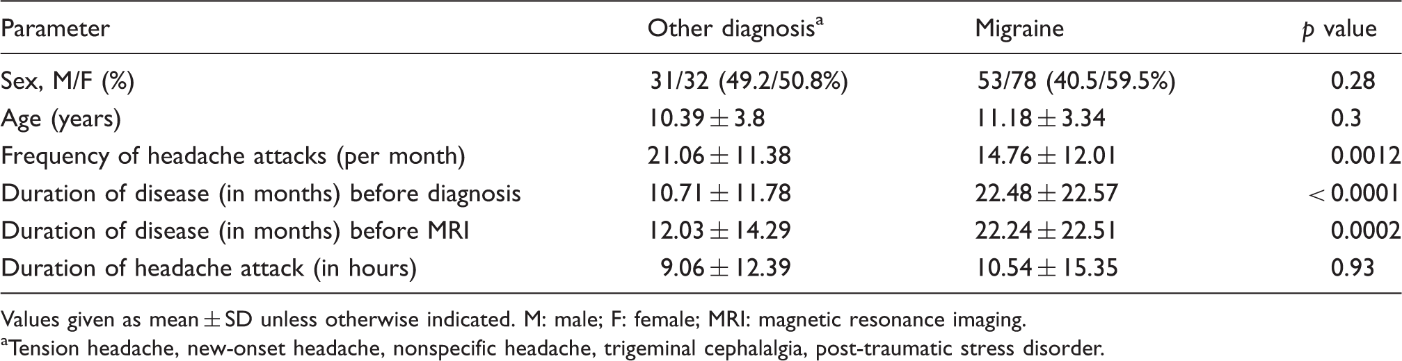
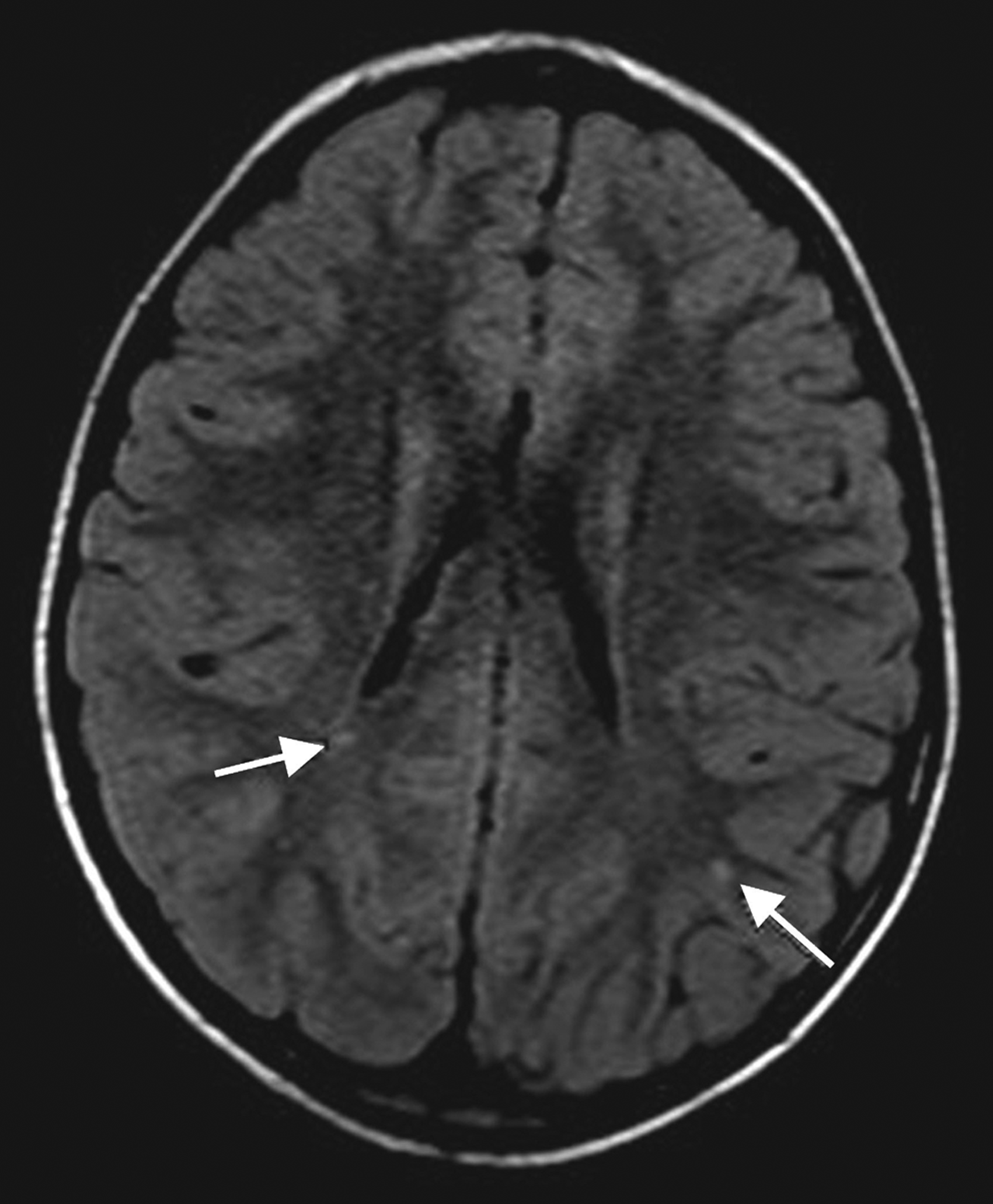
On comparison of the imaging parameters (Table 2), there was no statistically significant difference in the incidence of brain infarcts (cerebrovascular accident) between the headache group and the migraine group (1.5% and 1.6%, respectively). The only significant difference between the groups was the higher prevalence of white matter lesions in the patients with migraine (10.6% vs 0, p = 0.006). A total of 14 patients with migraine had white matter lesions; 13 had migraine without aura and one patient had migraine with aura. Thirteen patients had focal lesions and one patient with migraine without aura had confluent periventricular hyperintensities. All focal lesions were less than 3 mm in diameter. Thirteen patients had one to eight focal lesions (one patient had eight lesions, one patient had six, three patients had four, three patients had three, and five patients had one). Six patients had periventricular lesions; six, subcortical lesions; three, watershed zone lesions; and one, a thalamic lesion (Figures 1–3). The distribution of the lesions was variable, and some patients had lesions in more than one anatomical location.
FLAIR sequence showing tiny focal white matter hyperintensities in watershed zones. FLAIR: fluid-attenuated inversion recovery. FLAIR sequence showing tiny focal hyperintensities in subcortical white matter (left arrow) and periventricular white matter (right arrow). FLAIR: fluid-attenuated inversion recovery. T2-weighted image showing small hyperintense lesion in the left thalamus. Brain MRI findings in children and adolescents with migraine or other diagnoses. Values given as positive result numbers of magnetic resonance imaging (MRI) imaging in each group. Positive results also give as percentages. Incidental findings include all mentioned above except white matter lesions plus other incidental findings, four in each group.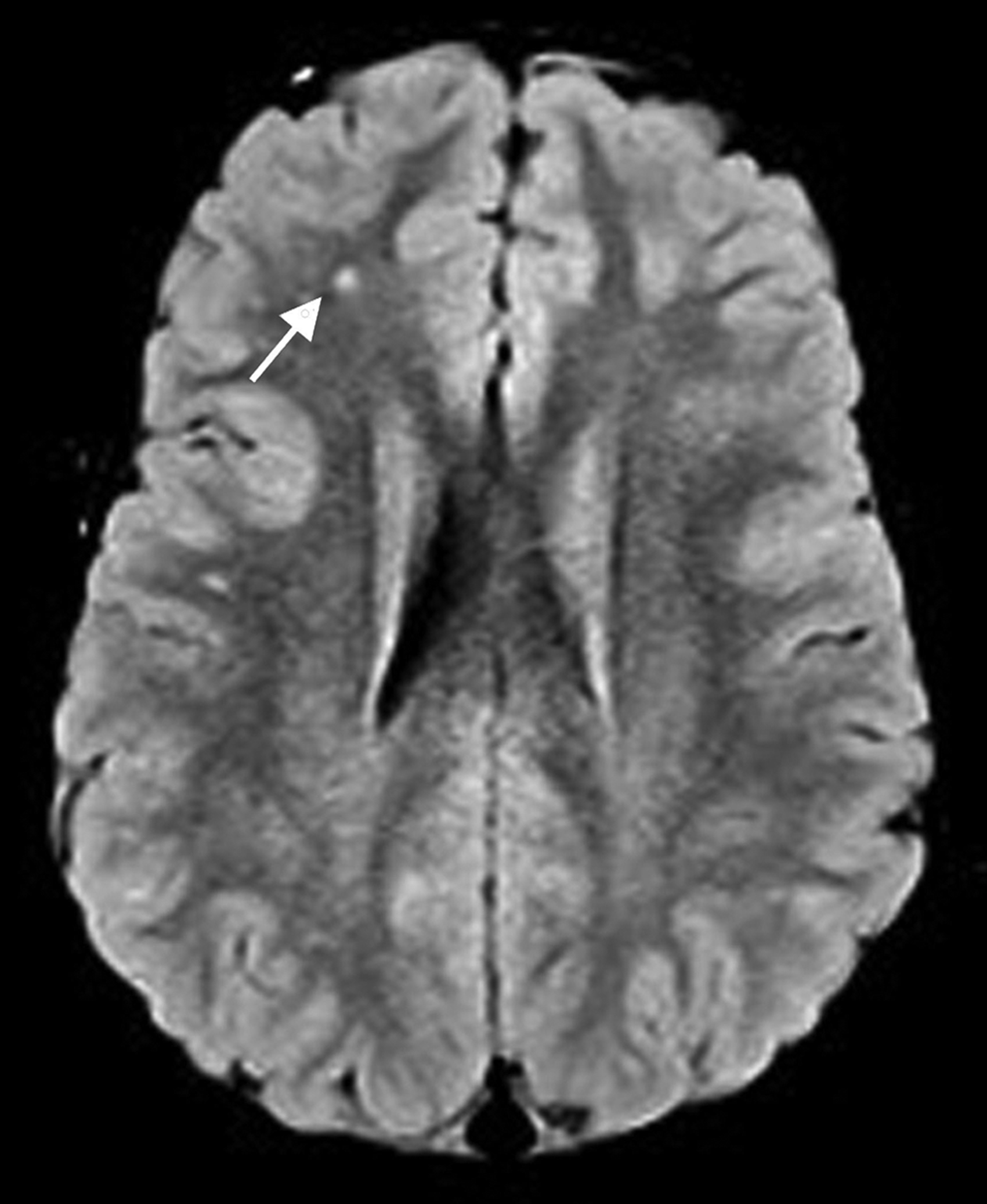
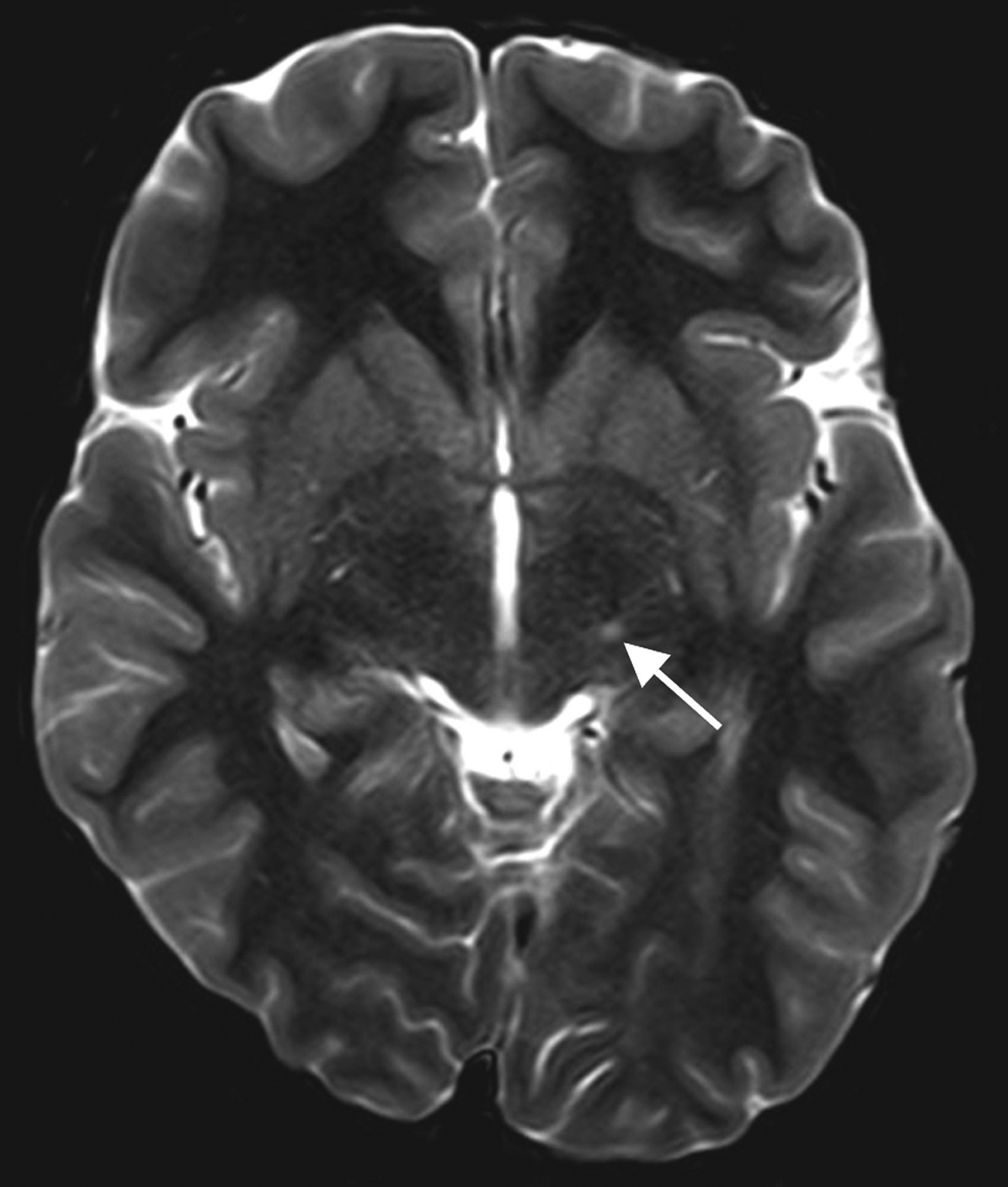

The sole patient with confluent white matter changes on T2 and FLAIR images (Figure 4) was not found to have any systemic disease after two years of follow-up. Preventive antimigraine treatment led to symptomatic improvement. Although the differential diagnosis of the imaging findings included periventricular leukomalacia due to old ischemia or infection, anamnesis did not reveal a past history of disease or perinatal insult.
FLAIR sequence showing confluent hyperintensity in periventricular white matter. FLAIR: fluid-attenuated inversion recovery.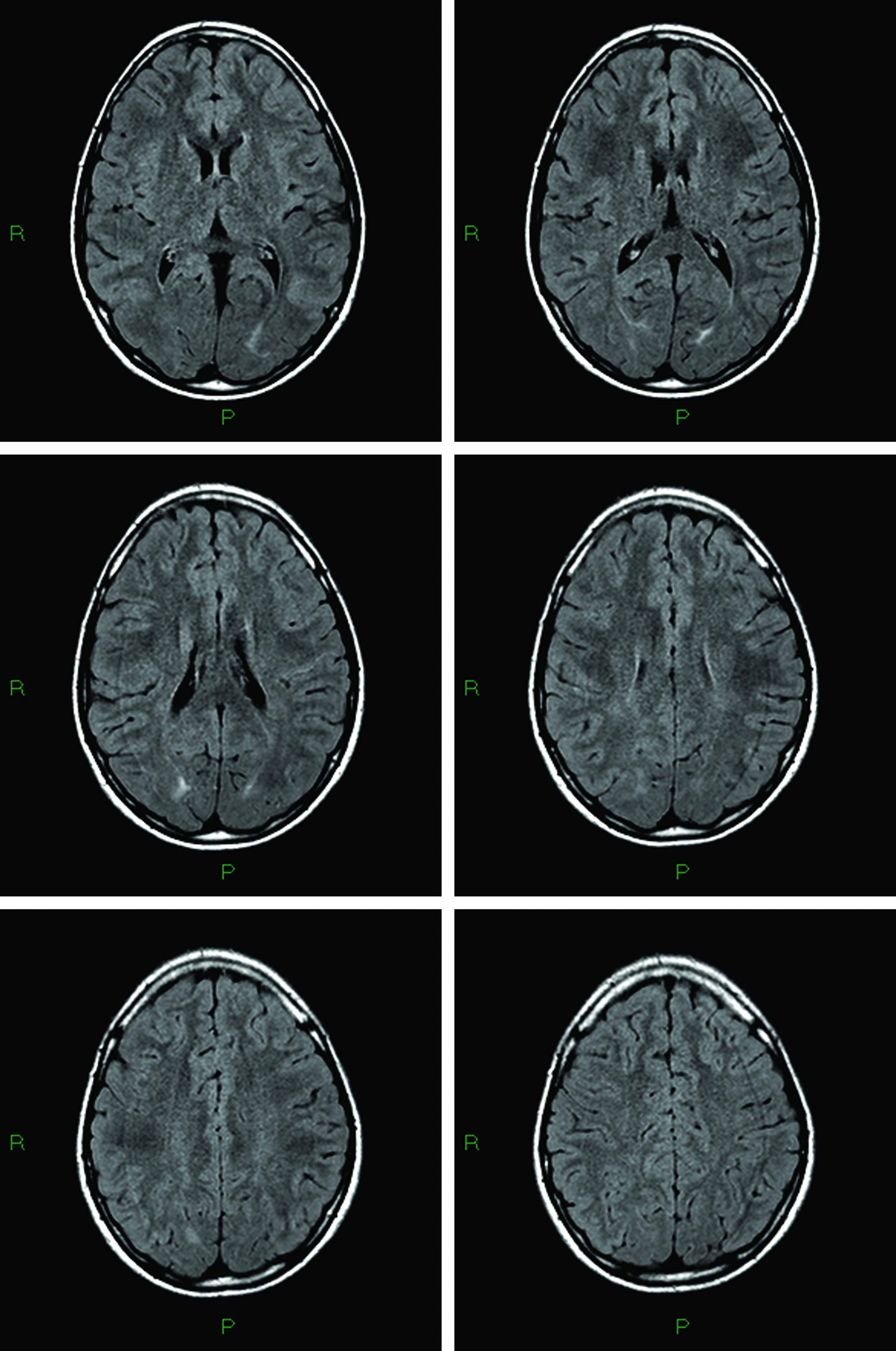
Within the migraine group, there were no statistically significant differences between the patients with and without white matter lesions in sex distribution, migraine-related clinical parameters, or rates of phonophobia, photophobia, osmophobia, allodynia, awakening pain, nausea, vomiting, dizziness, or migraine type (with/without aura), frequency of migraine attack per month, duration of attack in hours duration (in months) before MRI study, or duration of disease before diagnosis. A difference of borderline significance was noted in the duration of migraine attack between patients with and without white matter lesions (14.1 ± 14.23 versus 10.5 ± 15.5 hours; p = 0.065).
Prevalence of other MRI parameters was not different between those with and without white matter lesions.(Vircow Robin space, cerebrovascular accidents, brain atrophy paranasal inflammatory changes, or incidental findings).
Discussion
Neuroimaging performed to rule out underlying disease in children and adolescents with headache may yield benign findings related to the headache or incidental findings unrelated to the headache (1). In both cases, no change in patient management is warranted. The results of the present study indicate, in agreement with the literature, that imaging studies in young patients with chronic headache usually do not reveal treatable brain lesions. Previous studies of pediatric patients with headache yielded white matter lesions in only 2.9%–3.8% of imaging studies (9,14). None of the children had underlying abnormalities. However, the authors did not clarify the number of patients specifically with migraine headache. Data on the mean duration of headache and the mean interval from presentation to MRI scanning were also missing.
A correlation between migraine and structural brain damage has been suggested in several studies performed over the last decade in adults (3–7,14–17). The brain lesions typically appeared on T2WI or FLAIR images as small, multiple punctate, or discrete hyperintensities in the periventricular or deep white matter, with no mass effect, and with local loss of myelin and gliosis. Patients with and without aura were affected (15). According to a meta-analysis of seven case-control studies, adults with migraine were four times more likely to have white matter anomalies than those without migraine (16). Colombo et al. (15) proposed the loss of myelin and gliosis may be associated with brain aging and cerebrovascular disorders. Scher et al. (14) and Kruit et al. (17) reported that migraine with aura in midlife was associated with the presence of cerebellar infarct-like lesions, although this finding was statistically significant only in women.
A vascular etiology was suggested by the study of Wolf et al. (18), who used diffusion-weighted MRI (DWI) to examine adult patients with acute migraine and acute cerebellar ischemia. MRI studies were performed from two hours to seven days after the clinically acute attack, comparing to other studies that were performed not immediately after the acute migraine attacks (7,10). Particular attention was addressed to the vascular territory affected and the number and size of the lesions. They found that the majority of patients presented with prolonged aura and had acute ischemic lesions in the posterior circulation. The lesions had a multiple distribution and were located in distinct arterial territories. Although the headache protocol in our clinic does not include DWI, the characteristics of the lesions described by Wolf et al. (18) (posterior cerebellar artery territory or cerebellar infarction, either cortical based or large) are distinct from the small, multiple white matter lesions with a deep periventricular location seen in our patients, which more closely resemble those reported in the meta-analysis of Swartz and Kern (16). The finding that the lesions were not located in a particular arterial territory negates the theory of vascular infarcts. The absence of infratentorial lesions or cerebellar symptoms disagrees with the Cerebral Abnormalities in Migraine, an Epidemiological Risk Analysis (CAMERA) study (6), which reported that patients with migraine have a higher risk of silent infarct lesions. However, it is possible that small cerebellar infarcts may have been missed on thicker slices.
Gentile et al. (19) described an 18-year-old woman with two episodes of status migrainosus and recurrent, hyperintense, and reversible brain MRI findings. They suggested that these changes may be explained by vasogenic leakage and vasogenic edema, due perhaps to an alteration in the blood-brain barrier during the migraine attack.
It is suggested that white matter lesions may represent ischemic lesions secondary to repeated attacks of migraine-induced focal cerebral hypoperfusion or multiple microemboli (1). Several authors proposed that attack frequency and disease duration may serve as indicators of brain damage (20). Accordingly, the lower rate of white matter lesions in our pediatric cohort (10.6%) (16) is lower than reported in adults with migraine (22% range 16%–32%) by the meta analysis, since disease duration in pediatric patients is shorter.
The characteristics of the lesions in our study were similar to those reported by meta-analysis in adults (16) and may suggest a prolonged process of progressive brain damage starting from childhood and extending into adulthood. The lesions are most probably not reversible, since in none of our patients was the MRI performed during the acute phase of pain, and the mean time from symptom onset to imaging was 22 months. It is possible that additive, later-appearing comorbidities found mostly in adults (atherosclerosis, diabetes, hypertension, etc.) aggravate the brain damage. Along the same lines, the lack of a statistically significant difference in disease duration between our migraineurs with and without white matter lesions, and the only borderline significance of the association of these parameters with the duration of migraine attacks (in hours), may be at least partly explained by the 30-year-long follow-up in the earlier study (20) compared to two years in ours. The lower rate of white matter lesions in children may also be related to the apparent association between migraine with aura and white matter lesions in most reports of adult patients (14) whereas in our pediatric study all but one of the patients with white matter lesions had migraine without aura. Thus, pediatric migraineurs with white matter lesions may represent a distinct patient subgroup. A larger study with longer follow-up is needed to clarify the etiology and possible comorbidity of this finding in children. We found a higher prevalence of incidental findings than reported in the literature (1). This discrepancy may be due to the use of different MRI technologies with different sensitivities and differences among neuroradiologists. We detected three cerebrovascular accidents in our patients (one in the left middle cerebral artery territory, one in the right middle cerebral artery territory, and one in the right cerebral peduncle), all of which were old asymptomatic infarcts with no association to the diagnosis of migraine or other headache type. A similar prevalence (0.016) was reported by Schwedt et al. (1).
The present study is limited by the small sample size and the restriction of patients to those attending a dedicated headache clinic in a tertiary medical center who may have more severe disease than community-derived patients. This may explain the high frequency of headache attacks per month, and consequently, may have affected the prevalence of white matter lesions.
In conclusion, this is to our knowledge the first report of white matter abnormalities specific to pediatric migraineurs. The findings indicate that about 10% of pediatric patients with migraine have white matter lesions on MRI. The lesions are unrelated to background diseases or comorbidities. Further studies, with assessments of structural brain changes and brain function, are needed to clarify the pathogenesis of white matter lesions in children and adolescents with migraine.
Clinical implications
About 10% of pediatric patients with migraine have white matter lesions on MRI. Contrary to those found in adult patients, the pediatric white matter lesions in this study are unrelated to background diseases or comorbidities. The high frequency of headache attacks per month in our patients may have affected the prevalence of white matter lesions.
Footnotes
Acknowledgment
The authors thank Pnina Lilos for the statistical analysis.
Funding
This research received no specific grant from any funding agency in the public, commercial, or non-profit sectors.
Conflict of interest
None declared.