
Abstract
Background:
Fluctuations in limb volume degrade prosthesis fit and require users to accommodate changes using management strategies, such as donning and doffing prosthetic socks.
Objectives:
To examine how activities and self-report outcomes relate to daily changes in residual limb fluid volume and volume accommodation.
Study design:
Standardized, two-part laboratory protocol with an interim observational period.
Methods:
Participants were classified as “accommodators” or “non-accommodators,” based on self-report prosthetic sock use. Participants’ residual limb fluid volume change was measured using a custom bioimpedance analyzer and a standardized in-laboratory activity protocol. Self-report health outcomes were assessed with the Socket Comfort Score and Prosthesis Evaluation Questionnaire. Activity was monitored while participants left the laboratory for at least 3 h. They then returned to repeat the bioimpedance test protocol.
Results:
Twenty-nine people were enrolled. Morning-to-afternoon percent limb fluid volume change per hour was not strongly correlated to percent time weight-bearing or to self-report outcomes. As a group, non-accommodators (n = 15) spent more time with their prosthesis doffed and reported better outcomes than accommodators.
Conclusion:
Factors other than time weight-bearing may contribute to morning-to-afternoon limb fluid volume changes and reported satisfaction with the prosthesis among trans-tibial prosthesis users. Temporary doffing may be a more effective and satisfying accommodation method than sock addition.
Clinical relevance
Practitioners should be mindful that daily limb fluid volume change and prosthesis satisfaction are not dictated exclusively by activity. Temporarily doffing the prosthesis may slow daily limb fluid volume loss and should be investigated as an alternative strategy to sock addition.
Keywords
Background
The quality of socket fit is commonly acknowledged by both patients and prosthetists as the most important aspect of a prosthesis.1 –3 Socket fit is influenced by changes in the residual limb, most notably limb volume. 3 Trans-tibial sockets oversized by as little as 1.0% have been shown to induce clinically detectable changes in socket fit. 4
Activity may accentuate limb volume loss. 5 Pressures and shear stresses applied to residual limb soft tissues during ambulation may drive fluid out of the residuum, decreasing limb volume. Thus, users who spend more time weight-bearing, that is, standing and walking, would be expected to experience greater limb volume losses over the day than people who spend less time weight-bearing.
Prosthesis users who routinely gain or lose fluid volume are instructed by practitioners to adjust their prosthesis, typically by adding or removing socks, when they feel a change in socket fit. Users who spend much time weight-bearing would be expected to perform more sock accommodations compared with people who spend relatively little time weight-bearing. Those users who experience limb volume changes and do not make said changes would be expected to report reduced satisfaction, comfort, and perceived mobility compared to those who accommodate volume changes and maintain quality of their socket fit.
The purpose of this research was to explore whether trans-tibial prosthesis users’ morning-to-afternoon fluid volume change was associated with percentage time weight-bearing and user-reported satisfaction, comfort, and perceived mobility. We also evaluated whether persons who accommodated to daily limb volume change by adjusting sock thickness spent more time weight-bearing and reported greater satisfaction, comfort, and perceived mobility than those who did not accommodate.
Methods
Study design
A standardized, two-part laboratory protocol with an interim observational period was conducted to assess the correlation of residual limb fluid volume change and volume accommodation with prosthesis users’ health-related function, health, and quality-of-life. Residual limb fluid volume, activity, and self-report health evaluations of people with unilateral, trans-tibial amputation were collected during a single, three-part assessment (i.e. morning test session, between-sessions unrestricted activity, and afternoon test session) performed on a single day. Data were collected from October 2013 to June 2014. All procedures were approved by a University of Washington institutional review board and all participants provided informed consent.
Participants
Volunteers with trans-tibial amputation were recruited to be in this study. Individuals were recruited from regional prosthetics and orthotics clinics using posted flyers. Inclusion criteria included age 18 years or older, at least 9 months post-amputation with a stable residual limb, Medicare Functional Classification Level (K-level) 2 or higher, daily use (6 h or more per day) of a prosthesis with a definitive socket, and ability to walk on a treadmill for at least 90 s at a comfortable speed. Participants also needed to have a residual limb at least 9.0 cm in length to accommodate placement and spacing of bioimpedance electrodes. Exclusion criteria included skin breakdown (or other conditions that would preclude prosthetic ambulation) or presence of metal implants that would adversely affect the quality of bioimpedance data.
Instrumentation
A custom, semi-portable bioimpedance analyzer was used to measure participants’ residual limb fluid volume. Details of the bioimpedance system are described elsewhere. 6 Briefly, the instrument injected short packets of alternating current (~300 µA peak-to-peak, 5 kHz to 1 MHz) to electrodes positioned on the participant’s proximal thigh and distal inferior residual limb. Voltage was sensed by electrodes positioned on the anterior and posterior aspects at the level of the patellar tendon, mid-limb, and distal tibia, producing four regions of measurement: anterior-distal, anterior, posterior-distal, and posterior. We measured from anterior and posterior regions separately because the interosseous membrane between the tibia and fibula is a natural conduction barrier thus helps to isolate measurements from the two regions. A prior study suggested clinically meaningful differences between proximal and distal regions. 7 For short residual limbs (<12 cm), only the patellar-tendon-level and distal-tibia-level voltage-sensing electrodes were used; thus, there were only two regions of measurement: anterior and posterior. The electrodes and wires connecting the electrodes to the analyzer were less than 1.0 mm diameter and did not inhibit normal use of the prosthesis or movements of the limb. The electrodes were fabricated using conductive tape (ARCare 8881; Adhesives Research Inc., Glen Rock, PA), multi-stranded silver-plated copper wire with an aramid core and poly vinyl chloride insulation (New England Wire Technologies, Lisbon, NH), a flattened metal crimp to strain relieve wires as they exited the insulation within the electrode, and a thin layer of hydrogel (KM10B; Katecho, Inc., Des Moines, IA) between the electrode and skin. This electrode configuration was used successfully in previous studies.5,8 –10 Sensed voltages were transmitted via a 3-m cable back to a personal computer (PC) for subsequent analysis (Figure 1(a) and (b)). Evaluation tests conducted on the custom bioimpedance analysis system demonstrated minimal signal drift (0.02%/h), root-mean-square (RMS) noise (0.026%), and measurement error across the sensing range of the instrument (−0.4%). 6
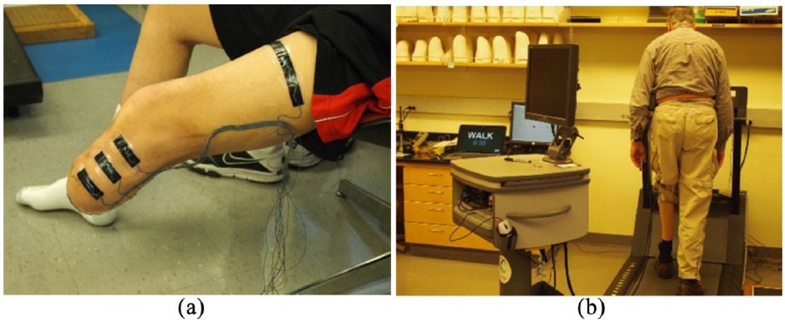
Residual limb fluid volume monitoring. (a) Residual limb instrumented with electrodes (distal current-injection electrode not visible). (b) Limb fluid volume monitoring test session.
Self-report measures
A self-report survey was used to solicit information about participants’ prosthesis-related function, health, and quality of life over the past 2 weeks. The survey included several standardized measures, including the Socket Comfort Score (SCS) 11 and the Ambulation, Residual Limb Health, Utility, and Well-Being subscales from the Prosthesis Evaluation Questionnaire (PEQ). One additional question from the PEQ that is not included in the PEQ subscales (i.e. “Over the past 2 weeks, rate how happy you have been with your current prosthesis”) was included to assess participants’ overall satisfaction with the prosthesis. 12 The SCS and PEQ were developed to evaluate prosthesis-related health outcomes in people with lower limb amputation, and both instruments have been reported to be valid and reliable when used for this purpose.11,12 Measures were presented to participants on paper using the developers’ recommended instructions and response options.11,12
Procedures
Participants first attended an in-person screening session to verify eligibility criteria and determine their comfortable walking speed. Before leaving the laboratory, participants were provided a 2-week sock log 13 to record their normal, daily use of prosthetic socks. A sock log was used to characterize participants’ sock use, because self-report (i.e. asking participants, “Do you change socks throughout the day?”) was previously found to be an unreliable indicator of accommodation in people with trans-tibial amputation. 13 Participants were also instructed to eat a normal breakfast and avoid caffeine or alcohol (diuretics) during the morning before their next visit. Participants returned to the laboratory after 2 weeks for an assessment that included a morning (AM) test session, a between-sessions activity monitoring period, and an afternoon (PM) test session.
Upon arrival at the AM test session, participants met with the study prosthetist to have their residual limb inspected and to discuss recent changes in their health, prosthetic fit, or activity that might affect limb fluid volume, limb fluid volume changes, or other studied outcomes. Participants were then administered the self-report survey. Upon finishing the survey, participants sat for 10 min with both feet on the floor to establish a stable limb fluid volume baseline. After resting, participants doffed their prosthesis and were instrumented for limb fluid volume monitoring with the bioimpedance analyzer. 6 Participants’ prostheses were also equipped with an activity monitor (GT3X+; ActiGraph, Pensacola, FL). The GT3X+ is a small (4.6 × 3.3 × 1.5 cm), lightweight (19 g), accelerometer-based sensor (±6 g dynamic range, 0.0023 g resolution) that has been used previously to monitor prosthesis users’ activity.14–17
Participants then performed a standardized, “sit–stand–walk” protocol intended to reflect typical daily activities. Participants first sat in a stationary chair for 90 s with their prosthesis donned. Then they rose and stood for 90 s. Weight-bearing through the prosthetic leg was monitored with a digital scale (349KLX Health o meter; Pelstar, McCook, IL) and participants were prompted to adjust their weight allocation as needed to achieve equal weight-bearing. Participants next stepped onto the treadmill and walked at their previously determined preferred walking speed for 90 s. Finally, participants stepped off the treadmill and stood on the scale with equal weight-bearing for 10 s. This sit–stand–walk protocol was repeated four more times. Upon completion of the last cycle, participants remained sitting for 10 min. The system was then turned off and the electrode wires were disconnected from the analyzer. The wires were coiled neatly and taped to the prosthesis so that the participant could move without restriction.
Participants were instructed to leave the laboratory for at least 3 h and go about their normal activities to the extent possible. Participants were informed that they could doff their prosthesis or change socks to accommodate volume changes as desired, but were asked not to doff their liner in order to avoid damaging the electrodes.
Participants returned to the laboratory for the PM test session and were re-administered the limb fluid volume test using the same sit–stand–walk protocol described for the AM test session. Upon conclusion of the test, participants doffed the prosthesis and the instrumentation was removed.
Analysis
Sock logs and self-report measures were scored by research staff according to developers’ instructions.11 –13 Participants were classified as “accommodators” if they reported adding or removing socks during any day in the 2-week monitoring period. They were classified as “non-accommodators” if no daily sock changes were made over the monitoring period.
Collected bioimpedance data were post-processed to determine extracellular fluid impedance using De Lorenzo’s 18 form of the Cole model. To convert extracellular fluid impedances to extracellular fluid volumes, a limb segment model was used.19,20 In this study, we investigated only extracellular fluid volume changes because extracellular changes were expected to be most relevant to the relatively short-term limb fluid volume fluctuations of interest in this study.
The rate of limb fluid volume change between AM and PM sessions expressed in %/h was calculated for each of the four channels. Residual limb fluid volumes measured during the brief 5−10 s stand after the fifth walk cycle during the AM and PM test sessions were used. Residual limb fluid volume change was expressed as a percentage difference
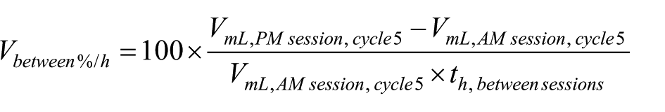
Differences in distributions between accommodators and non-accommodators were assessed with Fisher’s exact test (for sex and activity level) and Mann–Whitney U test (for age, time since amputation and body mass index (BMI)). Pearson correlation was used to assess the strength of linear association between outcomes and residual limb fluid volume change. Non-parametric test of medians (percent time doffed, sitting, standing, walking, and weight-bearing; SCS; PEQ satisfaction, ambulation, residual limb health, utility, and weight-bearing) was used for comparisons between “accommodators” and “non-accommodators.”
Results
Participant characteristics
Twenty-nine people with trans-tibial amputation participated in the study. On average, participants were male (82.8%), middle-age (mean = 56.7 (standard deviation (SD) = 14.8) years old), and established amputees (mean = 15.3 (14.4) years post-amputation). Most participants (93.1%) were unlimited or active community ambulators. About half of the participants (48.3%) used socks to manage daily limb volume changes and were classified as “accommodators.” Four accommodators started days with different sock ply. All of the non-accommodators started their days with the same sock ply. There were no significant differences between accommodators and non-accommodators according to sex (p = 0.65), age (p = 0.95), BMI (p = 0.56), or activity level (K-2 vs higher level, p = 1.0). However, non-accommodators had their amputation significantly longer than accommodators (median 12.9 years vs 5.5 years, respectively, p = 0.01). Individual participant characteristics are listed in Appendix 1.
Morning-to-afternoon fluid volume changes
Of the 29 participants, 21 (72.4%) lost fluid volume between sessions in most regions of the residual limb (3 of 4, 4 of 4, or 2 of 2 regions), 5 (17.2%) gained fluid volume in most regions, and 3 (10.3%) gained in half of the regions and lost in the other half (Appendix 2).
Participants spent most of their time between sessions sitting with their prosthesis donned (mean = 62.7% (18.0%), median = 65.4%), and spent less time walking (mean = 19.0% (13.0%), median = 13.0%) or standing (mean = 11.6% (7.0%), median = 10.4%). Participants doffed their prosthesis a mean of 6.5% (12.9%) and median of 3.4% of the time between sessions.
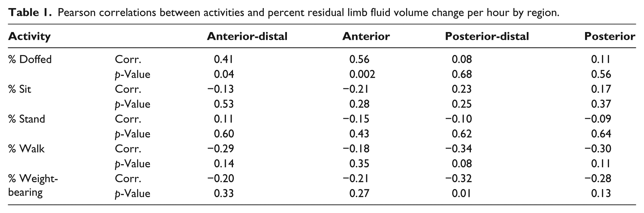
Morning-to-afternoon percent fluid volume change per hour generally increased with decreased percent time walking, standing, and weight-bearing (weight-bearing was the sum of standing and walking). However, the correlations were weak (Table 1; Figure 5 in Appendix 3). Correlation coefficients for all channels ranged from −0.34 to 0.11 and p-values ranged from 0.01 to 0.64. The high correlations for percent fluid volume change per hour in the anterior and anterior-distal regions with percent time doffed were due largely to two individuals with long percent doff times (37.7% and 62.9%). The remaining participants had percent doff durations ranging from 0.0% to 11.1%. Similarly, for the posterior-distal region and weight-bearing, two individuals had high percent fluid volume changes per hour (5.3%/h and 10.8%/h), while the remaining ranged from −2.4%/h to 2.4%/h.
Pearson correlations between activities and percent residual limb fluid volume change per hour by region.
Morning-to-afternoon percent fluid volume change per hour was not strongly correlated with reported socket comfort, satisfaction, ambulation, residual limb health, utility, or well-being (Table 2). Correlation coefficients ranged from −0.20 to 0.24 and p-values ranged from 0.22 to 0.94.
Pearson correlations between self-reported health outcomes and percent residual limb fluid volume change per hour by region.
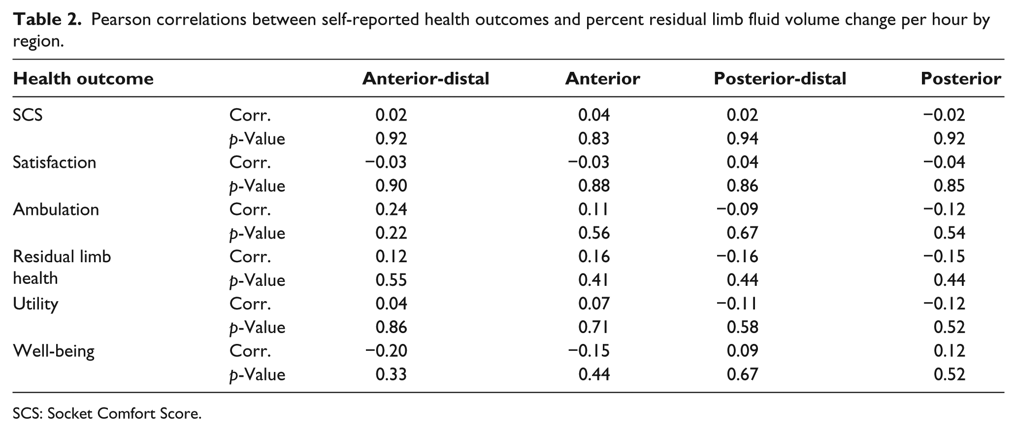
SCS: Socket Comfort Score.
Accommodators versus non-accommodators
Participants who accommodated to daily limb volume changes spent more time weight-bearing than those who did not accommodate, but the difference in medians was not statistically significant (p = 0.47) (Figure 2). Interestingly, participants who did not accommodate had a greater median percent time with their prosthesis doffed than accommodators, although the medians were not statistically different (p = 0.72). The distribution of percent time doffed for accommodators and non-accommodators showed that about a third of the participants were at the extremes: five accommodators and two non-accommodators spent 0.0%−0.9% time doffed, and three non-accommodators spent >10.0% time doffed.
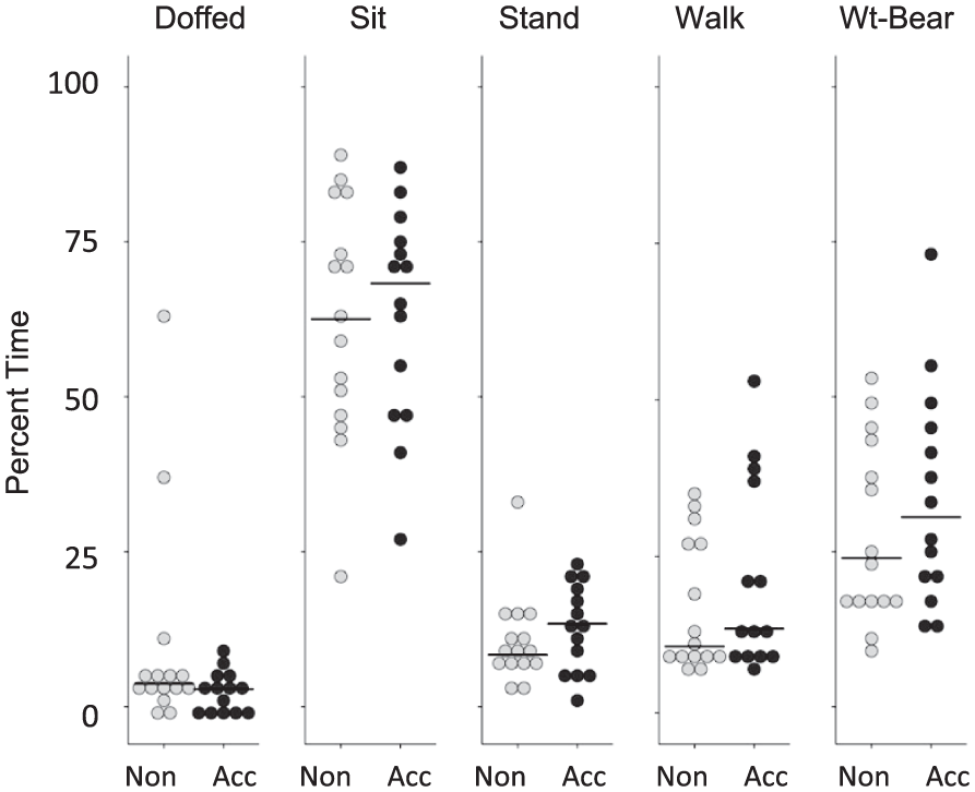
Activities between sessions. Accommodators (Acc) and non-accommodators (Non). Black horizontal lines are medians.
Median percent fluid volume changes per hour were not statistically different for accommodators versus non-accommodators (Table 3) with p-values ranging from 0.45 to 1.0.
Morning-to-afternoon percent fluid volume change per hour (accommodators and non-accommodators).
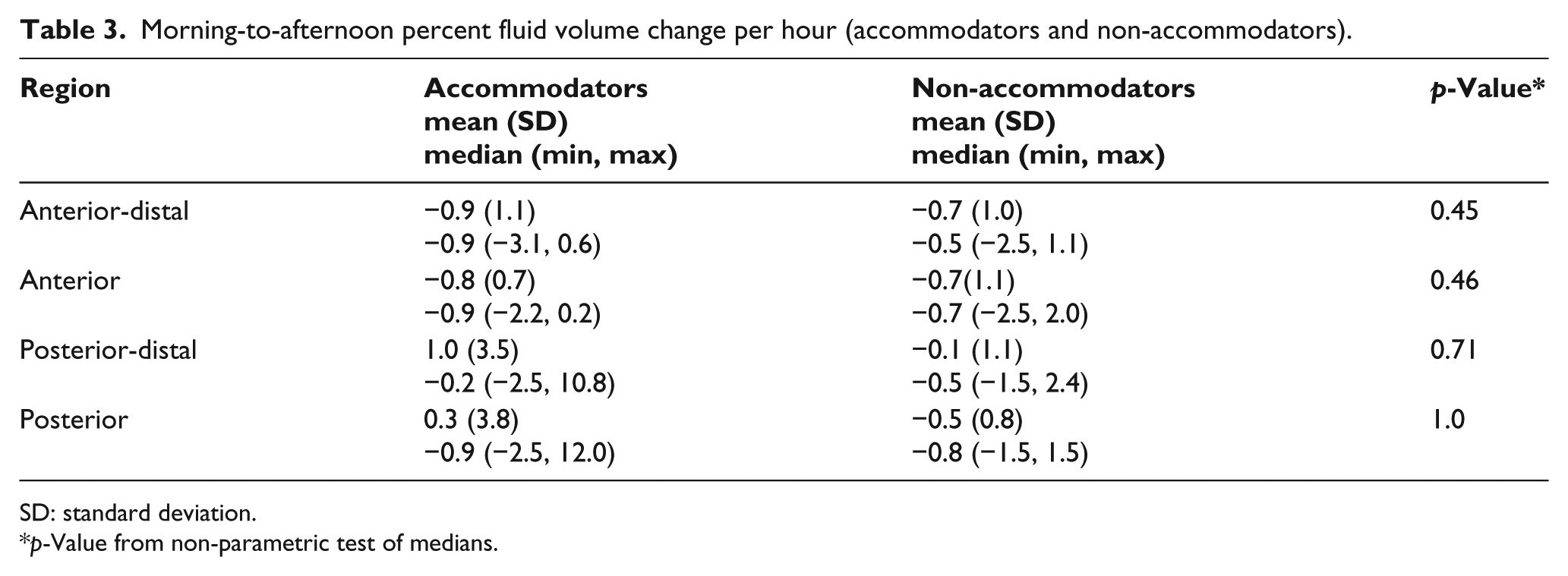
SD: standard deviation.
p-Value from non-parametric test of medians.
Across all self-report measures, persons who accommodated to daily fluid volume had lower median self-report scores than those who did not accommodate (Figure 3). Differences in medians for all PEQ subscales ranged from 5 to 11 (100-point scale) and was 1 for SCS (10-point scale). However, none of the differences were statistically significant Correlations between self-report measures and percent limb fluid volume change per hour were weak (Figures 6 and 7 in Appendix 3). P-values ranged from 0.27 to 0.72.
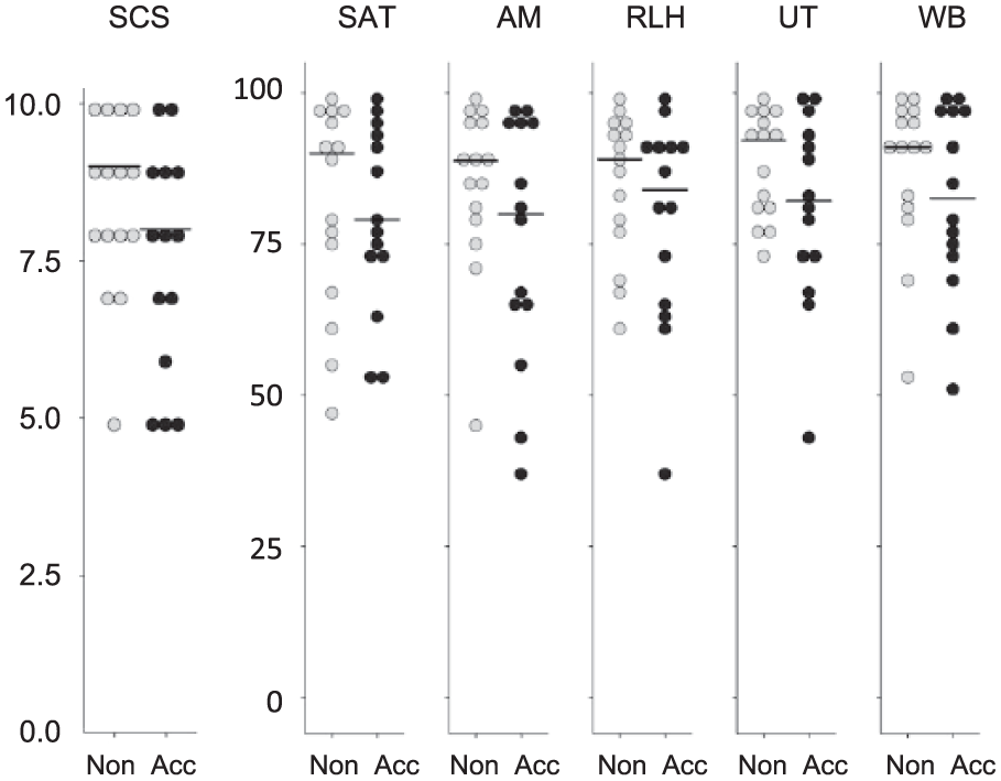
Self-report results. Accommodators and non-accommodators. Left: SCS. Right: PEQ subscales (SAT: satisfaction; AM: ambulation; RLH: residual limb health; UT: utility; WB: well-being). Black horizontal lines are medians.
Discussion
Results from the present study do not support the hypothesis that prosthesis users who spend much time weight-bearing, that is, standing and walking, experience greater percent limb fluid volume losses over the day compared with people who spend little time weight-bearing. Part of the reason this expectation is not supported may be because of the influence of doffing. Doffing the prosthesis periodically during the day, even for short periods, may help the user to counter fluid volume losses experienced during weight-bearing. 10 Also, previous research showed that walking did not necessarily cause a fluid volume loss, particularly if preceded by standing. In a prior investigation, 21 fluid volume gains were measured in 16 of 24 (66.7%) participants during walking in a PM in-laboratory test sessions. In the present investigation, we calculated fluid volume changes within PM test sessions and found that during walking, 21 of 29 (72.4%) participants gained limb fluid volume in the majority of monitored limb regions (3 of 4, 4 of 4, or 2 of 2) (Appendix 4). Fluid volume gains from both periodic doffing and walking may have contributed to the relatively low correlation between percent time weight-bearing and percent limb fluid volume loss per hour in the present study.
Results do not support the hypothesis that prosthesis users who report low health outcomes experience greater rates of percent limb fluid volume loss from the morning to afternoon compared with people who report high health outcomes. This result may have been influenced by the short testing interval (half a day), the atypical environment for the participants (laboratory and nearby locations accessible during the between-session period), and the design of the self-report measures (they were not specific to the test day but instead reflected 2-week periods immediately prior to the test day).
Since they had more mature residual limbs, non-accommodators in the present study may have been less prone to daily limb fluid volume changes than accommodators. However, the result that, on average, non-accommodators spent more time doffed than accommodators suggests that non-accommodators may have regularly used temporary doffing as an accommodation method. Thus, they may have been “accommodating,” just not by adding socks. Temporary doffing facilitates limb fluid volume recovery and retention, enlarging the residual limb and helping to keep it within an acceptable volume range so as to maintain fit (Figure 4(a)). 10
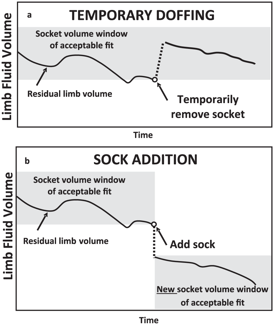
Accommodation strategies. Temporary doffing (a) compared with sock addition (b).
Sock accommodation affects limb fluid volume differently than temporary doffing. Prior research has shown that a prosthesis user’s limb fluid volume decreases when a sock is added. 22 By adding socks, accommodators shift the location of their acceptable volume window to lower volumes (Figure 4(b). It is unknown whether using periodic doffing instead of sock addition as an accommodation method reduces long-term residual limb volume loss (limb atrophy). If it did then regular periodic doffing would lengthen the duration a socket remained comfortable to the user. Interestingly, non-accommodators showed a tendency to report higher health outcome scores than accommodators (Figure 3). Maintaining limb fluid volume by temporary doffing rather than by accentuating limb volume loss with sock addition may be more satisfactory to prosthesis users. Future research efforts are needed to compare effectiveness of sock addition versus temporary doffing and to determine whether individual prosthesis users benefit using one method over the other. It would also benefit the field to know what characteristics of prosthesis users cause one method to provide more favorable user outcomes.
Research studies designed to assess other factors that may influence residual limb fluid volume loss such as diet, hydration, and medications need to be conducted and their impact compared with activity. By providing the clinical community with insight into the relative influence of these and other factors, researchers will help practitioners treat patients who experience poor socket fit.
Conclusion
Factors other than time weight-bearing (standing and walking) contribute to the rate of morning-to-afternoon limb fluid volume change on trans-tibial prosthesis users. Further investigation is needed to determine whether temporary doffing is a more effective and satisfying accommodation method than sock addition.
Footnotes
Appendix 1.
Appendix 2.
Appendix 3
Appendix 4.
Author Contribution
All authors contributed equally in the preparation of this manuscript.
Declaration of conflicting interests
The author(s) declared no potential conflicts of interest with respect to the research, authorship, and/or publication of this article.
Funding
The author(s) disclosed receipt of the following financial support for the research, authorship, and/or publication of this article: This work was funded by National Institute of Child Health and Human Development (grant/award number: “R01HD060585”).