
Abstract
Background
Orthodontic bonding materials are required to provide adequate adhesion throughout treatment while also permitting safe debonding upon completion. Recently, the incorporation of nanoparticles into composite resins has gained attention due to their antimicrobial properties and their potential to reduce enamel decalcification around orthodontic brackets. However, such modifications may influence the mechanical and physical characteristics of adhesives, particularly the shear bond strength. Limited evidence is available regarding the effect of zinc oxide nanoparticles on the bond strength of orthodontic brackets.
Aim
This study aimed to evaluate and compare the shear bond strength of orthodontic brackets bonded using an adhesive containing zinc oxide nanoparticles with that of a conventional orthodontic adhesive.
Materials and Methods
A total of 32 extracted human premolars with intact structure were selected for this in vitro investigation and randomly assigned into two equal groups (n = 16). Stainless steel brackets (McLaughlin–Bennett–Trevisi (MBT) 0.022″ × 0.028″) were bonded to the buccal surfaces using two different adhesive systems: one incorporated with 1% zinc oxide nanoparticles and the other without nanoparticle addition, serving as the control. After 24 h, shear bond strength was measured using a universal testing machine (UTM). The collected data were statistically analyzed using IBM Statistical Package for Social Sciences version 24, and an independent t-test was applied to determine the significance of differences between groups.
Results
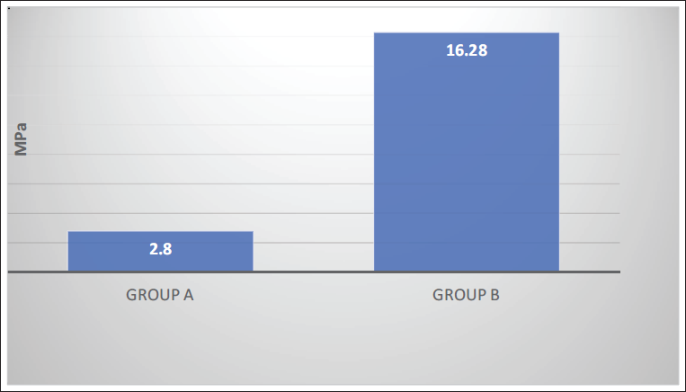
A statistically significant difference in shear bond strength was observed between the two groups. The experimental group demonstrated a mean value of 2.8 MPa, whereas the control group showed a considerably higher mean value of 16.28 MPa.
Conclusion
The addition of zinc oxide nanoparticles to orthodontic adhesives has a significant impact on shear bond strength, which may restrict its effectiveness for clinical use.
Introduction
Decalcification of the enamel adjacent to fixed orthodontic appliances is a frequently observed and clinically significant adverse effect associated with orthodontic therapy. 1 The prevention of white spot lesions primarily depends on the maintenance of effective oral hygiene measures, particularly regular brushing with fluoridated toothpaste. However, in patients with poor compliance, additional preventive strategies become necessary. One such approach involves the use of antimicrobial bonding agents around the bracket interface to minimize microbial activity. 2 Fluoride plays a crucial role in enhancing enamel resistance by promoting the formation of fluorapatite, which is less susceptible to acid dissolution. It can be administered through various delivery systems, such as varnishes, mouth rinses, gels, dentifrices, and fluoride-releasing orthodontic adhesives or luting cements. 3
Contemporary approaches for the prevention of white spot lesions focus on the use of advanced materials such as bioactive glass, casein phosphopeptide–amorphous calcium phosphate (CPP–ACP), and the incorporation of nanoparticles with anticariogenic properties into orthodontic appliance surfaces, as well as bonding adhesives and cements.4–6 The process of enamel decalcification is primarily initiated by the persistence of specific bacteria on the tooth surface, which produce organic acids over time, leading to mineral loss. Therefore, the use of bonding agents with antibacterial properties may play a significant role in reducing demineralization around orthodontic brackets. The application of nanotechnology in resin-based composites represents a notable advancement in the development of modern dental materials. 2
Nanotechnology involves the controlled manipulation of materials at the nanoscale, typically within the range of 1-100 nm, which imparts unique properties and enables diverse applications. 7 In dentistry, nanotechnology has been widely utilized across various fields, including implantology, restorative dentistry, radiographic techniques, periodontal therapy, and orthodontics. The incorporation of nanoparticles into composite resins has been shown to improve mechanical properties and enhance the overall performance of these materials. 8
Lee et al. reported that dental resins incorporated with silver nanoparticles demonstrate notable antimicrobial properties. 9 Previous investigations have also indicated that experimental composites containing silver nanoparticles provide effective antibacterial action while maintaining adequate shear bond strength (SBS). 10 However, the use of silver nanoparticles may result in discoloration of the composite material, and concerns regarding their biocompatibility still persist. 11 Furthermore, evidence from both in vitro studies and animal models suggests that nanoparticles based on copper and zinc may exhibit considerable toxic effects. 12
Streptococcus mutans has been shown to be sensitive to nanoparticles of silver, zinc oxide (ZnO), and gold, and their incorporation into orthodontic adhesives can reduce bacterial colonization as well as the development of white spot lesions. 13 However, the inclusion of such nanoparticles may also affect the physical and mechanical characteristics of adhesives, particularly SBS.
To date, orthodontic research on this subject has been largely confined to the antibacterial effects of different nanoparticles. Cronquist et al., 14 in a recent study, demonstrated that incorporating bioactive nanoAg-ACP microparticles into an orthodontic adhesive significantly enhanced enamel resistance to demineralization without adversely affecting SBS. The purpose of this study is to assess and compare the SBS of orthodontic brackets bonded with an adhesive containing 1% weight/weight (w/w) ZnO nanoparticles versus a conventional orthodontic adhesive. Jatania and Shivalinga 15 demonstrated that lower ZnO concentrations resulted in improved bond strength, and thus a concentration of 1% ZnO was selected in the present study. The study hypothesized that incorporating ZnO nanoparticles into orthodontic composites would not result in a statistically significant difference in SBS compared to conventional composites.
Materials and Methods
The Dr. HSRSM Dental College and Hospital’s Institutional Ethics Committee approved the study protocol (IEC Approval No: HDCH/Ethics/2025/68), and laboratory testing was carried out at Praj Metallurgical Laboratory, using a calibrated Universal Testing Machine (Computerized, Software-based) Company: ACME Engineers, India. Model No. UNITEST-10. The machine had an accuracy of ±1% and operated at a crosshead speed of 0.5 mm/min.
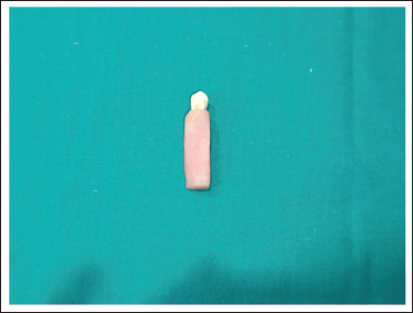
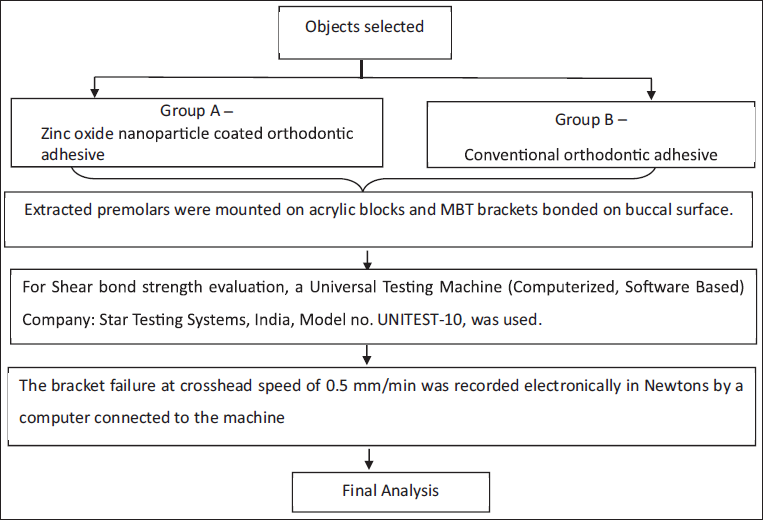
Based on a previous study, 2 and considering the minimum standardized difference, a type I error rate of 0.05, and a study power of 0.80, a total of 32 recently extracted, noncarious, nonfluorosed human premolars with intact buccal surfaces were selected, with 16 teeth assigned to each group (Figure 1). These teeth, extracted for orthodontic reasons, were acquired from the Department of Oral and Maxillofacial Surgery. Only teeth with sound buccal surfaces, free from restorations, attrition, abrasion, erosion, fractures, or prior chemical treatments were included in the study.
Extracted Premolar Mounted in Acrylic Block.
Group A: ZnO nanoparticle 1% (w/w) coated orthodontic adhesive (N = 16).
Group B: Conventional orthodontic adhesive (N = 16).
Method of Preparation of Nano Adhesive
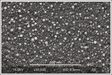
ZnO nanoparticles averaging 40-60 nm in diameter and a concentration of 1% by weight were utilized. ZnO nanoparticles were incorporated into Transbond XT (3M Unitek) orthodontic composite at a concentration of 1% (w/w) using a standardized mechanical dispersion protocol. The mixture was blended in a high-speed mechanical mixer (Model: LMHH160, Biotron Scientific, India) at 3,500 rpm for 5 min under dark conditions to ensure a homogeneous nanoparticle distribution and prevent premature polymerization. The presence and uniform distribution of ZnO nanoparticles in the adhesive were confirmed using scanning electron microscopy (SEM) imaging (×5,000 magnification). ZnO nanoparticles appear as brighter spherical structures distributed throughout the darker adhesive matrix background. This contrast difference is due to the higher electron density of ZnO compared to the resin matrix (Figure 2).
Scanning Electron Microscopy (SEM) Micrograph of 1% ZnO Nanoparticle-modified Adhesive.
Procedure
To avoid desiccation, each sample was washed for 5 s with pumice and water, rinsed for 10 s, and then allowed to air dry.
A 37% phosphoric acid was used for 30 s of etching, followed by a 10-s water rinse.
The application of the primer on the teeth was performed according to the manufacturer’s recommendations.
Using a light-emitting diode curing light, the teeth were cured for 15 s at a distance of 1 mm from their buccal surfaces. 16
Using a bracket holder and the corresponding group composite, stainless steel MBT brackets were adhered to the middle of each tooth’s buccal surface.
Finally, each tooth was cured for 40 s. 17
SBS was measured using an Instron machine (Figure 3) with a 0.5 mm/min speed and a 0.3 mm thick blade. Between the bracket base and the tooth, a shear force was applied parallel to the tooth’s longitudinal axis until the bracket was separated (Figure 4).
Acrylic Blocks Mounted on Universal Testing Machine (UTM) for Shear Bond Strength Testing.
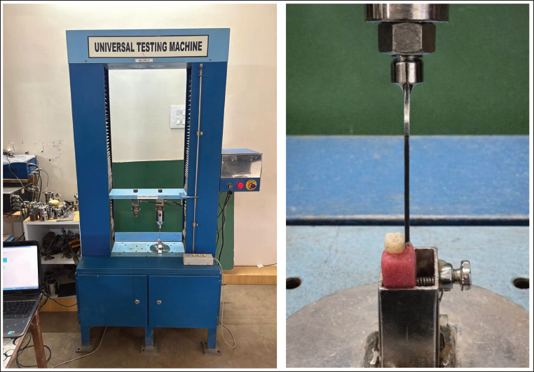
Shear Bond Strength Testing.
Statistical Analysis
The distribution of the data was assessed for normality using the Shapiro–Wilk test, yielding P values of .305 for group A and .261 for group B. As the data satisfied the assumption of normality, differences in SBS between the two groups were evaluated using an independent t-test.
Results
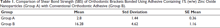
Table 1 provides the descriptive statistics for SBS in both experimental groups. Brackets bonded with adhesive containing 1% (w/w) ZnO nanoparticles (Group A) exhibited a mean SBS of 2.8 ± 1.44 MPa, whereas those bonded with conventional orthodontic adhesive (Group B) showed a markedly higher mean value of 16.28 ± 4.62 MPa.
Comparison of Shear Bond Strength (SBS) of Orthodontic Brackets Bonded Using Adhesive Containing 1% (w/w) Zinc Oxide Nanoparticles (Group A) with Conventional Orthodontic Adhesive (Group B).
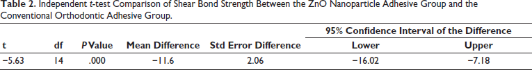
An independent t-test was conducted to compare the mean SBS between the two groups. The analysis revealed a statistically significant difference, with a mean difference of 11.6 MPa and a 95% confidence interval ranging from −16.02 to −7.18 MPa. Incorporation of 1% (w/w) ZnO nanoparticles into the adhesive resulted in a substantial reduction in SBS compared to the conventional adhesive, as reflected by a t-value of −5.63 with 14 degrees of freedom (P < .001) (Table 2, Figure 5).
Independent t-test Comparison of Shear Bond Strength Between the ZnO Nanoparticle Adhesive Group and the Conventional Orthodontic Adhesive Group.
Comparison of Shear Bond Strength Between Groups A and B.
Discussion
Orthodontic adhesives are known to retain cariogenic streptococci more readily than the materials used for brackets. To enhance the antibacterial properties of these composites, various nanoparticles—such as silver, titanium dioxide, ZnO, and ACP—can be incorporated. 13 In the present study, however, the focus was on assessing how the addition of nanoparticles affects the SBS of orthodontic adhesives rather than their antimicrobial efficacy. This distinction is crucial, as the primary function of orthodontic adhesives is to provide sufficient SBS to maintain stable bracket adhesion and prevent premature debonding, even if nanoparticles offer potential benefits in reducing enamel demineralization.
Owing to its antibacterial properties, ZnO has been applied in various medical contexts, including topical creams and ointments for treating burns, foot ulcers, and other traumatic skin injuries. Similarly, in dentistry, these antimicrobial characteristics have been incorporated into materials such as endodontic sealers and selected dental cements. 18
Several studies have suggested that the antibacterial activity of ZnO is mainly achieved by disrupting the function of bacterial cell membranes. Additionally, the production of intracellular reactive oxygen species, including hydrogen peroxide, a strong oxidizing agent, may contribute to bacterial cell damage.19, 20
Moorer and Genet 21 proposed that the antimicrobial activity of ZnO operates through a “reservoir” effect, whereby higher zinc concentrations in the bonding material can provide increased availability of zinc ions for gradual release. While these findings align with the current study in elucidating zinc’s antibacterial mechanism, the present research emphasizes a practical limitation: enhancing the reservoir effect with ZnO nanoparticles may compromise SBS, potentially restricting clinical use unless the adhesive formulation is carefully optimized to balance antimicrobial efficacy with mechanical performance.
In addition to providing antibacterial effects, it is essential that nanoparticles incorporated into orthodontic composites maintain sufficient SBS or ideally improve it. With this in mind, the present study evaluated the impact of incorporating 1% (w/w) ZnO nanoparticles into a composite on the SBS of orthodontic brackets. The results demonstrated that the composites containing ZnO nanoparticles exhibited a markedly lower mean SBS compared to the nanoparticle-free control composites.
Spencer et al. reported that higher concentrations of ZnO are associated with decreased SBS, observing mean bond strengths of 5.04 MPa for a 13% ZnO mixture and 4.56 MPa for a 23.1% ZnO mixture. 18 Similarly, Jatania and Shivalinga found that lower ZnO concentrations were linked to improved bond strength, supporting the choice of a 1% ZnO concentration for the current study. 15
The reduction in SBS observed in the nanoparticle group may be attributed to agglomeration of nanoparticles within the resin matrix, which disrupts polymer chain formation and compromises mechanical integrity. Furthermore, nanoparticles may interfere with light transmission and depth of cure, resulting in incomplete polymerization. Altered stress distribution and poor resin infiltration into enamel microporosities may also contribute to reduced bond strength.
The finding aligns with recent in vitro evidence from Prasad et al., 22 who evaluated experimental adhesives containing ZnO, silver (Ag), and resveratrol nanoparticles and reported that the ZnO nanoparticle group exhibited the lowest SBS among all experimental groups, with values notably lower than the control group. Although Ag and resveratrol nanoparticle groups maintained clinically acceptable SBS, the ZnO group demonstrated a clear reduction in adhesive performance, reinforcing concerns regarding the mechanical impact of certain nanoparticles when incorporated into adhesives.
In a similar study, Saeed et al. 23 reported that increasing the concentration of ZnO nanoparticles in Transbond XT adhesive improved antibacterial activity against S. mutans but caused a gradual decline in SBS, with the highest ZnO concentration exhibiting the lowest SBS. These findings highlight a general trend: higher levels of ZnO nanofillers may compromise adhesive performance despite enhanced antibacterial properties. Conversely, recent research indicates that careful adjustment of nanoparticle concentration can minimize these negative effects. For instance, Ajeli Bazkiayi et al. 24 found that an adhesive containing 1.5% ZnO nanoparticles achieved improved antibacterial activity and biofilm inhibition without a significant reduction in SBS compared to Transbond XT, suggesting that small modifications in nanoparticle content and distribution can maintain mechanical integrity while providing antimicrobial benefits.
However, there is currently no standardized protocol for incorporating ZnO nanoparticles into orthodontic adhesives, including the optimal w/w concentration or whether they should be added to the adhesive or primer; moreover, most existing studies are in vitro. Long-term clinical trials are therefore essential to evaluate the effectiveness, safety, and potential advantages of incorporating ZnO nanoparticles into orthodontic adhesives, ensuring that anticaries benefits can be achieved without adversely affecting SBS.
Conclusions
Adding small amounts of nanoparticles to adhesive materials can influence SBS, potentially causing failure of the bracket or adhesive.
In this study, the presence of ZnO nanoparticles in the adhesive notably weakened SBS, suggesting that careful consideration is needed when adding nanoparticles to maintain clinical performance.
Footnotes
Authors Contribution
Radhika Khandelwal: Conceptualization, study design, sample preparation, bonding procedures, shear bond strength testing, data analysis, and manuscript writing.
Sagar Mapare: Overall supervision, critical review, and final approval of the manuscript.
Arjun Karra: Supervision of the experimental procedures, study design, and methodology.
Ram Mundada: Guidance, critical review of the manuscript, and data analysis.
Kapil Fafat: Data validation and statistical input.
Vijay Yannawar: Literature review and manuscript editing.
Kanchan Wadekar: Data analysis and interpretation of results.
Declaration of Conflicting Interests
The authors declared no potential conflicts of interest with respect to the research, authorship, and/or publication of this article.
Ethical Approval
The study was approved by the ethical committee of Dr. HSRSM Dental College, Hingoli.
Funding
The authors received no financial support for the research, authorship, and/or publication of this article.
Informed Consent
The study utilized anonymized, previously extracted teeth. As per institutional guidelines, informed consent was waived by the ethics committee.