
Abstract
Cone-beam computed tomography (CBCT) imaging and computer-aided manufacturing were used to produce stereolithographic trays for indirect-direct bonding. The ability to align teeth considering both the crown and the root decreases the chances for post treatment relapse. Three-dimensional (3D) images for separate brackets in a bracket kit were obtained from CBCT scanning in DICOM format and then converted to stereolithography format using Mimics software. Another CBCT image was obtained for the patients’ dentition. The images were saved in DICOM format and then placed into the Mimics image processing software. The images were enhanced, and the teeth were isolated to gain a clear view of their roots. With both images in the Mimics image-processing software, each bracket was placed on its designated tooth and positioned accurately. After the brackets were placed, a 3D image of a U-shaped stent was added to the project. Then, the image of the stent was placed over the teeth and half of the brackets. In the Mimics software, the teeth and brackets were then subtracted from the image of the tray to have a negative replica. The subtracted image of the tray in stereolithography format was printed with a 3D printer to obtain a 3D printed bracket positioning tray with indentations for bracket seating. This allowed brackets to be seated on the tray and bonded using conventional bonding steps.
Keywords
Introduction
Orthodontic treatment should be able to provide predictable results at the final stage of the treatment in order to be considered successful. 1 The bracket positioning must be accurate to minimize further archwire bending and bracket repositioning in order to achieve the best possible treatment outcome in the shortest amount of time. 2 In order to maximize the treatment outcome and decrease the chances of post treatment relapse, it is important to align teeth both at the crown and at the root. 2 The roots of the tooth account for about half of its structure, which has been neglected.
Recent developments in imaging techniques allow complete visualization of hard and soft tissues in all three planes. 2 Digital orthodontics reduces chair side time, provides ease of operation for operator and patient, lowers the chances of distortion as seen with dental study models, provides ease of storing the records, and allows the access to previous data. 3 Post-treatment relapse is less likely when the crown and root of the tooth are aligned.2, 4
CBCT has proven to be an accurate imaging technique with excellent diagnostic values. 3D imaging of complete crown and root structures can be used along with intraoral scanning for digital bracket positioning, and designing of indirect bonding tray or jigs over it virtually, which can be 3D printed and used to transfer the digitally planned bracket positioning to the patients dentition without hampering the accuracy.1, 2 It has been mostly ignored that the roots make up about half the tooth. In order to achieve ideal whole tooth angulation and inclination, it might also be necessary to assess the roots. 4
Materials and Methods
Materials
Scanned DICOM format of metal bracket, CBCT imaging file of patient’s dentition saved in DICOM format, CBCT, Bonding material (Etchant, bonding agent, composite), Light curing unit, 3D printed trays for indirect bonding, CBCT imaging file of ongoing orthodontic treatment patient’s dentition saved in DICOM format.
Methodology
CBCT imaging and intraoral scanning was done to produce stereolithography (STL) files for indirect bonding in patients undergoing orthodontic treatment. Three-dimensional (3D) images for all brackets in a bracket kit were obtained from 3shape bracket library. CBCT imaging file of patient’s dentition was obtained and saved in DICOM format. The images of teeth were enhanced, and the teeth were isolated to gain a clear view of crown and root. Superimposition of intraoral scanning and enhanced images of teeth was done. Each bracket was placed on its designated tooth and positioned accurately. The ability to clearly visualize the crown and root of every tooth allowed for accurate positioning of the brackets with particular attention to the long axis of the tooth.
After the brackets were placed, a U-shaped shell was added over it. This U-shaped shell was further customized to be used as a transfer stent. The image of the stent placed over the teeth covered the incisal portion till half of the bracket. The teeth and bracket were then subtracted from the image of the tray to have a negative replica. The subtracted image of the tray in S.T.L. format was printed with a 3D printer to obtain a 3D printed bracket-positioning tray with indentation for bracket seating. This allowed brackets to be seated on the tray and bonded to the tooth using conventional bonding steps and indirect technique.
Conclusion
CBCT imaging and digital bracket positioning have revolutionized precise bracket placement.
CBCT Scan (Figure A & B)
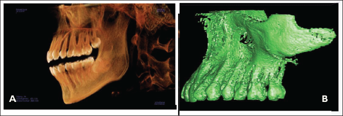
Cone-Beam 3D Images of Coronal, Sagittal and Axial Views of Maxillary and Mandibular Bone.
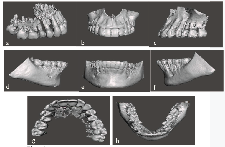
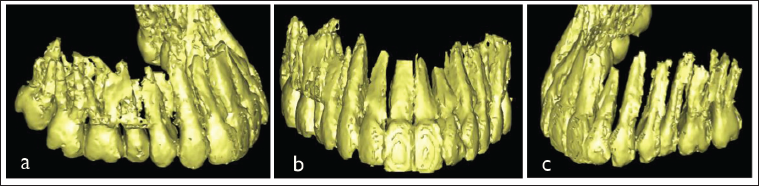
CBCT Image with Enhancement.
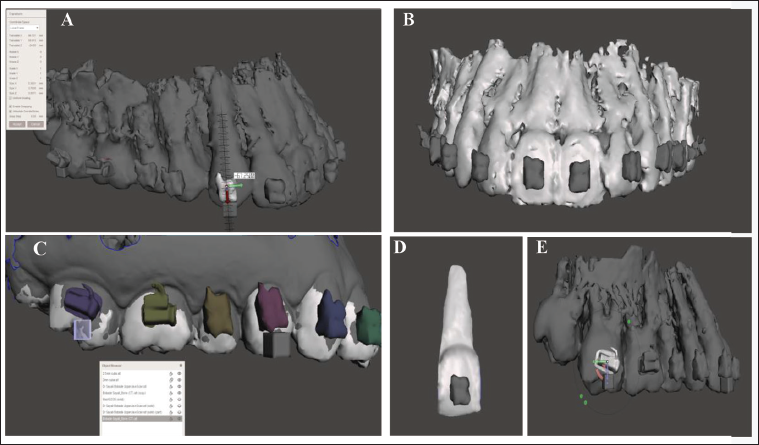
(A) Marking the Ideal Position and Bracket Placement on the Long Axis of the Tooth; (B) Bracket Placement on Virtual Model Frontal View; (C) Bracket Placement Using Blocks; (D) Single Tooth with Bracket; (E) Molar Tube Placement.
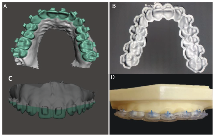
(A) Occlusal View of Bracket Placement and Checking of Each Bracket; (B) Steriolithographic Tray Ready for 3D Printing; (C) Virtual Tray Fabrication; (D) 3D Printed Tray with Brackets.
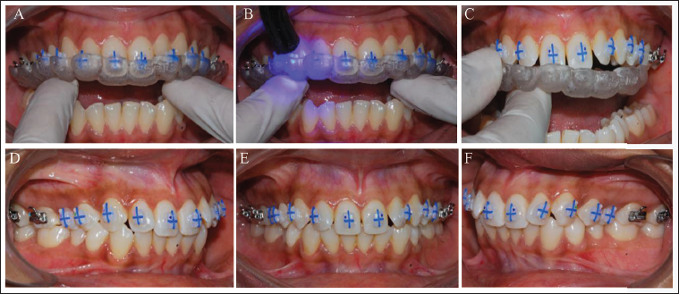
(A) Insertion of Tray; (B) Light Curing of the Brackets; (C) Removal of the Tray after Curing of the Bracket; (D–F) Intraoral Photographs.
Footnotes
Declaration of Conflicting Interest
The authors declared no potential conflicts of interest with respect to the research, authorship and/or publication of this article.
Ethical Approval
The study is approved by the Ethical committee of Institute and MUHS Research Committee.
Funding
The authors received no financial support for the research, authorship, and/or publication of this article.
Informed Consent
This is to request you to provide/give your consent to participate in a study titled, “CBCT Imaging for Bracket Positioning with Consideration to Root Axes.” As a part of the study, brackets will be placed and CBCT will be done. There are no risks involved during the study. No extra expenses will be incurred to you for the study.