
Abstract
Introduction
Assessment of growth is crucial in the diagnosis and treatment planning in orthodontics. Skeletal development can be assessed by using hand-wrist radiographs and lateral cephalograms. The advantage of the panoramic radiograph over hand wrist radiograph is that patient exposure is reduced following the ALARA principle.
Aim
The aim of this study was to investigate the correlation between third molar calcification stages and skeletal maturity using the CVM stages in North Maharashtrian population.
Methods
Dental panoramic and lateral cephalograms of subjects ranged in age from 9 to 20 years were selected. Demirjians method was used to assess the dental maturation stages of third molars on both the sides. Hassel and Farman classification was used for classifying into cervical vertebral maturation indicator stages. The collected data were statistically analyzed.
Result
There was a statistically significant correlation between cervical maturation stages and third molar calcification stages in North Maharashtrian population.
Introduction
Growing age is most effective and favorable for orthodontic treatment. 1 Assessment of the maturational stages can have a major influence on the diagnosis and treatment planning of orthodontic treatment. 2
The concept of biologic or physiologic age was developed in response to an observation that even children of the same chronological age might differ significantly in their level of development. 3 Physical, sexual, skeletal, and dental development are used to approximate an individual’s physiological age. 2 Skeletal maturity is assessed by visual inspection of the changes in the size and shape of developing bone during initial appearance and their resultant ossification process. 2, 3
Tooth calcification stages is a reliable method to assess the dental maturity. 3 The third molar has an advantage over the other molars since it continues to grow gradually and at a later age than the other molars. It continues to grow throughout adolescence, providing a different reference point from the other teeth. 2 Assessment of skeletal maturity through panoramic radiograph has an advantage over hand wrist radiographs that extra patient exposure can be avoided following ALARA (as low as reasonably achievable) principle. 3 Various studies of Lauterstein et al. have found a strong correlation between dental maturation stages and skeletal maturation stages.4–6 Third molar could be used as a growth maturity indicator if a positive correlation between third molar development and skeletal growth is found.
Therefore, this study aims to evaluate the third molar calcification stage as indicators of skeletal maturity in North Maharashtrian population.
Methods
The present study consists of 101 patients reported to the department of orthodontics and dentofacial orthopedics. The ethical committee had reviewed and approved all the sample size and selection criteria for the study. The OPG and lateral cephalograms of 45 boys and 56 girls with age range between 9 and 20 years were selected. All the selected subjects were informed and written consent was obtained.
Inclusion criteria included the following:
The radiographs having all third molars present in it. Subjects between 9 and 20 years of age.
Exclusion criteria considered were as follows:
Radiographs representing congenital anomalies of the cervical vertebrae. Radiographs of subjects having any systemic diseases or on any medication that could affect growth (such as endocrine disorders and syndromes). Radiographs lacking distinctness of structures.
The following assessment was done.
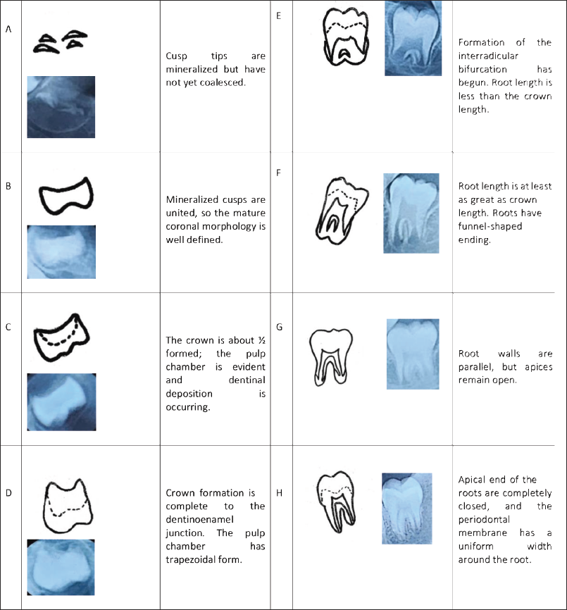
Dental maturity evaluation: Demirjian et al.
7
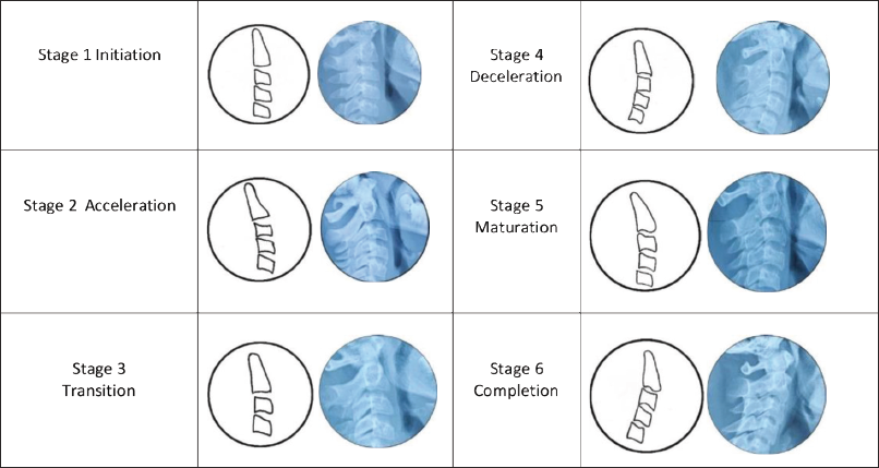
calcification stages (A to H) were used to classify the third molar tooth on panoramic radiograph as shown in Figure 1. Cervical vertebral maturity evaluation: Hassel and Farman classification
8
was used for cervical vertebral maturation staging on lateral cephalogram as shown in Figure 2. Assessment of sample: All the staging of cervical vertebral maturation and third molar calcification were done by the same orthodontist without knowing the age or gender of the subject.
Developmental Stages of Third Molar Formation Using Demirjian Classification.
Cervical Vertebral Maturation Index (CVMI) Stages Using Hassel and Farman’s Classification.
Statistical Analysis
Analysis was performed using the SPSS statistical V 21.0 (version) for Windows (SPSS Inc, Chicago, Illinois, USA). Descriptive quantitative data were expressed in mean and standard deviation, respectively. Upper and lower third molar calcification stages and cervical vertebral maturation stages were described with frequency distribution. Age of subjects and calcification of upper and lower third molar stages were correlated with cervical vertebral development stage using Spearman’s correlation coefficient. The level of significance was set as p ≤ .05.
Result
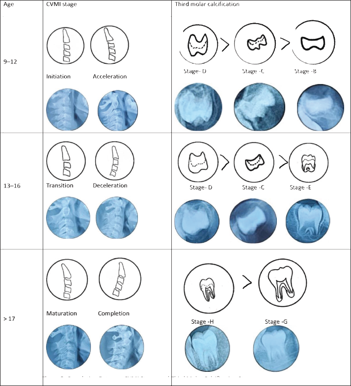
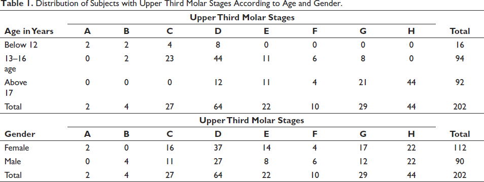
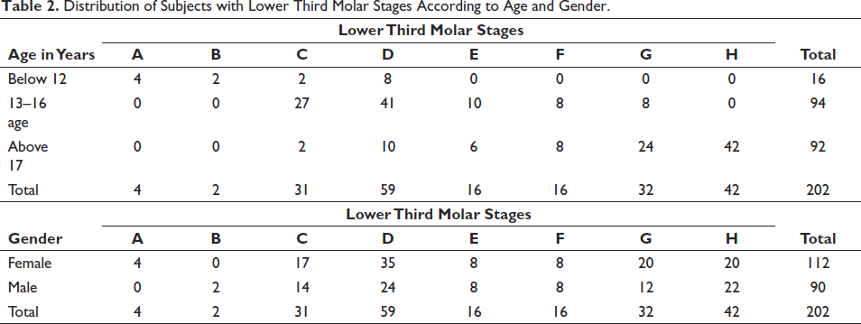
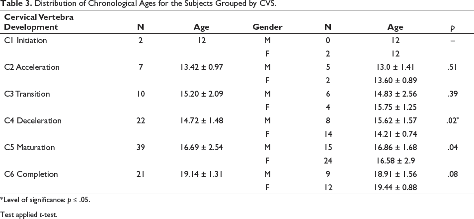
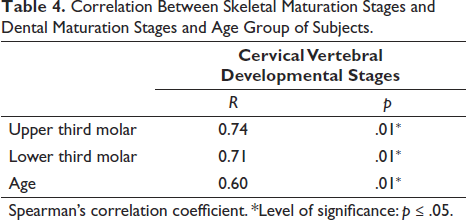
Tables 1 and 2 illustrate the distribution of upper and lower third molar stages according to age and gender. In 9–12 years group, upper third molar stages observed in a descending order were D, C, B, respectively, while the lower third molar stages were D, C, B, respectively. There were no E, F, G, H stages in both the groups. In 13–16 years group, the calcification stages observed in a descending order were D, C, E, G, respectively, in upper and lower third molar stages. In the age group of more than 17 years, most observed H stage followed by G stage of calcification in upper and lower third molars (Figure 3). In both the male and female group of this sample, the stages of upper and lower third molar in the descending order were D followed by H. Table 3 shows statistically significant difference in the distribution of chronological age between males and females in C4 stage of cervical vertebral maturation stages. The females were younger than males in the C4 stage. There is a significant relationship between cervical vertebral maturation stages and third molar calcification stages of subjects in both upper and lower third molars (Table 4). As CVMI increases according to age, the third molar calcification stage also increases in both the genders (Figure 3).
Correlation Between CVMI Stages and Third Molar Calcification Stages.
Distribution of Subjects with Upper Third Molar Stages According to Age and Gender.
Distribution of Subjects with Lower Third Molar Stages According to Age and Gender.
Distribution of Chronological Ages for the Subjects Grouped by CVS.
Test applied t-test.
Correlation Between Skeletal Maturation Stages and Dental Maturation Stages and Age Group of Subjects.
Discussion
Skeletal age can be estimated by cervical vertebral maturity indicators, hand wrist maturation, and dental development. 10 Researchers have shown that cervical vertebra in lateral cephalogram can also be used to predict the growth of the mandible.11, 12 In addition, few researchers have shown a slight correlation between skeletal and dental maturation.13, 14
According to Demirjian, dental maturity calcification stages are an important biologic factor. 15 Few authors have shown that the developmental stages of canine and second molars have an association with skeletal maturity.3, 16 However, there are highest variations seen in the timing of third molar development as compared to the other teeth. 17 Therefore, third molar was included in this study.
This study aimed to evaluate the correlation between cervical vertebrae maturation and third molar dental maturation in North Maharashtrian population.
Table 3 illustrates the distribution of the chronological ages of subjects according to CVM stages. The mean chronologic age of girls was slightly lower than boys in CS4 and CS5 stages. However, it was more lower in CS4 as compared to CS5. The CS4 stage being earlier in females than in males. In stage CS4, the mean chronologic age was 14.72 ± 1.48 (Table 3). In CVM method, CS1, CS2 represents the pre-pubertal period, CS3–CS5 shows the pubertal growth spurt period, while CS6 is the post-pubertal growth spurt period (Table 3). These results are in accordance with the previous studies.18, 19 The correlation coefficients between skeletal maturation and dental maturation were 0.74 and 0.71 for upper and lower third molars, respectively (Table 4). Few studies have found increased correlation coefficient between dental and skeletal maturation due to a lesser number of sample size and errors.14, 20 The relationship between the dental and skeletal maturity also differs among different geographic areas and ethnic groups. 21 In this North Maharashtrian study, a statistically significant correlation was found between dental maturation stage and cervical vertebra maturation stage (Table 4). Krailassiri et al. 22 in their study found a weak correlation, while another study of Engstrom et al. 23 found a strong association.
Few researchers found a strong association between the mandibular canine with the pubertal growth spurt, 24 while few researchers found a strong correlation of skeletal maturation with that of second premolar. 22
Skeletal maturity increased simultaneously with the increase in dental ages for males and females. An earlier occurrence of CS4 stage was observed in females. All correlations between skeletal and dental ages were statistically significant.
Conclusion
There is a statistically significant correlation between cervical vertebral maturation stages and third molar calcification stages in North Maharashtrian population.
Footnotes
Declaration of Conflicting Interests
The authors declared no potential conflicts of interest with respect to the research, authorship, and/or publication of this article.
Ethical Approval
Ethical clearance has been approved by the ethical committee for this study. The reference no. is EC/NEW/INST/2021/1810.
Funding
This study is self-funded. The authors received no financial support for the research, authorship, and/or publication of this article.
Informed Consent
The participant has consented to the submission of the article to the journal.