
Abstract
Orthodontic management of ectopic canines is quite challenging and time consuming due to the presence of thin buccal cortical bone. Sectional mechanics provide distal and extrusive force on canine but without any torque control. So, palatal root torquing during canine retraction is needed to increase the buccal cortical bone thickness and to avoid bone dehiscence and gingival recession. This article describes an innovative spring which provides 3-dimensional control by simultaneous retraction, extrusion, and torquing of ectopic canine.
Introduction
Ectopic eruption of permanent canines is most commonly seen in orthodontic practice. The prevalence of ectopically erupted teeth is 1.0% to 2.2%. 1 2 Orthodontic management of such ectopic canines is challenging, time consuming, and mandates 3-dimensional control. Segmental mechanics are preferred than continuous mechanics to avoid adverse effects on adjacent teeth like flattening of arch and open bite. 3
Sectional loops and springs such as closing loop, T-loop, T-loop with helicoid spring, and cantilever spring generate distal and extrusive force on canine but without any torque control. As the ectopic canine root has close proximity to the buccal cortical plate, palatal root torquing during canine retraction will center the root and ensure optimal cortical thickness, thereby preventing dehiscence and gingival recession. 4 So, in order to achieve a simultaneous 3-dimensional correction, we have designed a simple and versatile spring abbreviated as “MASTER spring” (mini-implant assisted simultaneous torquing extrusion retraction spring) for correction of ectopic canines.
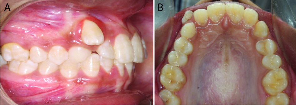
The technique is demonstrated in a 21-year-old female patient with highly placed maxillary right canine along with retained deciduous maxillary right canine (Figure 1A, B).
Steps in Fabrication and Activation of MASTER Spring
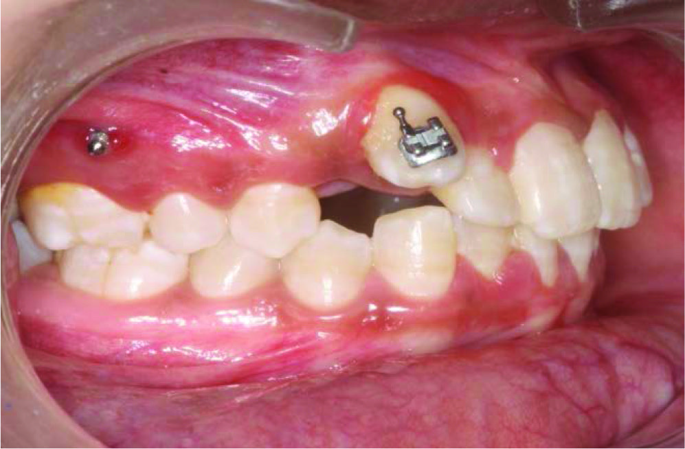
Initially, the deciduous maxillary right canine was extracted, ectopic canine bracket is bonded (MBT 022 slot), and mini-implant (1.2 × 8 mm) is inserted between first molar and second premolar (Figure 2).
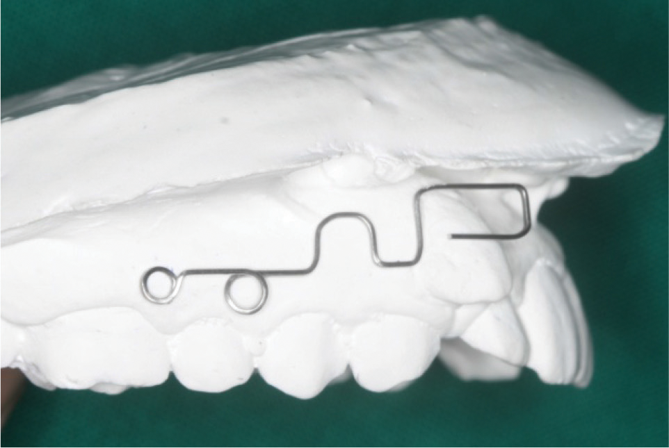
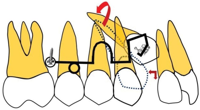
MASTER Spring is fabricated with 0.017 × 0.025″ TMA wire, which is initially bent to encircle the mini-implant head.
A helix of 3-mm internal diameter facing occlusal is fabricated 4-mm mesial to implant which is followed by apically directed U-loop of 4 mm height and 5 mm width.
Finally, a box loop of 6 mm height and 8 mm width is made facing gingivally with free end of which engages into the canine bracket slot (Figure 3).
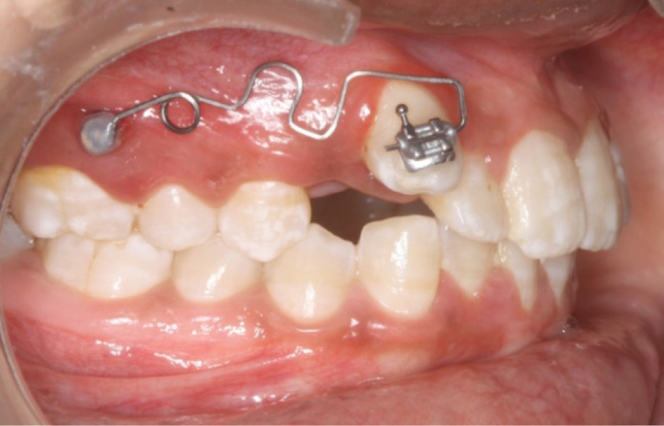
The spring is activated by closing the helix by 1 to 2 mm which produces an extrusive force, compression of U loop results in retraction, and the box loop is twisted to produce palatal root torque (Figures 4 and 5).
Spring is stabilized on the mini-implant head with Transbond XT composite following activation.
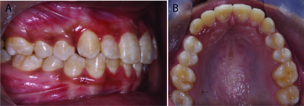
Spring is activated for every 6 weeks and the ectopic canine was aligned in 4 months (Figure 6A, B).
Pretreatment Intraoral Photograph.
Ectopic Canine Is Bonded and Mini-Implant Inserted.
Fabrication of MASTER Spring on Model.
Implant-Supported MASTER Spring After Activation and Insertion.
Schematic Diagram of MASTER Spring Design: Mini-Implant-Assisted Simultaneous Torquing, Extrusion, and Retraction of Ectopic Canine.
After Alignment of Maxillary Right Canine.
Advantages of MASTER Spring
Ease of fabrication
Minimal soft tissue irritation
Simultaneous torquing, extrusion and retraction without adverse effects on the adjacent teeth
Reduced treatment duration
Conclusion
MASTER spring is more effective and versatile in correcting the ectopic canines with simultaneous torquing extrusion and retraction.
Footnotes
Declaration of Conflicting Interests
The authors declared no potential conflicts of interest with respect to the research, authorship, and/or publication of this article.
Funding
The authors received no financial support for the research, authorship, and/or publication of this article.
Statement of Informed Consent
Written informed consent was obtained from the subject for the use of photographs for publication.