
Abstract
Background:
Though studies have been conducted on the PowerScope, not many researches are available in the literature which compare its effects with other fixed functional appliances. Therefore, the aim of our study was cephalometric evaluation and comparison of the skeletal, dentoalveolar, and soft tissue changes brought about by the Forsus Fatigue Resistant Device and PowerScope appliance.
Materials and Methods:
Pre and Posttreatment cephalometric records of 20 patients with Class II Division 1 malocclusion treated with fixed functional appliances (Forsus Fatigue Resistant Device and PowerScope) were compared. Values of various cephalometric parameters were used to evaluate the skeletal, dental, and soft tissue changes. Wilcoxon signed-rank test (intragroup comparison) and Mann–Whitney U test (intergroup) were used to see significant differences in the parameters (P ≤ .05).
Results:
Both the appliances were successful in correcting the Class II discrepancy. Skeletal changes were seen significantly in both the groups, though they were more in the Forsus patients. Dentoalveolar changes were predominantly seen in PowerScope patients. Also, an improvement in soft tissue profile was seen in both the groups.
Conclusion:
From our study, we concluded that the PowerScope and the Forsus Fatigue Resistant Device are equally good options for the correction of skeletal Class II malocclusion due to a retrognathic mandible, as they achieve changes in all the three aspects: skeletal, dentoalveolar and soft tissue.
Introduction
Malocclusion, which is defined as a change in teeth position and skeletal growth, is a worldwide public health problem.
1
Malocclusion in human populations is not uncommon, and attempts made to correct it date back to at least 1000
Studies have revealed that protrusion of the maxilla caused only 20% of Class II Division 1 malocclusion cases, while most patients reported with an etiology of retruded mandible. These findings led researchers to resort to the use of functional appliances that helped stimulate the growth of the mandible to treat skeletal Class II malocclusion. 4 The functional appliances can further be grouped into removable and fixed variants.
Fixed functional appliances used are of the rigid, flexible, or hybrid type. The most commonly used rigid fixed functional appliances are the Herbst appliance and the mandibular advancement repositioning appliance (MARA). The popular flexible devices are the Jasper Jumper, and Scandee tubular jumpers, while the most preferred hybrid appliance is the Forsus Fatigue Resistant Device (FFRD). 5 The ForsusTM FRD is a novel three-piece telescoping spring for Class II correction and is moderately well tolerated by patients who may encounter some initial discomfort, which normally reduces with time. 6
Over the period of time, various fixed functional appliances have been introduced into the branch of orthodontics, with PowerScope being the latest one. It is a direct derivative of the Herbst Type II appliance and was developed by Dr. Andy Hayes in conjunction with American Orthodontics in 2016. The appliance consists of a telescopic mechanism consisting of an inner shaft/push rod, middle and outer tubing. There is a nickel–titanium (NiTi) spring delivering a constant 260 g force. PowerScope is available as a one-size-fits-all machine preassembled with connection nuts for speedy chairside application. 7 Since not many studies have been done to assess the effects of PowerScope so our study was carried out for the cephalometric evaluation of it’s skeletal, dental and soft tissue effects and the subsequent comparison with changes brought about by Forsus.
Materials and Method
Pretreatment and posttreatment cephalometric records of 20 patients (12 females and 8 males, aged 12 to 15 years) reporting to the department of Orthodontics and Dentofacial Orthopaedics of Maharishi Markandeshwar College of Dental Sciences and Research, having Class II Division 1 malocclusion, overjet more than 4 mm, no missing or extracted permanent teeth treated with fixed functional appliances (Forsus FRD[10] and PowerScope[10]) were evaluated. The pretreatment growth status was CVMI 3/4. The total duration of treatment with the appliance was 6 to 9 months. Values of various skeletal, dental, and soft tissue cephalometric parameters were used to compare the changes. Wilcoxon signed-rank test (intragroup comparison) and Mann–Whitney U test (intergroup) were used to see significant differences in the parameters (P < .05).
Results
Comparison of Pre and Posttreatment Values in the Forsus Group.
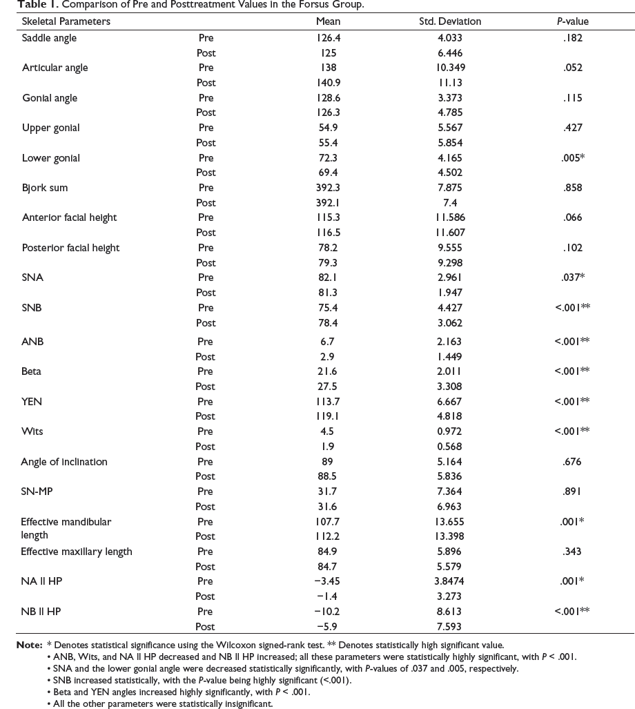
ANB, Wits, and NA ll HP decreased and NB ll HP increased; all these parameters were statistically highly significant, with P < .001.
SNA and the lower gonial angle were decreased statistically significantly, with P-values of .037 and .005, respectively.
SNB increased statistically, with the P-value being highly significant (<.001).
Beta and YEN angles increased highly significantly, with P < .001.
All the other parameters were statistically insignificant.
Comparison of Pre and Posttreatment Values in Forsus Group.
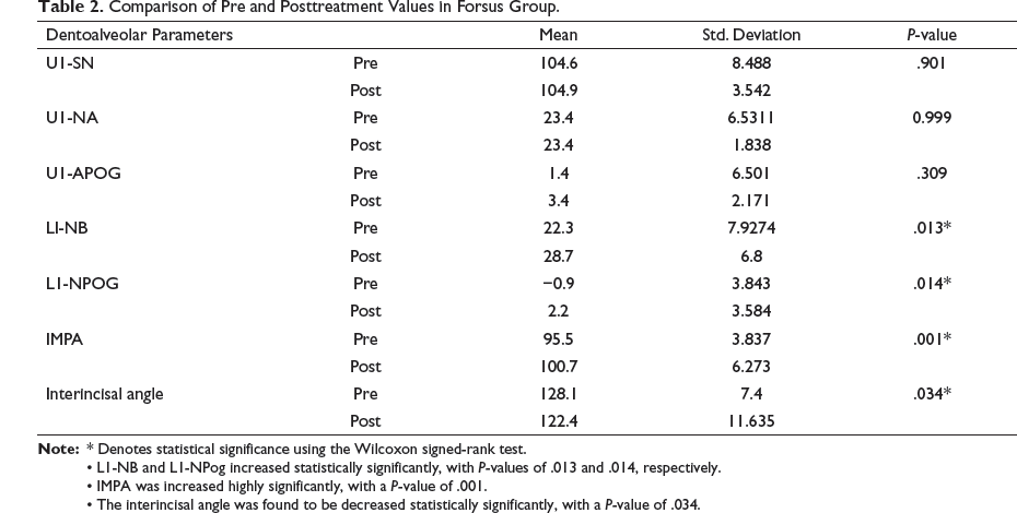
L1-NB and L1-NPog increased statistically significantly, with P-values of .013 and .014, respectively.
IMPA was increased highly significantly, with a P-value of .001.
The interincisal angle was found to be decreased statistically significantly, with a P-value of .034.
Comparison of Pre and Posttreatment Values in Forsus Group.
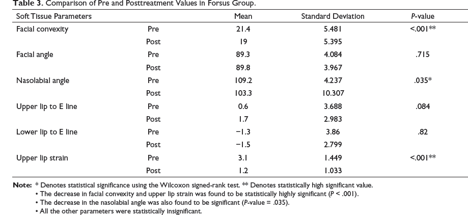
The decrease in facial convexity and upper lip strain was found to be statistically highly significant (P < .001).
The decrease in the nasolabial angle was also found to be significant (P-value = .035).
All the other parameters were statistically insignificant.
Overjet and Overbite Changes in Forsus Group.
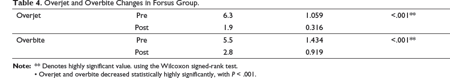
Overjet and overbite decreased statistically highly significantly, with P < .001.
Comparison of Pre and Posttreatment Values in the PowerScope Group.
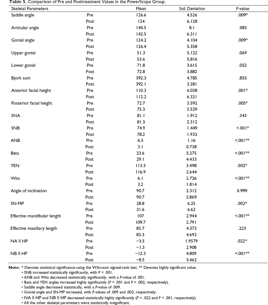
SNB increased statistically significantly, with P < .001.
ANB and Wits decreased statistically significantly, with a P-value of .001.
Beta and YEN angles increased highly significantly (P < .001 and P = .002, respectively).
Saddle angle decreased statistically, with a P-value of .009.
Gonial angle and SN-MP increased, with P-values of .009 and .002, respectively.
NA ll HP and NB ll HP decreased statistically highly significantly (P = .022 and P < .001, respectively).
All the other skeletal parameters were statistically insignificant.
Comparison of Pre and Posttreatment Values in PowerScope Group.
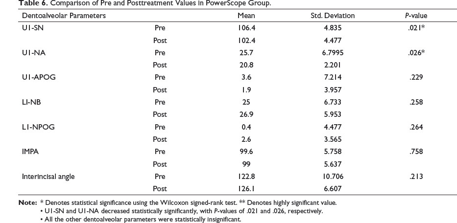
U1-SN and U1-NA decreased statistically significantly, with P-values of .021 and .026, respectively.
All the other dentoalveolar parameters were statistically insignificant.
Comparison of Pre and Posttreatment Values in PowerScope Group.
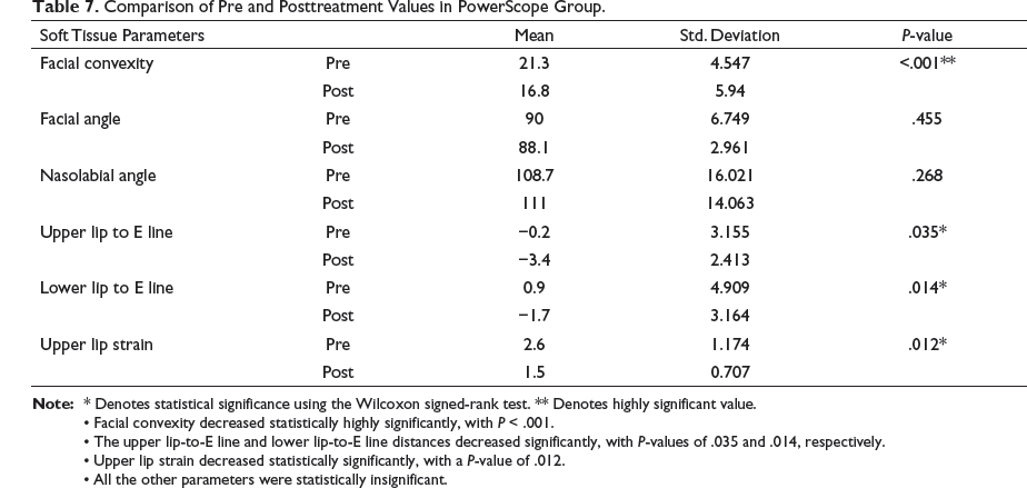
Facial convexity decreased statistically highly significantly, with P < .001.
The upper lip-to-E line and lower lip-to-E line distances decreased significantly, with P-values of .035 and .014, respectively.
Upper lip strain decreased statistically significantly, with a P-value of .012.
All the other parameters were statistically insignificant.
Overjet and Overbite Changes in PowerScope Group.
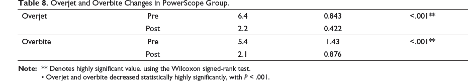
Overjet and overbite decreased statistically highly significantly, with P < .001.
Comparison between the Forsus and PowerScope Groups—Difference between Pretreatment and Posttreatment Values.
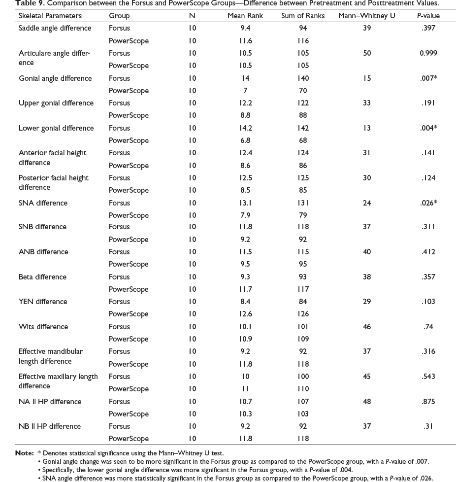
Gonial angle change was seen to be more significant in the Forsus group as compared to the PowerScope group, with a P-value of .007.
Specifically, the lower gonial angle difference was more significant in the Forsus group, with a P-value of .004.
SNA angle difference was more statistically significant in the Forsus group as compared to the PowerScope group, with a P-value of .026.
Comparison Between the Forsus and Powerscope Groups. Difference Between the Pre and posttreatment Values.
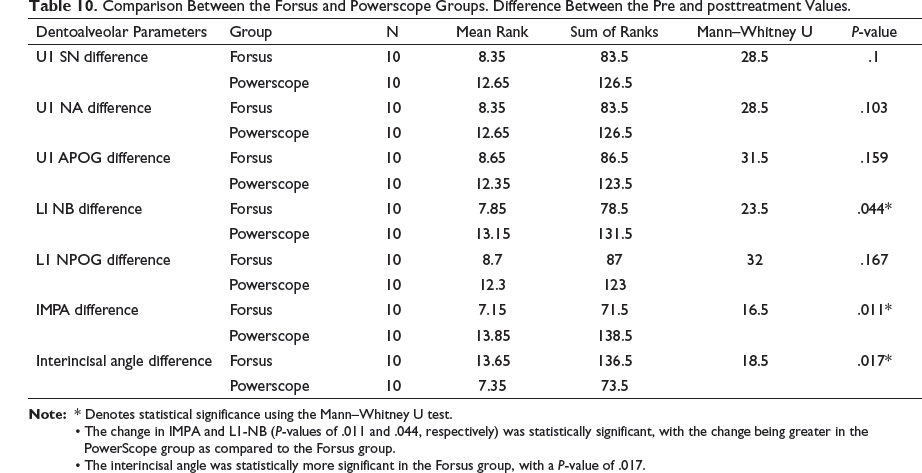
The change in IMPA and L1-NB (P-values of .011 and .044, respectively) was statistically significant, with the change being greater in the PowerScope group as compared to the Forsus group.
The interincisal angle was statistically more significant in the Forsus group, with a P-value of .017.
Comparison Between the Forsus and Powerscope Groups. Difference Between the Pre and Posttreatment Values.
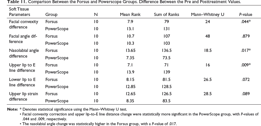
Facial convexity correction and upper lip-to-E line distance change were statistically more significant in the PowerScope group, with P-values of .044 and .009, respectively.
The nasolabial angle change was statistically higher in the Forsus group, with a P-value of .017.
Comparison Between the Forsus and Powerscope Groups. Difference Between the Pre and Posttreatment Values.
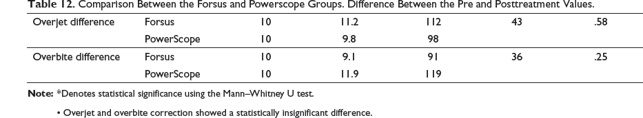
Overjet and overbite correction showed a statistically insignificant difference.
Discussion
Forsus Group
Skeletal Changes (Table 1)
The skeletal parameters like lower gonial angle and SNA decreased significantly, with P-values of .005 and .037, respectively, representing “headgear effect,” thus creating an ideal situation for the correction of skeletal Class II discrepancy as described by Trenouth. 8 The SNB angle increased, with the P-value being highly significant, that is, less than .001. This change contributes to the change in the position of the mandibular base by bringing the position of point B forward. Previous studies done by Franchi et al, 9 Heinig and Goz, 10 and Karacay et al 11 showed the same changes. Wits, beta angle, and YEN angle increased with P < .001, which was statistically highly significant. The ANB angle also showed a highly significant change with P < .001. NA‖HP decreased with P-value of .001, which was statistically significant. This measurement describes the apical base of the maxilla in relation to the N point. Thus, the decrease is attributed to the retrusion of the apical base of the maxilla, thus substantiating the “headgear effect” exerted by the appliance. NB‖HP increased with a statistically highly significant P < .001.This measurement describes the horizontal position of the apical base of the mandible in relation to the N point. Thus, the increase in the value is attributed to the horizontal advancement of the mandible. Effective mandibular length increased with P < .001.
Dentoalveolar Changes (Table 2)
L1-NB and L1-NPog increased significantly, with P values of .013 and .014, respectively. This increase is attributed to the fact that the lower incisors were proclined. Previous studies done by Franchi et al, 9 Heinig and Goz, 10 Karacay et al,[11] Darda et al,[12] Aras et al,[13] and Linjawi and Abassy 15 also documented the same increase. The incisor mandibular plane angle (IMPA) increased statistically with a P-value of .001 that was highly significant. The interincisal angle decreased significantly with a P-value of .034.
Soft Tissue Parameters (Table 3)
Facial convexity decreased statistically highly significantly, with P < .001. The nasolabial angle increased statistically significantly with a P-value of .035. The change in the nasolabial angle and upper lip strain in the absence of significant changes in the inclination of incisors can be attributed to the “maxilla growth restriction and distalisation of molars effect” exerted by the push action of Forsus, which is in concordance with the study conducted by Franchi et al 9 Also, as the wires used in upper arch were 0.019″ × 0.025″ stainless steel, incisor inclination could be preserved without alterations.
Upper lip strain was decreased with a P-value statistically highly significant, that is, less than .001. The upper lip strain improvement showed better lip competency at the posttreatment stage as supported by the studies of Heinig Goz 10 and Karacay et al. 11
Overjet and overbite showed a highly significant reduction, with P-values less than .001 (Table 4).
PowerScope Group
Skeletal Changes (Table 5)
The saddle angle decreased with a P-value of .009, which was statistically significant, as also seen in the study by Savana et al. 14 This decrease depicts the anterior positioning of the mandible, which contributes to the correction of facial convexity as observed in this study.
The gonial angle increased by a mean value of 2.2 ± 1.2 with a P-value of .009, which was statistically significant. The anterior and posterior facial height significantly increased, with P-values of .001 and .005, respectively. The SNB angle increased, with the P-value statistically significant at less than .001. The beta angle and YEN angle increased statistically, with P-values of .001 and .002, respectively. This increase is attributed to the correction of the sagittal jaw relationship between the maxilla and mandible. Wits and the ANB angle showed a highly significant change, with P < .001. NA‖HP decreased and NB‖HP increased, with P-values of .022 and <.001, respectively, which were statistically significant.
Dentoalveolar Changes (Table 6)
U1-SN and U1-NA decreased statistically (P-values of .021 and .026, respectively). The mean reduction is attributed to inclination correction because of fixed mechanotherapy and the distalizing effect of PowerScope on maxillary dentition.
Soft Tissue Parameters (Table 7)
Facial convexity decreased statistically significantly, with P < .001. The upper lip-to-E line and lower lip-to-E line distances increased significantly, with P-values of .035 and .014, respectively. The increase in the upper lip-to-E line distance in PowerScope is attributed to inclination correction (as shown by the decrease in U1-SN and U1-NA), thereby resulting in backward lip movement and the consequent improvement in profile.
Upper lip strain was decreased, with a P-value of .012, showing better lip competency at the posttreatment stage.
Overjet and overbite showed highly significant reduction, with P < .001 (Table 8).
Comparison Between Forsus Fatigue Resistant Device and PowerScope (Tables 9 to 12)
Skeletal Changes (Table 9)
In our study, Forsus FRD and PowerScope both showed statistically significant differences. The SNA difference between Forsus FRD and PowerScope had mean ranks of 13.1 and 7.9, respectively, in which the P-value was statistically significant (.026). This change was better in the Forsus group, suggesting that it exerts a higher distalizing effect on the maxillary arch in comparison to PowerScope. The gonial angle difference was higher with Forsus, showing better effect than PowerScope. The decrease in the gonial angle in the Forsus group is attributed to its intrusive effect on molars—a fact supported by the meta-analysis done by Linjawi and Abbassy 15
Dentoalveolar changes: (Table 10)
The IMPA difference between Forsus FRD and PowerScope had means of 7.15 and 13.85, respectively. PowerScope was more effective than Forsus on the lower anteriors, with a significant P-value of .011. The L1-NB difference and IMPA value difference has come out to be significant and more in Powerscope while calculating pre and post comparison value is because the data is not normally distributed in the difference table and a non-parametric test has been used (compared to the parametric test used for individual groups) which can find significance even when mild changes are present.
Also, the mean rank difference is higher in the PowerScope group due to the data point distribution.
All the other dentoalveolar parameters, such as U1-SN, U1-NA, U1-APog, L1-NB, and L1-NPog, showed almost similar effects across both the groups, and therefore the comparison values were statistically insignificant.
Soft Tissue Changes (Table 11)
Facial convexity in PowerScope was more effectively corrected as compared to Forsus (P-value = .044). Forsus showed greater effects on the nasolabial angle than the Powerscope group, with means of 13.65 and 7.35, respectively, having a significant P-value of .017. The upper lip-to-E line distance difference between the two groups showed that PowerScope was more effective compared to the Forsus group, with the P-value being statistically significant at .009.
Overjet and Overbite parameters also showed statistically insignificant difference in comparison value of both the groups (Table 12).
Thus, in our study, we observed that both the Forsus FRD and PowerScope appliances are efficient in correcting Class II problem in growing patients with a retrognathic mandible. Skeletal, dentoalveolar, and soft tissue changes are brought about by both appliances, although the Forsus group showed more skeletal changes 16 and PowerScope showed more dentoalveolar and soft tissue effects. 17
Limitations
A follow-up of patients few years post retention to evaluate the long-term stability of the results of the two appliances was not taken into consideration.
Though the intent of this study was to comprehensively evaluate the changes with both appliances in all three aspects, that is, skeletal, dental, and soft tissue, occlusal plane parameters were not focused on, and so any changes pertaining to these could not be established.
Summary and Conclusion
The present study was conducted to compare the effects of two appliances, namely, PowerScope and Forsus FRD, used for the correction of Class II discrepancy. The comparison between the two groups showed that both appliances were successful in correcting Class II discrepancy. Skeletal alterations were seen to be significant using both devices, though they were higher with the Forsus. Dentoalveolar and soft tissue changes were predominantly seen in PowerScope patients.
Thus, it can be concluded from our study that both appliances are effective in bringing about skeletal, dentoalveolar, and soft tissue changes.
Footnotes
Declaration of Conflicting Interests
The authors declared no potential conflicts of interest with respect to the research, authorship, and/or publication of this article.
Funding
The authors received no financial support for the research, authorship, and/or publication of this article.
Statement of Informed Consent and Ethical Approval
Necessary ethical clearances and informed consent was received and obtained respectively before initiating the study from all participants
This is to confirm that the manuscript was neither presented as part of a meeting nor previously published.