
Abstract
The branch of orthodontics has had an interest in the cervical vertebrae wherein cervical spine is used as a reference structure for natural head position, so skeletal age was evaluated by studying variations in the cervical vertebral morphologies. Among all evaluations, very limited data is available wherein comparison between cervical vertebral body volumes between the different malocclusions has been done. This study aimed to compare the differences in the volumes of cervical vertebral bodies of C2, C3, and C4 between skeletal class I and class II malocclusions of both horizontal and vertical growth patterns. In class I the volume was significantly lesser as compared to class II. It was seen that there was statistically no significant difference in the volume between the horizontal and vertical growers. It can be concluded from this study that cervical vertebral body volume has no effect on growth pattern. However, variations in cervical vertebral body volume are seen with different malocclusions.
Introduction
The head is supported by the cervical vertebral column, consisting of 7 cervical vertebrae. 1 The branch of orthodontics has had an interest in the cervical vertebrae for such reasons including natural head positions where cervical spine was designated the reference structure and skeletal age was evaluated by studying variations in the cervical vertebral morphologies. 2 These can be useful in determining a treatment protocol in patients with remaining growth. Length of the mandible was seen to be associated with cervical vertebral column straightness. 3
There also exists an association between craniofacial and cranio-cervical morphology according to literature.2, 3 As an orthodontist deals with varying growth and development of craniofacial structures and modifications in form and function are required, hence there is a need for better understanding of the morphology of cervical vertebrae between the different craniofacial patterns in the sagittal plane without any interfering effects of growth. 4
Regarding skeletal class II accompanied with increased over jet, there was a 52.9% prevalence of fusion anomalies in cervical vertebrae observed, where the associations were statistically significant linking morphology of cervical column with cephalometric measurements. 5
A study by Choi et al. conducted in 2016 concluded that there is a positive co relation between the skeletal maturation age and the volume of bodies of second, third, and fourth cervical vertebrae. 6 Three dimensional methods were chosen due to the reason that, apart from giving an image similar to lateral cephalograms in the sagittal plane, it also presents the craniofacial skeleton in all 3 dimensions and enables the assessment of volume.
A limited number of studies have compared the cervical vertebral body volumes between the different malocclusions such as skeletal class I and class II malocclusions. Volume was chosen as a measurable parameter to assess if differentiation of skeletal patterns could be done based on volumetric analyses as the CVM method only tells us about the skeletal age of the patient and not the pattern of malocclusion it may lead to. Therefore, this study aimed to find a co-relation between the cervical vertebral body volume and class II vertical and horizontal cases, while subjects with class I malocclusion were taken as the control group.
Objectives
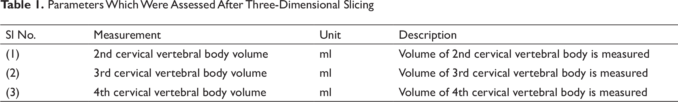
To evaluate the cervical vertebral body volumes of second, third, and fourth cervical vertebrae (C2, C3, and C4) in orthognathic cases of skeletal class I and cases of skeletal class II vertical and horizontal patterns (Table 1).
To compare the cervical vertebral body volumes C2, C3, and C4 in cases of skeletal class II vertical and horizontal patterns with orthognathic cases of skeletal class I.
Materials and Methods
This observational study was conducted in the Department of Orthodontics and Dentofacial Orthopaedics. Ethical clearance was obtained from the institutional ethics committee (Ref ID: IEC/2018-19/7640). Data was collected from the previous research already done in the department which was used for evaluation of volume of body of cervical vertebrae.
The samples consisted of 30 cases aged between 16–24 years. The samples were divided into 3 groups based on ANB angle, molar relationship, and Frankfort Mandibular Plane Angle (FMPA) with 10 samples in each group.
Group 1: Orthognathic skeletal class I (ANB 0–4°, class I molar relation, FMPA 25±2°) Group 2: Skeletal class II vertical pattern (ANB >4°, class II molar relation, FMPA > 27°) Group 3: Skeletal class II horizontal pattern (ANB >4°, class II molar relation, FMPA < 23°)
Digital Volume Tomography (3D-DVT) scanned images which were taken using Phillips Allura Xper FD20 3D RA, Digital subtraction angiography unit (Netherlands) with exposure parameters of 80 kvp, 10 Mpa, and 4–5 s with field of view 12″270° rotation and radiation dose of 1.8 mSv were analyzed. Images were obtained with patients in a supine position such that the Frankfort horizontal plane was perpendicular to the floor. Patients were also instructed not to swallow or move their head.
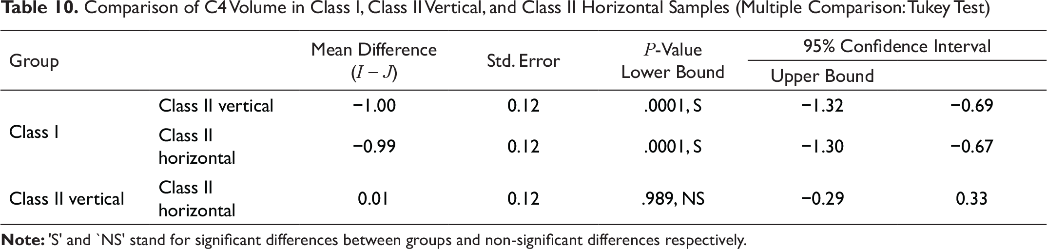
Once the images were obtained, the cervical spine was isolated using the “cut” function in the software. Each cervical vertebra (C2, C3, and C4) to be assessed volumetrically was then sequentially isolated by slicing. The body of the vertebrae was then identified and sliced. Once the body of the vertebrae was separated the spherical volume parameter was used from the software. The cut body of the vertebra was placed inside this sphere which then assessed the volume of the body (Figure 1). The volume measurement was done in ml (1000 mm3 = 1 ml)
Result
Tables 2–10 show the statistical analysis that was done by using descriptive and inferential statistics using one-way ANOVA and multiple comparisons were done by using post hoc Tukey test. The software used in the analysis was SPSS 24.0 version and P < .05 was taken as the level of significance.
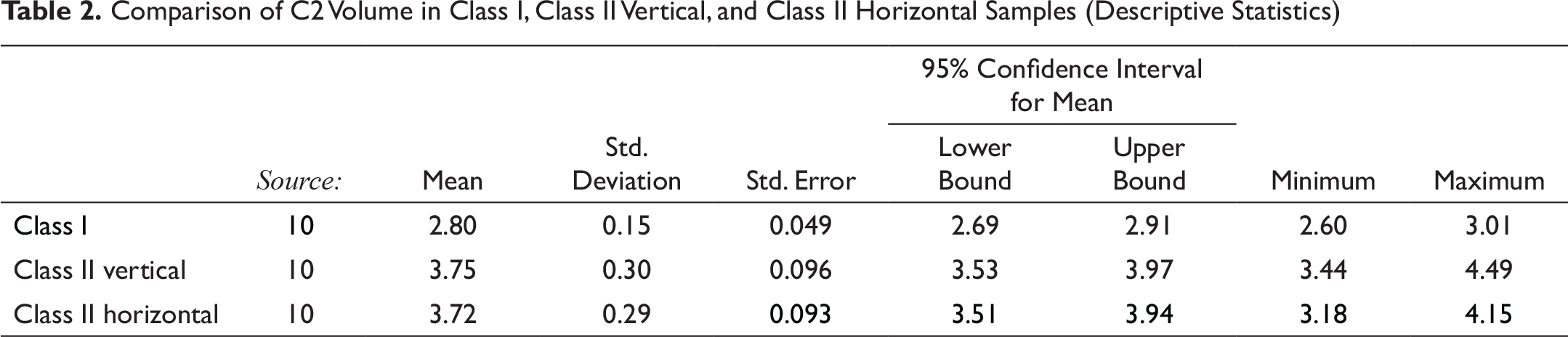
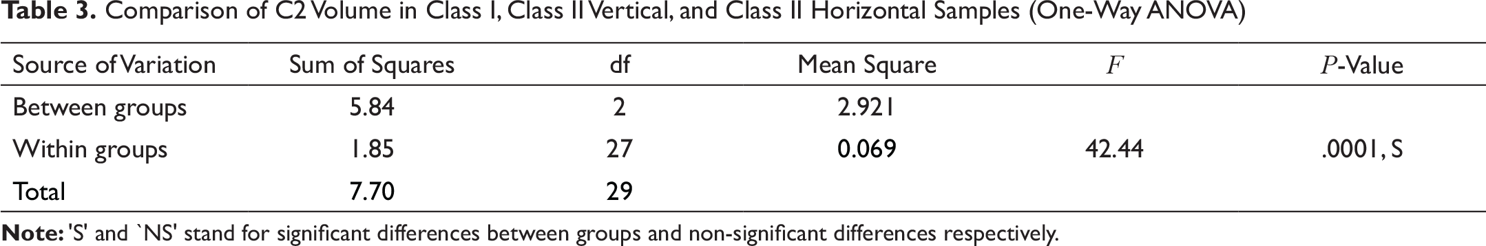
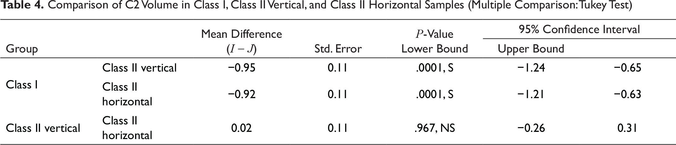
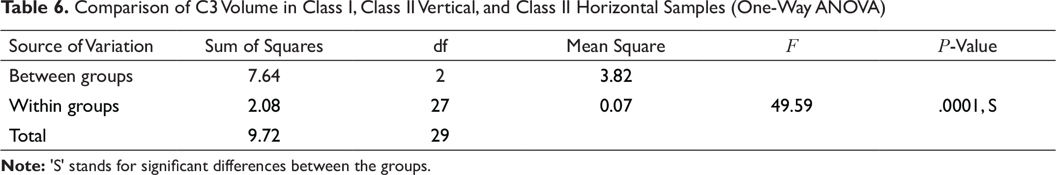
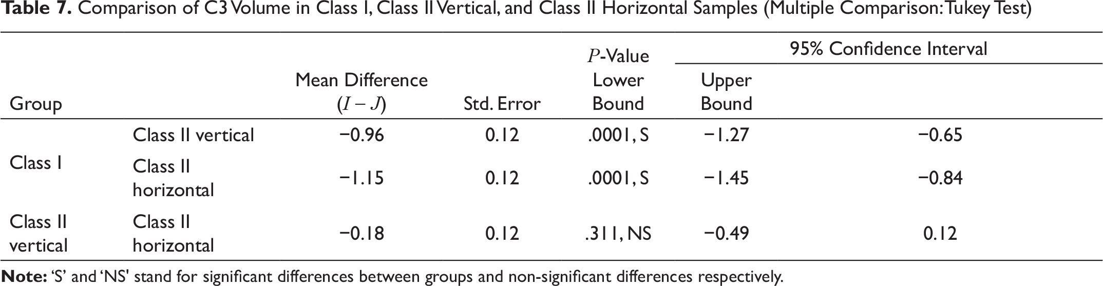
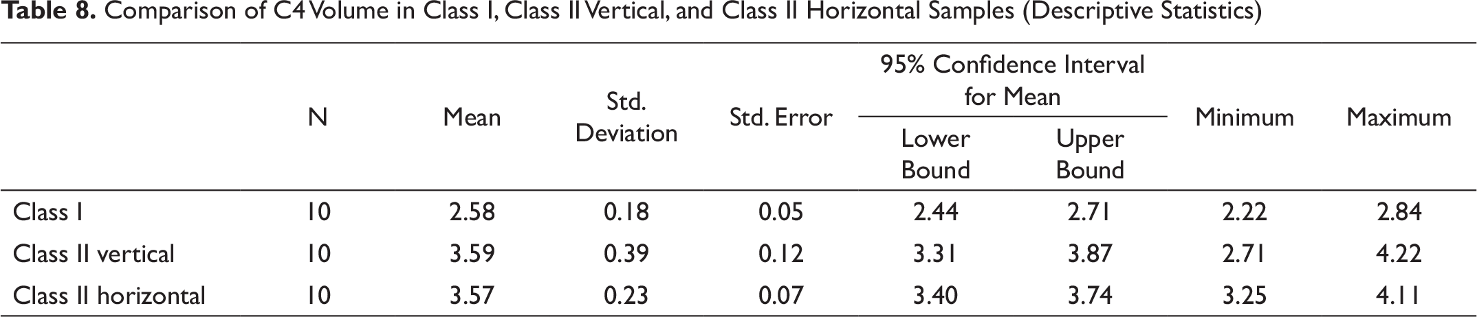
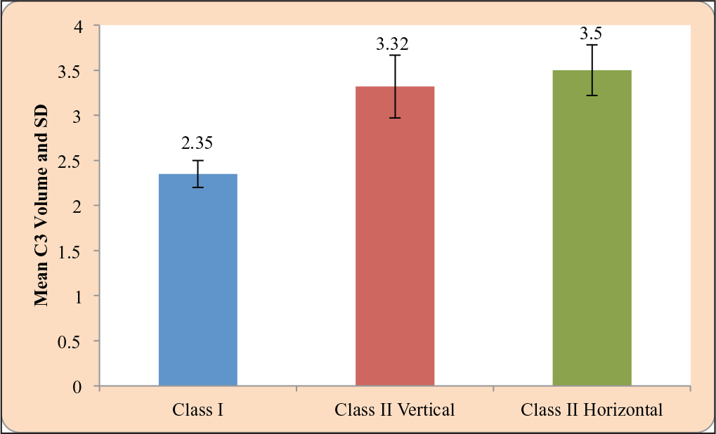
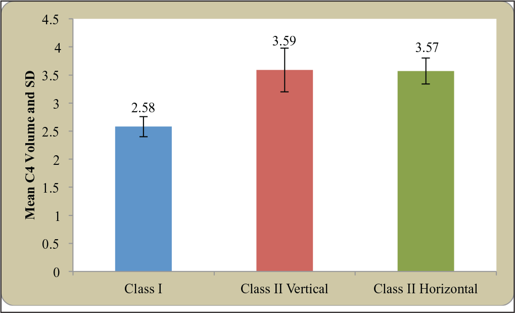
Statistically significant differences were observed between groups 1 and 2 and groups 1 and Group 3 regarding all 3 parameters. Groups 2 and 3 had mild differences but were not statistically significant. The C3 volume parameter showed the greatest difference between groups 2 and 3; however, it was statistically insignificant(Figures 2–4).
Discussion
There is an existing association present between craniofacial and cranio-cervical morphology according to literature.2, 3, 7 A previous study has concluded that there is a co relation between the skeletal maturation age and the volume of bodies of second, third, and fourth cervical vertebrae. In a study by Ousama et al. in 2018, it was seen that there was a moderate to strong positive co-relation between the cervical vertebral volume parameter and the MP3 stages, specifically of the fourth cervical vertebrae. 7
Parameters Which Were Assessed After Three-Dimensional Slicing
Comparison of C2 Volume in Class I, Class II Vertical, and Class II Horizontal Samples (Descriptive Statistics)
Comparison of C2 Volume in Class I, Class II Vertical, and Class II Horizontal Samples (One-Way ANOVA)
Comparison of C2 Volume in Class I, Class II Vertical, and Class II Horizontal Samples (Multiple Comparison: Tukey Test)
Comparison of C3 Volume in Class I, Class II Vertical, and Class II Horizontal Samples (Descriptive Statistics)
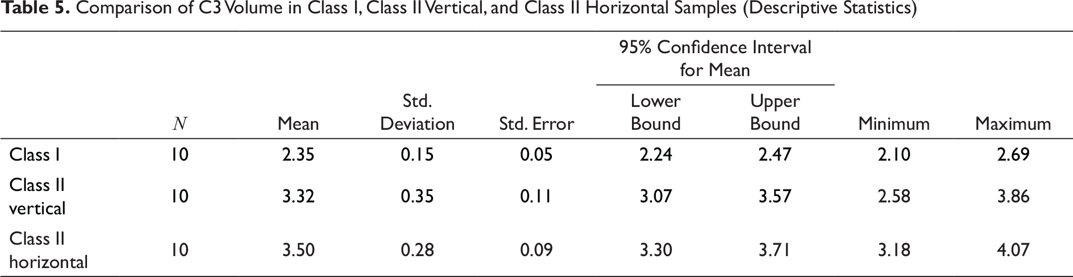
Comparison of C3 Volume in Class I, Class II Vertical, and Class II Horizontal Samples (One-Way ANOVA)
Comparison of C3 Volume in Class I, Class II Vertical, and Class II Horizontal Samples (Multiple Comparison: Tukey Test)
Comparison of C4 Volume in Class I, Class II Vertical, and Class II Horizontal Samples (Descriptive Statistics)
Comparison of C4 Volume in Class I, Class II Vertical, and Class II Horizontal Samples (One-Way ANOVA)
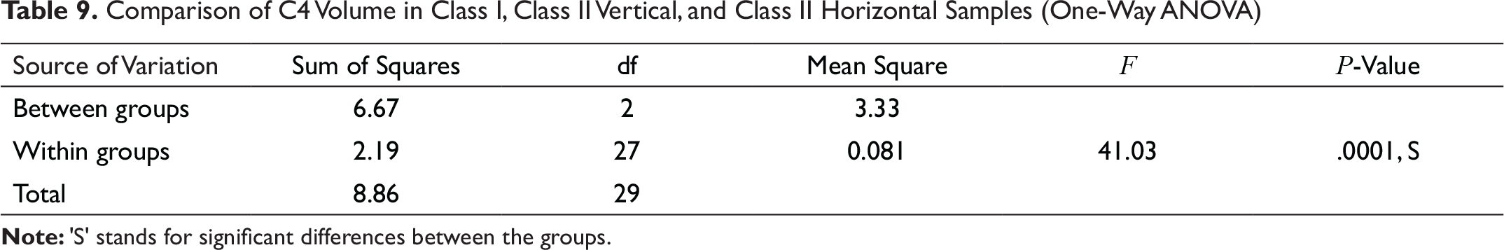
Comparison of C4 Volume in Class I, Class II Vertical, and Class II Horizontal Samples (Multiple Comparison: Tukey Test)
It was also noted that there was only slight deviation in the volume parameter between the horizontal and vertical growers in class II malocclusions. From this study, a positive co-relation was found between cervical vertebral body volume and class II malocclusions. Thus, it can be concluded from this study that growth pattern has no verifiable effect on the volume of cervical vertebrae. However, variations in cervical vertebral body volume are seen with different malocclusions.
Although the CVMI can determine the skeletal age of the patient, it cannot be utilized for predicting future malocclusions. Similar studies are required in the future to evaluate the applicability of volumetric evaluation to predict the future growth of the jaws and craniofacial structures. Additionally, if the volume of the cervical vertebrae can be evaluated at an early age and correlated with patterns of malocclusions, it can be used as a diagnostic tool in the future to diagnose and preferably intercept developing malocclusions.
Conclusion
Cervical vertebrae have long been used to assess the skeletal age of individuals. Since this study was performed in non-growing age groups, further studies are required in subjects of the growing age. However, with the advent of 3D scanning methods such as Cone Beam Computed Tomography (CBCT), 3D-DVT which have significantly less exposure as compared to conventional 3D scanning methods, it may now be possible not only to ascertain skeletal age of the patient but also to predict the pattern of malocclusion that may occur. This knowledge can be incorporated to take appropriate interceptive measures in any developing patterns of malocclusion in younger age groups.
Isolation of cervical vertebrae to be assessed; (2) vertebrae to be assessed sliced; (3) body of cervical vertebrae identified; (4) slicing done to obtain cervical vertebral body; (5 and 6) sphere volumetric parameter selected and body placed inside to assess volume.
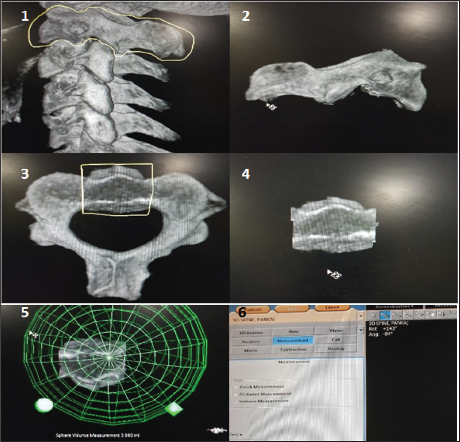
Comparison of C2 volume in class I, class II vertical and class II horizontal
Comparison of C3 volume in class I, class II vertical and class II horizontal
Comparison of C4 volume in class I, class II vertical and class II horizontal
Footnotes
Declaration of Conflicting Interests
The authors declared no potential conflicts of interest with respect to the research, authorship, and/or publication of this article.
Funding
The authors received no financial support for the research, authorship, and/or publication of this article.