
Abstract
Abstract
There is a plethora of literature available on miniscrew implant placement since they have been used for anchorage since 1997. Inappropriate usage of a micro-implant could lead to the fracture of the micro-implant, but literature has inadequate documentation probably due to insufficient reporting of these cases. A simple technique for the retrieval of fractured mini-implant is presented here.
Introduction
In contemporary orthodontics, the increased use of mini-implants is established. Inadvertent and irresponsible use of micro-implants has highlighted implant fracture as a major drawback. Fracture of miniscrew implants depends on the amount of insertion torque during placement. 1 It frequently occurs in the mandible where the cortical bone density is significantly high, thereby increasing insertion torque. Accepted torque value of self-drilling mini-implants is 3-10 N cm. 1 Self-drilling screws are ideal for D2 and D3 bone (Misch classification). Although greater anchorage is achieved when mini-implants are inserted in D1 bone, mini-implants are more vulnerable to fracture due to increased resistance by D1 bone.2, 3
Simple steps for retrieval are as follows:
Patient is informed about mini-implant fracture and retrieval procedure is also explained.
Patient is cleaned and draped, and the area of the mini-implant fracture is anesthetized locally using 2% lignocaine with adrenaline (1:200 000).
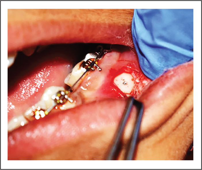
After mini-implant fracture, there is usually no bleeding at the site and the abraded gingiva makes it difficult to localize the site of implant fracture. To localize it, a small incision is made at the site with BP blade no. 15. Sufficient exposure of site is essential to allow the retrieval procedure (Figure 1[a]).
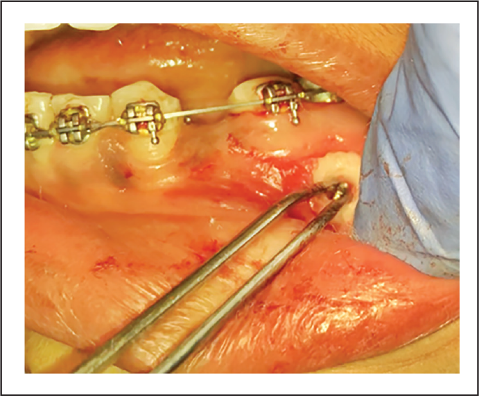
With the use of contra-angled micro-motor hand piece, with tungsten carbide burs (RA#1/4 Round bur and RA# 700 taper fissure cross cut bur) at low speed and copious saline irrigation, a circumferential trough around the implant is created, thereby loosening it (Figure 1[b]).
Care is taken to have a centrifugal force (outward toward the bone) rather than centripetal (inward toward the implant), which ensures that the implant is not thinned down further, which might make it more prone to fracture.
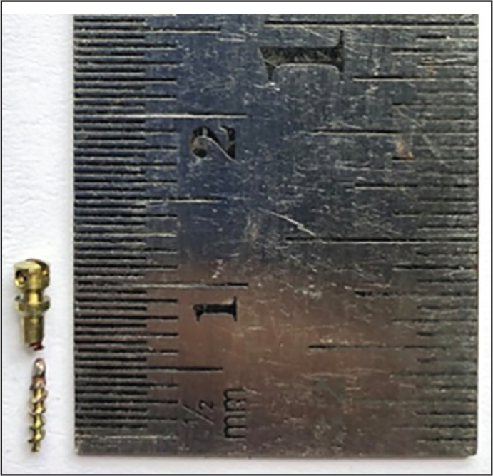
Extent of implant loosening is carefully evaluated by a thick straight probe after every round of circumferential bone cutting. Higher forces of lateral probing may result in implant fracture at lower length. Once it is sufficiently loosened, the tip of micro-implant is held with thin artery forceps and counter clockwise movement is given to loosen the screw.
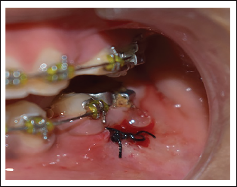
Fractured implant is removed and sutures are placed to close the area (Figure 1[c] and Figure 1[d]).
Post-surgical instructions are given to the patient. Patient is also prescribed antibiotics and analgesics for 3 days.
To confirm full retrieval, Intra-oral periapical X ray is taken.
Suture removal is done after a week
Exposed Site of Fractured Mini-implant
Bur Hole Around the Implant Periphery
Retrieved Fracture Implant
Closed Site with Suture
Conclusion
Miniscrews are valuable tools that increase the quality of orthodontic treatment if properly used. The orthodontist should keep in mind the possibility of fracture of the mini-implant during placement, and the above-mentioned armamentarium should always be available on the day the implant placement has to be performed.
Footnotes
Declaration of Conflicting Interests
The authors declared no potential conflicts of interest with respect to the research, authorship, and/or publication of this article.
Funding
The authors received no financial support for the research, authorship, and/or publication of this article.