
Abstract
Coelomic fluid surrounds the internal organs of asteroid echinoderms (asteroids, otherwise known as sea stars or starfish) and plays an essential role in the immune system, as well as in the transport of respiratory gases, nutrients, waste products, and reproductive mediators. Due to its importance in physiology and accessibility for nonlethal diagnostic sampling, coelomic fluid of asteroids provides an excellent sample matrix for health evaluations and can be particularly useful in disease and mortality investigations. This is especially important in light of recent increases in the number of affected individuals and species, larger geographic scope, and increased observed frequency of sea star wasting events compared with historic accounts of wasting. This review summarizes the current knowledge about coelomocytes, the effector cell of the asteroid immune system; coelomic fluid electrolytes, osmolality, acid–base status and respiratory gases, and microbiota; and genomic, transcriptomic, and proteomic investigations of coelomic fluid. The utility of coelomic fluid analysis for assessing stressor responses, diseases, and mortality investigations is considered with knowledge gaps and future directions identified. This complex body fluid provides an exciting opportunity to increase our understanding of this unique and ecologically important group of animals.
Keywords
Echinoderms derive their name from the Latin for “spiny skin,” a reference to the calcareous spines commonly found in their body wall. These exclusively marine invertebrates are found worldwide in a variety of aquatic ecosystems from the intertidal zone to the deep sea. 110 There are currently 5 extant classes in the Phylum Echinodermata: Crinoidea (crinoids; sea lilies and feather stars), Echinoidea (echinoids; sea urchins and sand dollars), Holothuroidea (holothuroids; sea cucumbers), Ophiuroidea (ophiuroids; basket stars and brittle stars), and Asteroidea (asteroids; sea stars, otherwise known as starfish). 77 This review will focus on the coelomic fluid of asteroid echinoderms.
The coelomic fluid of invertebrates, analogous to vertebrate blood, fills the body cavity and bathes the internal organs. In asteroids, there are 4 compartments to the coelomic system: the perivisceral coelom containing the organs; the genital coelom, a narrow space between the inner and outer epithelial region of the gonads; the hyponeural coelom surrounding the hyponeural (motor) nervous system; and the water vascular system. 73 The water vascular system is the hydraulic system responsible for asteroid locomotion. It contains a ring canal around the central disk, radial canals down the length of each arm, and perpendicular canals that terminate in ampullae and tube feet. 116 The madreporite is a calcareous structure on the aboral surface of the disk, which is presumed to play a role in the control of seawater entering the coelomic system. 47 As the perivisceral coelom is the largest and most accessible coelomic compartment for sample collection in asteroids and functions in whole body physiology, 22 this review will focus on fluid and cells of this coelom, unless otherwise noted.
As it surrounds the internal organs, the coelomic fluid plays an essential role in many physiological mechanisms including respiratory gas exchange, transport of nutrients and waste products, reproduction, and immune response - the latter a focus of this review. Respiratory gas exchange is accomplished through diffusion across papulae, evaginations of the coelomic lining, predominantly located on the aboral surface and across the tube feet.26,41,110,115 Sequences homologous to hemoglobin have been identified in the genomes of various sea star species; 24 however, little is known about the function of respiratory pigments in asteroids. Current research indicates that respiratory gases are carried in a dissolved state in coelomic fluid. 41 Nutrients are transported in coelomic fluid and flow nearly continuously between the organs and coelomic fluid. 45 Coelomic fluid also plays a role in providing amino acids and fatty acids to the ovaries during active reproductive phases. 9
The aim of this review is to summarize the current knowledge of coelomic fluid in asteroid echinoderms; to investigate the utility of coelomic fluid as a diagnostic tool in the evaluation of sea star stressor responses, diseases, and mortality investigations; and to highlight future perspectives.
Coelomic Fluid in Clinically Normal Asteroids
While health status evaluations of asteroids are currently challenging to conduct, the following sections summarize what is known about coelomic fluid from clinically normal sea stars who have not been intentionally exposed to any environmental stressors or disease agents. The components of coelomic fluid include cells, known as coelomocytes, and various uncharacterized proteins, mediators, and factors.
Coelomocytes
Coelomocytes are the circulating cells in the coelomic fluid and are involved in nutrient transport, 45 waste product removal, 36 clumping in response to wounding to prevent coelomic fluid loss, 12 and are the effector cell of the echinoderm immune system. 73 In their role within the immune system, coelomocytes are involved in phagocytosis80,86 and non-self-recognition, 32 and produce many bioactive molecules. Coelomocytes produce reactive oxygen species; 86 phenoloxidase; 118 Asterina pectinifera lectin (APL-R); 79 various hemagglutinins;15,48,125 proteins with interleukin-1-like activity; 8 and a protein called “sea star factor” that has immunosuppressive properties.84,105 Coelomocytes also express a gene homologous to C3, a member of the mammalian complement cascade. 86 To protect themselves from infection, asteroids also have antimicrobial activity including lysozyme-like and hemolytic activity in their cell-free coelomic fluid, coelomocytes, ova, pyloric ceca, and body wall. 57 Asterosaponins are a class of steroid oligoglycosides with diverse structures. These secondary metabolites have hemolytic, cytotoxic, antiviral, and antipredator defensive properties and have been identified in the cell-free coelomic fluid of sea stars.87,122,126
In echinoderms, the predominant coelomocyte type, and most commonly used term for this cell type, is the phagocyte.104,117 Phagocytes are similar in morphology to mammalian histiocytes and, using light microscopy and Romanowsky-type stains, are characterized as cells with a single round nucleus composed of clumped chromatin, low to moderate nuclear to cytoplasmic ratio, and moderate to abundant cytoplasm that infrequently contains phagocytized debris even in clinically normal sea stars.104,128,134 Various types of coelomocytes have been reported in asteroid coelomic fluid in 12 studies of 12 species representing 3 orders and 7 families (Table 1). Additional information including morphologic descriptions can be found in Supplemental Table S1. See Fig. 1 for representative images of coelomocytes as well as spermatozoa and ova as incidental findings when evaluating coelomic fluid cytologic specimens. The plethora of terms used for cell types in coelomic fluid of asteroids that are identified by various methodologies (e.g., light microscopy, electron microscopy, fluorescence microscopy, phase microscopy, flow cytometry) across classes, and even between species within the same class, prohibits extrapolation of cell identification across echinoderm classes. The types of coelomocytes for Echinoidea, 117 Holothuroidea,37,62 and both of these classes 23 have been reviewed, but there is a dearth of information on the coelomocytes of the Ophiuroidea and Crinoidea. Therefore, we suggest using “phagocytes” for the predominantly observed cell type in coelomic fluid of echinoderms and “coelomocytes” for those cell types with morphology different from phagocytes.
Overview of terminology used for identified cell types in coelomic fluid of asteroid echinoderms in various studies.
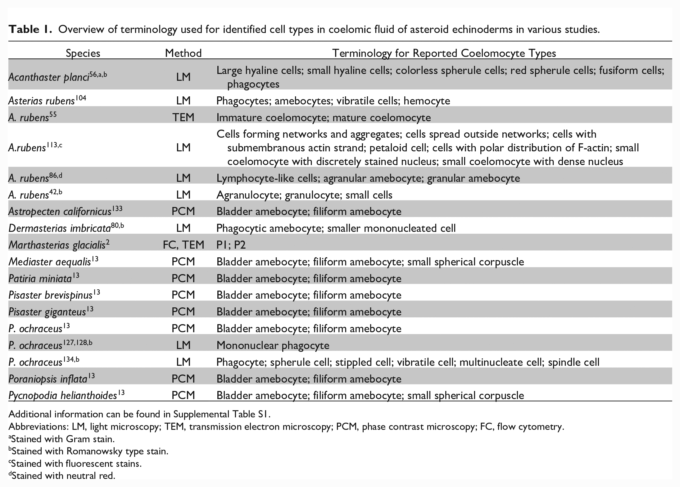
Additional information can be found in Supplemental Table S1.
Abbreviations: LM, light microscopy; TEM, transmission electron microscopy; PCM, phase contrast microscopy; FC, flow cytometry.
Stained with Gram stain.
Stained with Romanowsky type stain.
Stained with fluorescent stains.
Stained with neutral red.
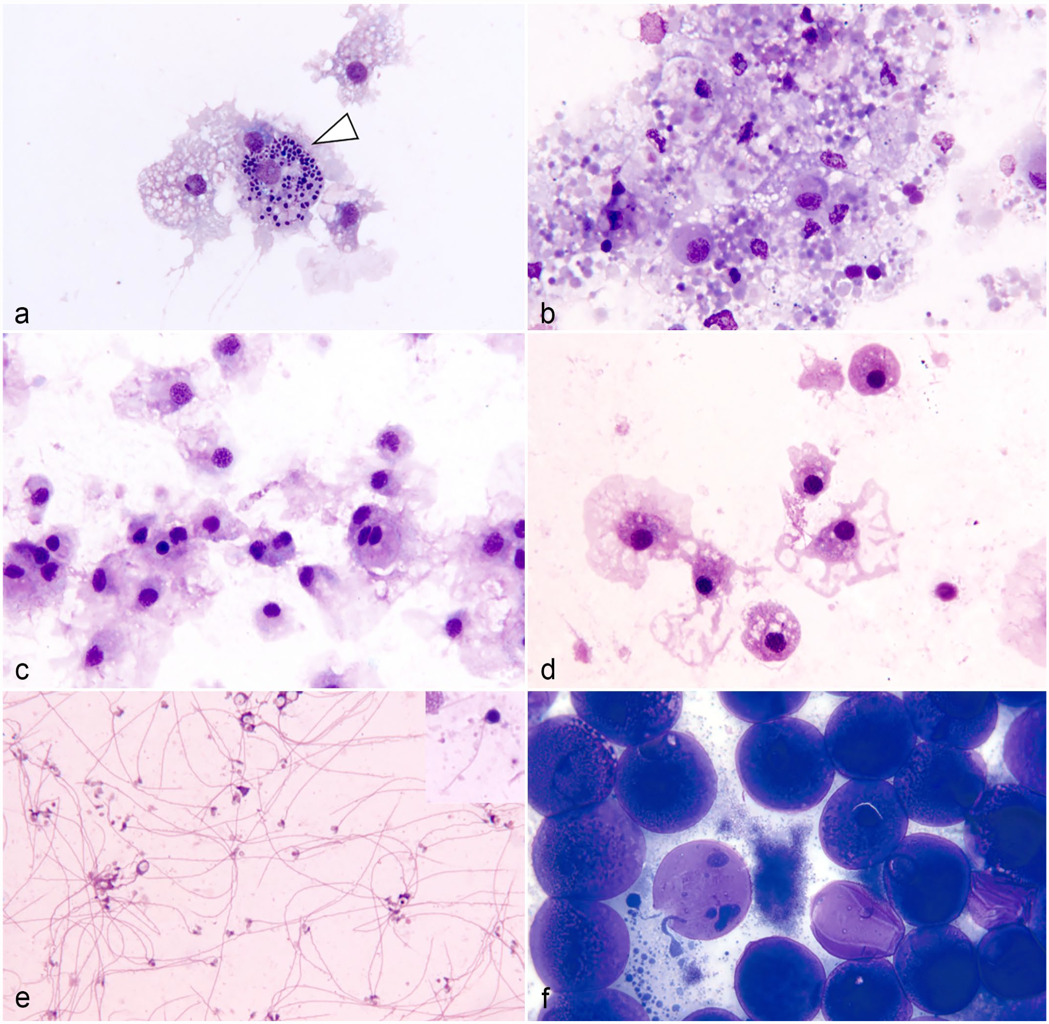
Photomicrographs of coelomic fluid preparations of sea stars. Methanolic Wright-Giemsa stain. (a) Clinically normal North Atlantic common sea star (Asterias rubens): coelomic fluid cytocentrifuge preparation with phagocytes and pigment-carrying coelomocyte (white arrowhead). (b) Mottled sea star (Evasterias troschelii) affected by sea star wasting (SSW): coelomic fluid direct smear with phagocytes containing variable amounts of phagocytized material of undetermined origin; the background contains proteinaceous debris presumably from cellular breakdown. (c) North Atlantic common sea star (Asterias rubens) coelomic fluid cytocentrifuge preparation collected from automized arm showing phagocytes. (d) North Atlantic common sea star (Asterias rubens) exposed to decreased tank water pH (pH 7.4) during a laboratory experiment: coelomic fluid cytocentrifuge preparation with phagocytes. (e) North Atlantic common sea star (Asterias rubens) after losing one arm: coelomic fluid direct smear containing mature spermatozoa at various degrees of degeneration (insert: intact mature spermatozoan) consistent with incidental finding. (f) Ochre sea star (Pisaster ochraceus) affected by SSW: coelomic fluid direct smear with mature oocytes consistent with an incidental finding, presumably following rupture of gonads associated with autotomy.
Coelomocyte morphology changes rapidly in vitro, especially if the cells are transferred onto a glass slide for evaluation, such as direct smears or wet-mount preparations.80,82,104,114 This must be considered when determining coelomocyte types. When placed on a solid surface, the cellular morphology can change markedly and rapidly. This also occurs in vivo when coelomocytes migrate from coelomic fluid to tissues.33,94 Morphological differences in vitro may indicate cellular activation or maturity, 55 rather than truly different cell types. In addition, it needs to be considered that variable terminology has been applied to morphologically similar cell types, which creates further confusion.
Immunoblotting, immunocytochemical labeling of coelomocytes with anti-toposome antibodies, and ultrastructural investigations have demonstrated that the coelomic lining epithelium is likely the source of circulating coelomocytes.14,55,104 Coelomic lining epithelial cells have flagella with a typical microtubule arrangement (9+2) and microvilli, as identified by electron microscopy.14,55 These flagellated coelomic epithelial cells can be visualized at high magnification using a 100× objective by light microscopy, or by electron microscopy. 55 Electron microscopy suggests that immature coelomocytes, which are flagellated, are released from the coelomic epithelium into the coelomic fluid where they mature. 55 While echinoids and some holothuroids have distinct coelomocytes called “vibratile cells” because of their flagellar motion, these cells have not been identified from coelomic fluid of asteroids.23,106,107 The role of the axial organ and Tiedemann bodies in the production or recycling of coelomocytes is unclear. Both of these structures are composed of meshwork of connective tissues and myocytes, and are postulated to have a role in filtration of body fluids and translocation of nutrients.39,43,46,130 Using radiolabeling and autoradiography, the axial organ and Tiedemann bodies of Asterias forbesi were not found to produce significant amounts of coelomocytes. 43 More recent work using 5-bromo-2′-deoxyuridine (BrdU) labeling and confocal microscopy showed that the axial organ, Tiedemann bodies, and coelomic epithelium had increased cellular proliferation in response to immune-stimulating molecules. 70 A study using phagocyte specific gene expression in echinoids concluded that coelomocyte production occurred in the axial organ and pharynx; however, this study did not investigate coelomic epithelium. 53
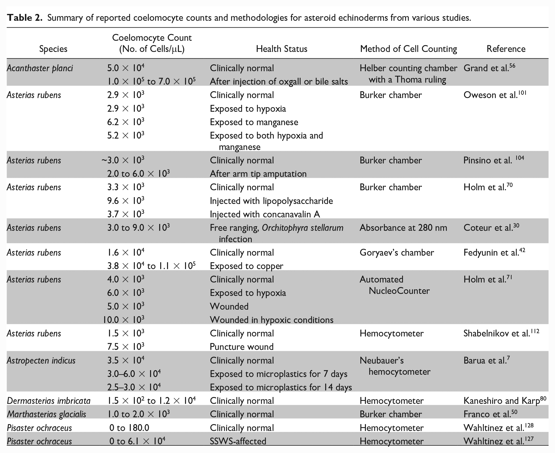
Coelomocyte counts can provide valuable information about the health status of asteroids. A wide range of methods for counting coelomocytes as well as coelomocyte counts have been published for asteroids (Table 2). Coelomocyte counts for clinically normal asteroids range from 0 to 5.0 x 10⁴ cells/μL.56,128 The variations in cell counts may be due to differences in sample collection techniques (e.g., cutting off the tip of an arm versus needle and syringe), the use of anticoagulants, and differences in methodology for cell counting. Caution should be used when using automated analyzers (e.g., a NucleoCounter70,71) as they are validated for the sizes of mammalian blood cells and may not be accurate when counting asteroid coelomocytes.
Summary of reported coelomocyte counts and methodologies for asteroid echinoderms from various studies.
Coelomocytes rapidly form “clumps” to prevent coelomic fluid loss in cases of penetrating body wall injury 81 which are also observed in vitro during sampling. These clumps are often referred to as “clots,” however, this term should be avoided in echinoderms as they do not possess thrombocytes; furthermore, the processes of cellular clumping are distinct from the coagulation cascade in vertebrates. Coelomocyte clumping has been observed in all extant classes of echinoderm. 78 In asteroids, phagocytes or their progenitor cells can initiate clumping, depending on the species, and the cells in the clumps appear to merge together.12,78 In in vitro experiments, clumping is inhibited by divalent cation reducing agents (e.g., potassium oxalate, sodium citrate, and ethylenediaminetetraacetic acid [EDTA]) and it was shown that a minimum concentration of divalent cations is required for clumping.12,81
To enable accurate coelomocyte counts, several types of anticoagulants have been used. Due to the involvement of calcium and magnesium in clumping, the most commonly used anticoagulants include chelating agents, calcium free salt solutions, or a combination of both. Some of the reported anticoagulants are calcium and magnesium free artificial sea water solution with EDTA, 42 EDTA,86,98 and coelomocyte culture medium containing ethylene glycol tetraacetic acid (EGTA). 59 Collection of coelomic fluid into diluted formalin has also been described;5,31,134 however, these samples can be used for cell counts but not for cytological evaluation as contact of cells with formalin or formalin fumes results in alterations of cell morphology. 49
Electrolytes and Osmolality
Electrolyte concentrations and osmolality of asteroid perivisceral coelomic fluid have historically been regarded as iso-ionic and iso-osmotic to seawater.10,27,34,128 However, this information comes from research with a limited number of individuals and few species, and has largely not been re-evaluated in decades. The coelomic fluid of clinically normal Asterias rubens, Luidia clathrata, Pisaster ochraceus, and Solaster endica have been found to be iso-ionic to seawater.10,27,34,128 While the differences were not statistically significant, the coelomic fluid of P. ochraceus had slightly lower magnesium and calcium and slightly higher potassium and chloride compared with seawater. 128 Similarly, the coelomic fluid of Marthasterias glacialis had lower magnesium and higher potassium compared with seawater. 108 Some studies have found perivisceral coelomic fluid to be slightly hypo-osmolar or hyper-osmolar to seawater, with variations between species.11,44,58 At least some species of sea star appear to be able to actively regulate their coelomic fluid volume through unknown mechanisms.38,102
Acid Base Status and Respiratory Gases
The pH of sea star coelomic fluid ranges from 0.5 to 1.5 units below the pH of seawater due to carbon dioxide production from metabolism.27,35,41,61 The buffering capacity of coelomic fluid predominantly results from equilibration of the bicarbonate buffer system contained in seawater (92%), with the remaining 8% due to coelomocytes. 29
Sea stars perform respiratory gas exchange through their papulae and tube feet.26,41,110,115 Respiratory rates for asteroids vary widely between species and studies, and are influenced by body size, feeding status, reproductive state, and activity of the individual. 88 There appear to be species-specific differences of the impact of water temperature on asteroid respiration.88,103 Respiratory gases diffuse across coelomic fluid, which has lower oxygen and higher carbon dioxide concentrations compared with surrounding seawater. 41 The published partial pressure of oxygen (pO2) in the coelomic fluid of asteroids ranges from 84 to 135 mm Hg;91,115 however, differences in methodologies and differences in the dissolved oxygen content and temperature of water would undoubtedly influence coelomic fluid pO2, so direct comparisons are difficult.
Microbiota
The microbiomes of coelomic fluid from multiple sea star species have been described.74,96 Sea stars have tissue-specific microbiomes and changes in microbial communities may inform the health status.69,74 Asteroids in aquaria had 2.55 × 10⁴ bacterial cells/mL in their coelomic fluid while coelomic fluid from free ranging asteroids had 1.27 × 10⁵ bacterial cells/mL, both of which were 1 to 2 orders of magnitude lower than the bacterioplankton abundance in the seawater surrounding the asteroid. 74 There were few shared microbial taxa between coelomic fluid and seawater, which suggests selection or enrichment of microorganisms by the host.74,96 However, this may be species dependent as Patiria pectinifera had a similar microbial community to surrounding seawater. 96 On cytological evaluation of coelomic fluid from P. ochraceus using Wright-Giemsa stain, no bacteria or other microorganisms were noted. 128 The virome of coelomic fluid in sea stars has not been described at the time of writing this manuscript.
There are few reports of successful bacterial culture from sea star tissues or coelomic fluid. In one report, bacteria from swabs of the integument of Astropecten jonstoni were successfully grown on trypticase soy agar or broth in aerobic conditions at 20°C ± 1°C for 3 to 4 days. 120 Samples for cultures from sea stars should be handled similar to the techniques developed for marine fish, including the use of media with comparable osmolality and/or salinity to seawater and incubation at ambient water temperature for at least 5 days to detect slow-growing microorganisms. 83 Due to the challenges associated with bacterial cultures, nonculture dependent techniques, such as next-generation sequencing, should also be considered.
Molecular Investigations of Coelomic Fluid
The majority of molecular investigations in echinoderms have been conducted in the purple sea urchin (Strongylocentrotus purpuratus), a model organism for developmental and systems biology from which the first echinoderm genome was published. 119 Genomics, the study of the genome of an organism, can provide information on development, evolution, population size, connectivity of populations, and adaptation to environmental changes.19,68 In an early evaluation, 1.52 pg of diploid DNA was isolated from the coelomocytes of Echinaster sepositus. 40 There are currently 8 genomes available for asteroids (Acanthaster planci, A. rubens, M. glacialis, Patiria miniata, Patiriella regularis, Pisaster brevispinus, P. ochraceus, and Plazaster borealis) with vastly different levels of annotation. 97 The genome of S. purpuratus was surveyed for genes that are homologues of known immune system components, yielding interesting insights into the echinoderm immune system. 67 No similar analyses have been done for asteroids.
Transcriptomic studies of gene expression provide insight on ribonucleic acid (RNA) activation and the functioning of cells. 1 Gene expression evaluations of asteroid coelomocytes have provided insights into their roles. These include evaluation of expression of homologues of the ArC3 gene which encodes the C3 component of the mammalian complement cascade. 86 Transcriptomes for the coelomic epithelium of Coscinasterias muricata and clinically normal P. pectinifera have been published.52,85 The C. muricata transcriptome was generated from a clonal individual, with RNA extracted from the coelomic epithelium of the normal arms rather than the regenerating arms. 52 The P. pectinifera transcriptome demonstrated that there are tissue specific differences in gene expression between the body wall, coelomic epithelium, tube feet, stomach, pyloric ceca, and gonad. 85
Proteomics involves the identification and quantification of proteins within a cell, tissue, or organism, and offers an opportunity for the evaluation of the functions, structure, interactions, and post-translational modifications of those proteins. 6 Since proteins can be considered the effector molecule of biological functions, proteomics provide another avenue to gain valuable information about physiology and interactions between animals and their environment. The proteome of coelomocytes from M. glacialis has been published and led to insights about the multiple roles of coelomocytes including cytoskeleton regulation, cellular adhesion, signaling, cell regulation, and cellular proliferation. 50 Coelomic fluid proteins are also involved in endocytosis, protein degradation, production of cytokines and growth factors through the AGE-RAGE signaling pathway, and extracellular matrix-receptor interactions. 54 Proteomic analyses of circulating coelomocytes and coelomic epithelial cells demonstrated the relationship between the coelomic epithelium and circulating coelomocytes. 113
Coelomic Fluid in Disease and Mortality Investigations
Due to the physiological importance of coelomic fluid to virtually every biological process in asteroids and the integral role of coelomocytes in the immune system, coelomic fluid is an ideal sample matrix for the evaluation of stressor responses, disease, and mortality investigations. Coelomic fluid can be nonlethally collected and evaluated using many techniques, including commonly used diagnostic tests in veterinary medicine and molecular approaches. The following sections summarize the body of knowledge about the coelomic fluid from asteroids that have been experimentally exposed to environmental stressors or free-ranging asteroids that suffered from disease.
Sea star wasting (SSW) refers to a suite of clinically evident changes that include varying degrees of epidermal lesions followed by autotomy and body wall disintegration, often leading to death, and has occurred in a variety of sea star species worldwide.66,99 While these reports describe epizootics of SSW, individual sea stars can also present with this suite of clinically evident changes. Although this term is often used to refer to the event that began along the Pacific coast of North America in 2013, SSW has been documented as far back as 1898. 92 The cause or causes of SSW remain unknown to date. Although early reports indicated a sea-star-associated densovirus may be implicated, 64 this virus has subsequently been found in tissues of clinically normal asteroids across the world and does not appear to play a role in SSW.63,65 Interestingly, quantitative polymerase chain reaction and reverse transcriptase polymerase chain reaction have not detected sea star-associated densovirus or Asterias forbesi-associated densovirus in coelomic fluid, even when virus is detected in the body wall, gonads, and pyloric ceca.75,76 The current understanding of SSW indicates that environmental stressors appear to play a central role in the pathogenesis, whether through direct effects or changes to body wall microbial communities.4,66,99
Coelomocytes
Coelomocyte counts have been widely used as a marker of sea star responses to stressors or disease. Table 3 summarizes coelomocyte responses of various species of asteroids exposed to stressors or disease. Compared with controls, coelomocyte counts tend to increase in sea stars that are exposed to environmental stressors.42,56,71,100 One notable exception are sea stars exposed to decreased water pH as lower coelomocyte counts were reported in A. rubens exposed to water pH 7.7 for 1 week or 6 months. 61
Summary of reported coelomocyte counts following exposure of various asteroid species to stressors or disease.
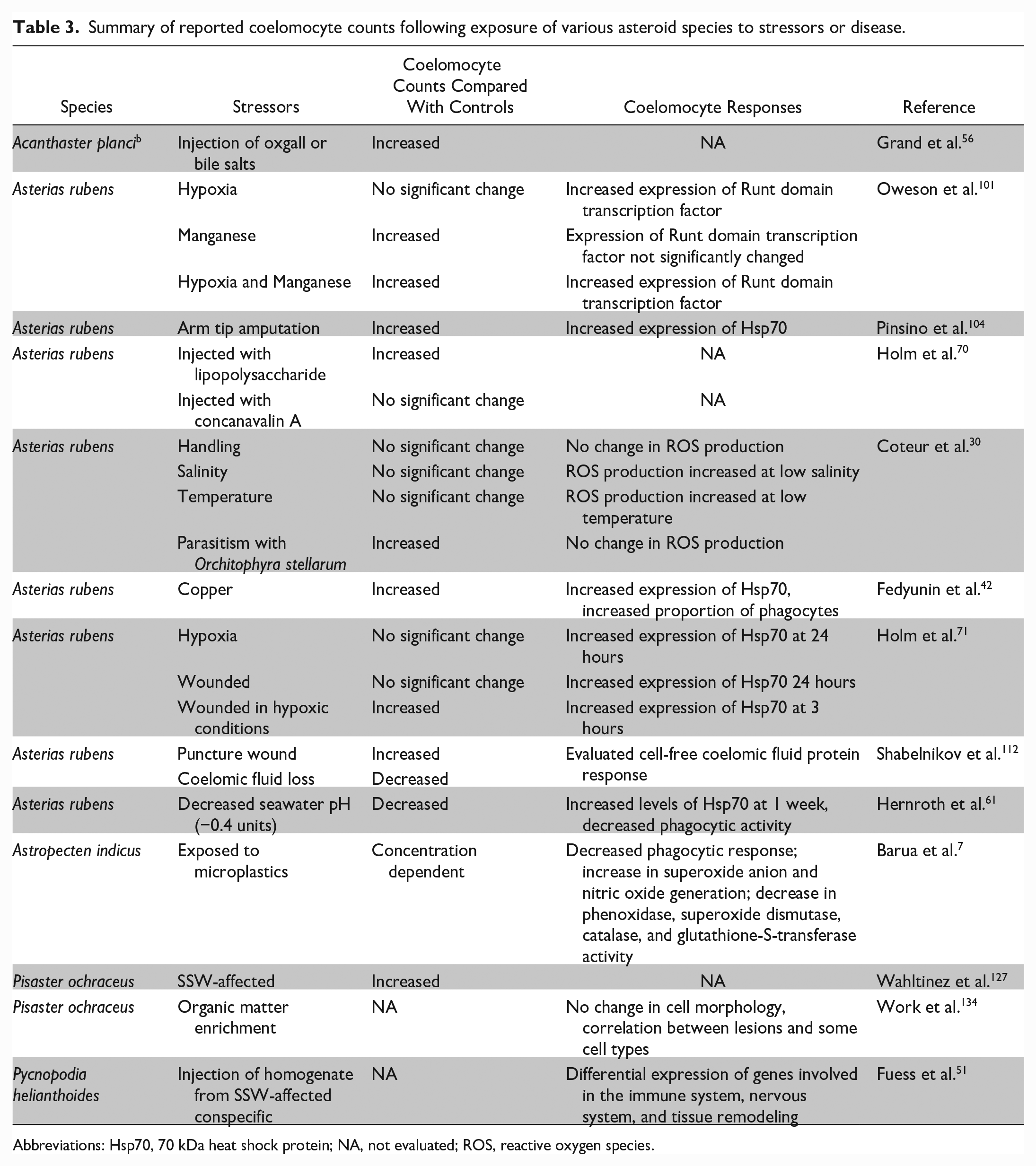
Abbreviations: Hsp70, 70 kDa heat shock protein; NA, not evaluated; ROS, reactive oxygen species.
Electrolytes and Osmolality
The majority of the research evaluating coelomic fluid electrolytes and osmolality has been conducted in relation to changes in salinity of surrounding water. Asteroids do not appear to be able to regulate their coelomic fluid electrolytes or osmolality when placed in diluted seawater. After being placed in 55% seawater diluted with isotonic glucose, coelomic fluid sodium, potassium, calcium, and chloride concentrations of A. rubens began to decrease within 1 hour; after 8 hours, only calcium was higher in coelomic fluid than compared with diluted seawater. 10 Similarly, when placed in diluted (15 ppt) or concentrated (35 ppt) seawater, coelomic fluid sodium, potassium, calcium, chloride, and magnesium of L. clathrata equilibrated to concentrations of the surrounding medium within 24 hours. 34 Concentrations of sodium, potassium, calcium, chloride, and magnesium in coelomic fluid of Evasterias troschelii sampled during a tidal cycle varied as the electrolytes in the seawater around them changed. 121 The coelomic fluid of A. rubens, L. clathrata, and P. ochraceus remains iso-osmotic to the medium, even when the concentration of the surrounding seawater is changed.11,34,58 However, L. clathrata appeared to be able to regulate coelomic fluid volume as measured by wet weight and whole animal volume.34,38
Changes in salinity are not the only osmoregulatory challenges faced by asteroids, as loss of water through evaporation during emersion (removal from water) also results in changes to chloride in coelomic fluid. For instance, A. rubens emersed for 3 hours lost 13% of their body weight with a 19.4% increase in coelomic fluid chloride, while those emersed for 6 hours lost 20% of their body weight with a 26.0% increase in chloride. 11
Sea stars affected by SSW had changes to their coelomic fluid electrolytes and osmolality, which provides information on the pathophysiology of SSW. SSW-affected P. ochraceus had higher coelomic fluid chloride, osmolality, and total protein, with lower calcium when compared with clinically normal conspecifics. 127 The coelomic fluid changes are suggestive of dysregulation of osmotic balance and calcium, which may indicate why sea stars often appear inflated in early stages of SSW and have decreased activity.
Acid Base Status and Respiratory Gases
Intertidal sea stars are faced with emersion, which challenges their ability to efficiently respire, especially at higher temperatures. Intertidal P. ochraceus were emersed for 6 hours at 3 different temperatures (5°C, 15°C, and 25°C; seawater 13°C). At the lowest temperature, coelomic fluid pO2 increased while pH and pCO2 did not change. At the temperature closest to seawater, there was a transient decrease in pO2, decreased pH, and increased pCO2. At the highest temperature, pO2 and pH decreased while pCO2 increased. Coelomic fluid bicarbonate levels increased at all temperatures. 91 These results provide insight into the physiological mechanisms that allow intertidal sea stars to survive in this challenging environment.
Laboratory exposures of sea stars to decreased seawater pH indicate that sea stars appear to have limited capacity to mount adaptive physiologic responses to stressor exposure, such as ocean acidification. The coelomic fluid pH of A. rubens exposed to seawater with a decreased pH (7.4–7.7) significantly decreases and remains decreased even after 6 months of exposure.3,28,61
Microbiota in Coelomic Fluid
There are very few reports of microbiota in asteroid coelomic fluid during environmental stressor exposures or disease. Free or phagocytized bacteria were observed in the coelomic fluid of 16 of 55 (29%) SSW-affected P. ochraceus, and were absent in clinically normal controls. 127 Coelomocytes are capable of rapidly responding to bacterial invasions. One hour after injection of Micrococcus lutens bacteria, coelomocyte counts increased and the number of actively phagocytizing coelomocytes also increased. 31
Molecular Investigations of Coelomic Fluid
Molecular techniques provide a powerful tool for understanding the responses and impacts of environmental stressors and disease. Genotyping and differential gene expression for pyloric ceca, body wall, and tube feet in SSW-affected sea stars have been published, but are beyond the scope of this review.21,109,129 Coelomocytes from 3 experimentally SSW-affected Pycnopodia helianthoides differentially expressed genes involved in nervous system processes, tissue remodeling, and immune system functions. The immune pathways that were enriched included the Toll pathway, complement cascade, melanization, and arachidonic acid metabolism. 51 This study demonstrated that P. helianthoides mounted a robust immune response to the injection of homogenate from 3 SSW-affected conspecifics and included the first gene-based report of melanin synthesis in an asteroid. In that study, coelomocytes showed upregulation of quinone oxidoreductase, an enzyme that is thought to be involved in melanin synthesis due to its homology with enzymes found in melanin synthesis in humans. 51 However, it is unknown if this pattern of differential gene expression holds true to SSW in free-ranging asteroids. In this research, cellular material was injected from conspecifics, and since coelomocytes are involved in recognition of self when exposed to allogeneic and xenogeneic material 32 and SSW appears to have complex, multi-factorial causes, care must be taken in the comparison of these data to other species. Nevertheless, the P. helianthoides study provides an excellent example of the utility of transcriptomic investigations of coelomic fluid.
Echinoderms have been used as bioindicators of heavy metal pollution in their environments.89,124 Without evaluating the entire transcriptome, a specific transcription factor can be quantified using real time polymerase chain reaction (RT-PCR) to evaluate differentiation of circulating coelomocytes and the coelomic epithelium. Expression of a transcription factor containing a Runt domain was evaluated using RT-PCR in A. rubens exposed to hypoxic conditions and both hypoxic conditions and manganese. 100 In hypoxic conditions, manganese reduces to the bioavailable Mn2+ state, which negatively impacts the immune responses of other marine invertebrates.60,101 Since Runt proteins are important in cellular proliferation and differentiation, 25 this represents an opportunity to evaluate coelomocyte functions. A. rubens exposed to hypoxic conditions and both hypoxic conditions and manganese had increased runt mRNA expression in coelomocytes and the coelomic epithelium, while sea stars exposed to only manganese did not have a significant difference in runt mRNA expression compared with controls. While this increased runt mRNA expression was not associated with increased numbers of circulating coelomocytes or proliferation of the coelomic epithelium, the coelomocytes had higher levels of runt mRNA than coelomic epithelium, which suggests that it may be more important in later stages of coelomocyte maturation. 100
Proteomic analyses have also been used to evaluate the responses of A. rubens coelomocytes to heavy metals. Immunoblotting revealed that heat shock proteins (Hsp70) in coelomocytes increase in response to experimental exposure to copper. 42 Chaperone proteins such as Hsp70 assist in protein folding, refolding misfolded proteins, moving proteins, and breaking up nonfunctional protein aggregates. 123 These proteins are often upregulated in animals experiencing physiological stress due to their role in preventing negative effects from environmental stressors. 123 Sea stars exposed to heavy metal gradients in nature show a correlation between Hsp70 expression in coelomocytes with cadmium and copper concentrations in soft tissues. There was no correlation with Hsp70 expression in coelomocytes and soft tissue lead and zinc concentrations. 90 Two-dimensional gel electrophoresis showed that 31 protein spots differed in relative abundance, which suggests differential expression; 90 however, the proteins were not identified or quantified.
There is considerable interest in the use of sea stars as a model system to study their remarkable regenerative capability,16–18 which has led to research on the coelomic fluid response to wounding. Following wounding, immunocytochemistry and immunoblotting techniques demonstrated an increase in Hsp70 in the coelomocytes of A. rubens.72,104 An increase in Hsp70 expression was also seen in nonwounded sea stars exposed to hypoxic conditions and those exposed to both wounding and hypoxic conditions. 72 It is worth noting that baseline heat shock proteins had high inter-individual variability. 105 The proteome of cell-free coelomic fluid from A. rubens has been characterized using mass spectroscopy. Both coelomic fluid loss and a puncture wound resulted in changes to the proteome, with eight and four differentially abundant proteins in the coelomic fluid loss and puncture wound groups, respectively. 112 The coelom appears to be a conduit for first line defense cells (e.g., phagocytes) and stem cells to access the wound site after injury.
Coelomic fluid contains a peptide called scalding factor, or autotomy promoting factor, which results in softening of the body wall and arm autotomy.20,93 This peptide was purified from the coelomic fluid of sea stars that were induced to autotomize arms through scalding or being dropped 2 meters. Within minutes of injecting the purified peptide into a resting sea star, the recipient sea star’s body wall softens and arm autotomy ensued. 93 This is likely due to an interaction between the peptide and mutable collagenous tissue, a tissue that is unique to echinoderms and is capable of rapid changes in tensile strength mediated by the nervous system. 131 This tissue is found in the body wall of asteroids.95,133 While chemicals and ions can induce changes in mutable collagenous tissue, current evidence suggests that this action is indirectly associated with the response mediated by juxtaligamental cells, the mechano-effector cells of mutable collagenous tissue.111,132 Since changes to the body wall are a common feature of SSW, 99 understanding the complex interactions between coelomic fluid, mutable collagenous tissue, and autotomy is imperative.
Future Perspectives of the Utility of Coelomic fluid in Health and Disease
Evaluation of coelomic fluid samples is a valuable, nonlethal diagnostic and research tool to evaluate the health, physiological changes, response to stressors, and disease processes in sea stars. Coelomic fluid with its cellular and acellular components is integral to nearly all biological processes from respiration to reproduction and immune functions. Coelomocytes, the circulating cell in coelomic fluid, are the effectors of the immune system and play an important role in clumping to prevent coelomic fluid loss, regeneration, and transport of nutrients and waste products. Despite its importance to asteroid biology, there are major gaps in the basic understanding of coelomic fluid. The underutilization of modern molecular techniques represents an opportunity to apply state of the art technologies to increase our understanding of coelomic fluid in health and in response to stressors and disease. Genomic surveys, similar to the one conducted for S. purpuratus evaluating immune gene homologues, 67 can inform about similarities and differences between the immune responses of different echinoderm classes. Similarly, coelomic epithelial lining transcriptomics represent an important field of further study, revealing new insights into coelomocyte development, maturation, and function.52,85 One major knowledge gap is the identification, quantitation, and characterization of the biological functions of the major proteins in coelomic fluid using proteomics or metabolomics.
One issue of utmost importance is the standardization of nomenclature for coelomocyte types to allow direct comparison of research results and to further elucidate the roles of coelomocytes in cellular responses. There is a need for standardized language when referring to echinoderm cell types while considering differences in morphological and functional cell types between classes and species. Cytochemical stains have been under-utilized in invertebrates and may prove to be a useful tool for determining coelomocyte types. Furthermore, standardization of recommended test methods for coelomic fluid, including pre-analytical (e.g., sample collection, anticoagulants, and any other specific requirements) and analytical methods (e.g., coelomocyte evaluation and enumeration, chemistry and protein analysis) will be useful for comparability of results in various clinical and research settings.
Another research priority is the evaluation of coelomic fluid and coelomocytes from more sea star species; current knowledge is based on less than 30 of the 1500 extant asteroid species. Further, the impact of seasonality and changes associated with reproductive activity will provide important information to accurately tease out effects from various stressors affecting populations in situ. While conducting future research, data obtained from asteroids should be compared with data collected from other asteroids using similar methodologies. An infusion of new approaches and investigations will help unlock the potential of this complex body fluid to increase our understanding of asteroid biology and pathophysiologic mechanisms. The ongoing mass mortality of sea stars along the west coast of North America emphasizes the importance of these investigations.
Supplemental Material
sj-pdf-1-vet-10.1177_03009858231176563 – Supplemental material for Coelomic fluid of asteroid echinoderms: Current knowledge and future perspectives on its utility for disease and mortality investigations
Supplemental material, sj-pdf-1-vet-10.1177_03009858231176563 for Coelomic fluid of asteroid echinoderms: Current knowledge and future perspectives on its utility for disease and mortality investigations by Sarah J. Wahltinez, Maria Byrne and Nicole I. Stacy in Veterinary Pathology
Footnotes
Declaration of Conflicting Interests
The authors declared no potential conflicts of interest with respect to the research, authorship, and/or publication of this article.
Funding
The authors received no financial support for the research, authorship, and/or publication of this article.
Ethics Statement
Since sea stars are not currently covered by research oversight guidelines in the United States, no Institutional Animal Care and Use Committee (IACUC) review was performed for the sea stars used to obtain images of coelomocytes. All sea stars were handled and housed in accordance with guidelines for aquatic species published in the Guide for the Care and Use of Laboratory Animals, 8th edition.
Supplemental material for this article is available online.
References
Supplementary Material
Please find the following supplemental material available below.
For Open Access articles published under a Creative Commons License, all supplemental material carries the same license as the article it is associated with.
For non-Open Access articles published, all supplemental material carries a non-exclusive license, and permission requests for re-use of supplemental material or any part of supplemental material shall be sent directly to the copyright owner as specified in the copyright notice associated with the article.