
Abstract
This report describes a unique pattern of alopecia in 8 American red squirrels (Tamiasciurus hudsonicus) from 2013 to 2021. All animals were juveniles; 6 were female and 2 were male. Seven presented between September and November, and one presented in April. All squirrels had widespread, bilaterally symmetric, noninflammatory, well-demarcated alopecia involving the entire trunk and legs and normal hair on their muzzle and dorsal surfaces of their paws. Six months later, a normal hair coat had grown on 2 of the animals, which were littermates. Hair fully grew 2 months later in another animal. Histopathology of the alopecic skin was performed in 7 of 8 animals. The following changes were noted: bent and coiled hairs, perforating folliculitis, melanin clumping, and distortion of hair shafts. Based on features of follicular dysplasia and apparent seasonality, this condition has some similarities to canine seasonal flank alopecia. A genetic etiology is suspected.
Alopecia in rodents is commonly described in a laboratory setting, with mice often serving as animal models for human alopecia. Alopecia in wild rodents, such as squirrels, is rarely reported. The most commonly reported causes of alopecia in squirrels are notoedric mange, dermatophytosis, and poxviruses, all of which cause dermatitis.1,2,6 Noninflammatory alopecia, as is observed in this case series, is poorly understood and rarely reported in wildlife. Noninflammatory, truncal alopecia reported in a series of captive flying squirrels was thought to be due to dietary deficiency when hair growth resumed after feeding a commercial mouse chow diet. 16 A single case report describing idiopathic, noninflammatory, total alopecia in a gray squirrel has also been published. 4 Outside of these case reports, scientific documentation of noninflammatory alopecia in squirrels, and wildlife in general, is extremely limited. This report describes severe widespread alopecia associated with hair follicle dysplasia (HFD) in a group of American red squirrels (ARS; Tamiasciurus hudsonicus).
From 2013 to 2021, 8 juvenile ARS presented for widespread, noninflammatory, bilaterally symmetric, well-demarcated alopecia involving the entire dorsal and ventral trunk and neck that variably extended down the legs (Fig. 1a, b). The exposed skin was normal in color, texture, and thickness with no overt signs of inflammation. Some of the animals had small to moderate numbers of short broken hairs remaining in the affected regions. The hair on the head, face, pinnae, and tail was variably absent. All animals had normal hair on their muzzle and dorsal surfaces of all paws. A small number of 1- to 2-mm-diameter crusts were noted on the dorsal trunk and tail of squirrels 1, 2, 7, and 8 (Fig. 1c). Crust cytology from squirrel 2 showed few cocci and degenerate neutrophils. All but one squirrel were found within the central and eastern portions of New York State between late September and early November, with one squirrel found in April (Table 1). Four of the squirrels in our study were found with or near other similar alopecic ARS (Table 1). Squirrel 4 and the two other alopecic squirrels found with it by a wildlife rehabilitator were euthanized out of the concern for an infectious disease spreading to other wildlife. Squirrel 7 was considered healthy but was euthanized out of concern that the animal would not survive if released because of the significant alopecia.
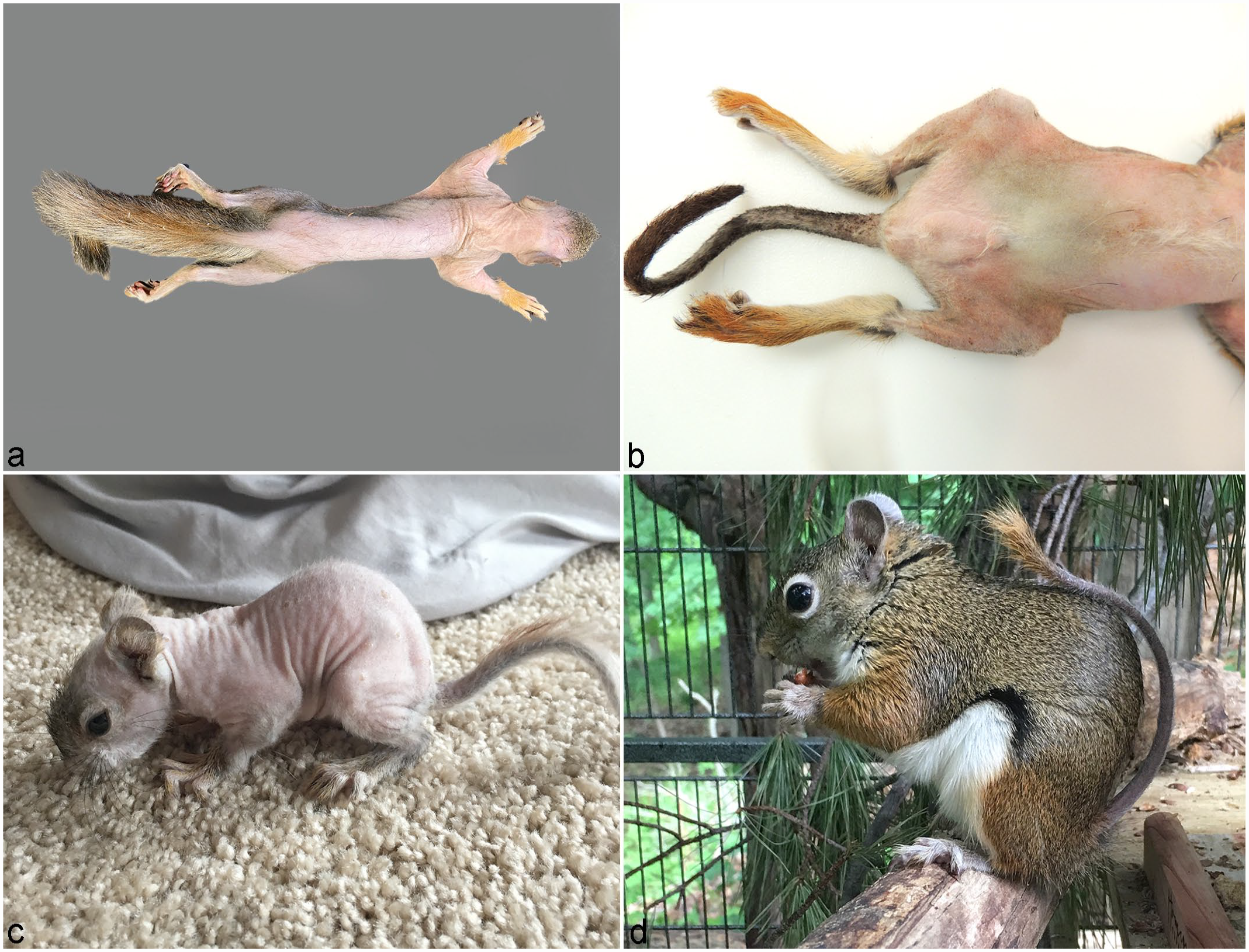
Hair follicle dysplasia, American red squirrel (Tamiasciurus hudsonicus). (a) Severe widespread alopecia with normal hair on the top of the head, paws, and tail; squirrel 6. (b) Severe widespread alopecia of the trunk, thighs, and tail with a sharp line of demarcation at the hock where a normal coat of hair remains distal to the hock; squirrel 1. (c) Juvenile American red squirrels (ARS) found in April 2018 with widespread alopecia of the trunk, neck, tail, and upper extremities. A few small crusts are present over the dorsal trunk; squirrel 8. (d) The same juvenile ARS as in (c) 2 months later. Normal-appearing hair covers the body with the exception of the tail.
Clinical data for 8 American red squirrels (Tamiasciurus hudsonicus) with hair follicle dysplasia.
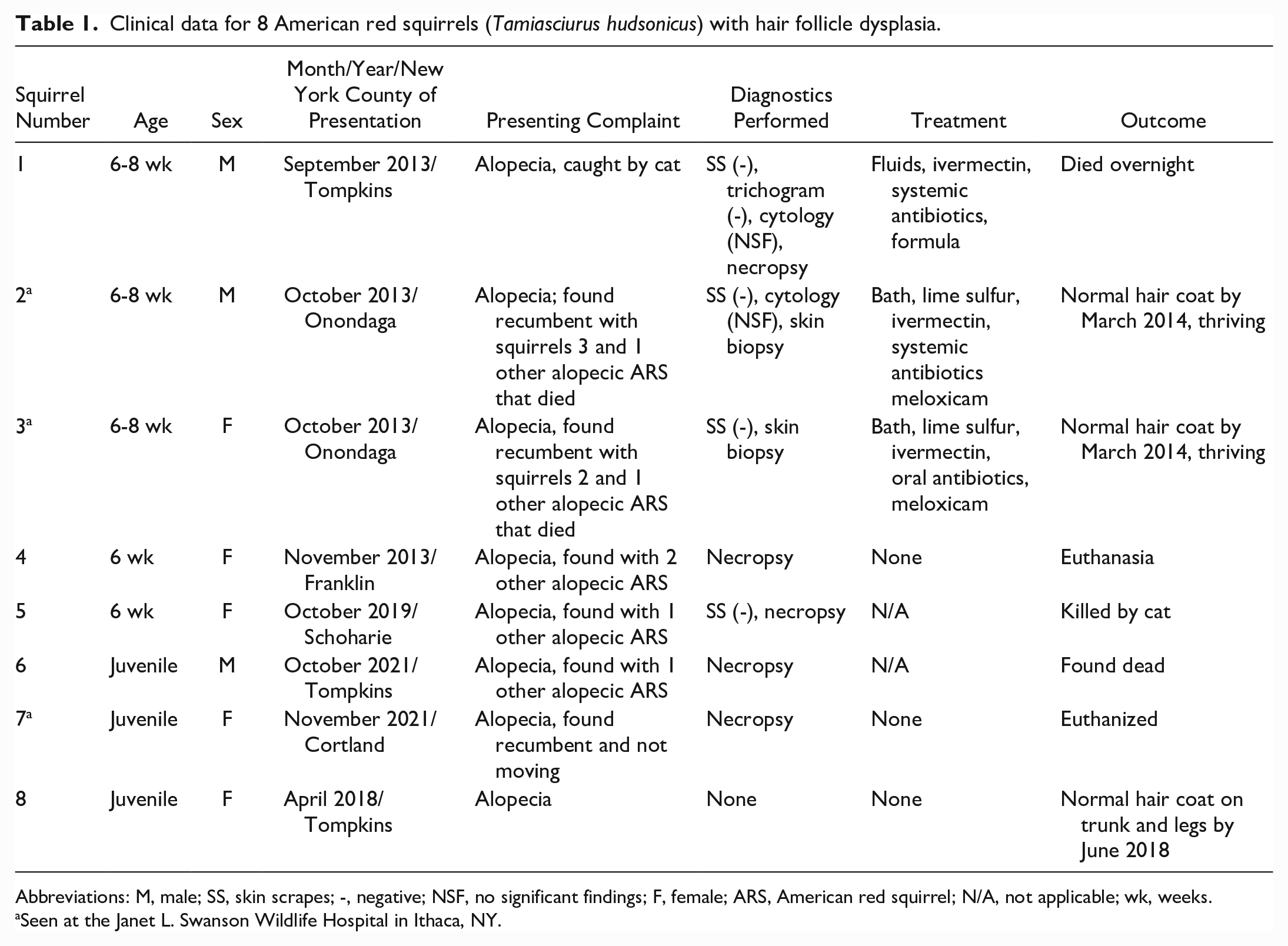
Abbreviations: M, male; SS, skin scrapes; -, negative; NSF, no significant findings; F, female; ARS, American red squirrel; N/A, not applicable; wk, weeks.
Seen at the Janet L. Swanson Wildlife Hospital in Ithaca, NY.
Complete necropsy examinations with histopathology were performed on squirrels 1 and 4-7. Necropsy findings for squirrel 1 include hepatic lipidosis, gastric mural abscess, and mild thymic atrophy. Squirrel 5 had a puncture wound to the skull and hemorrhage in the brain and thorax, consistent with cat predation. Both were in poor body condition. Other than the alopecia and hair shaft abnormalities, no other significant abnormalities were found on the gross or histologic examination of squirrel 4, 6, or 7. An eighth juvenile ARS was found in April (Fig. 1c), and by June, it had grown normal hair over its body with the exception of the tail (Fig. 1d). No skin biopsy specimens were obtained from this animal.
Haired skin from a normally haired ARS carcass was used to compare for the histopathologic features in these squirrels. Normal hairs from this animal had 1-µm diameter, round to 1 × 3-µm oval melanin granules in the medulla with smaller numbers of 1-µm-diameter granules in the cortex. In contrast, histopathologic evaluation of skin biopsies from 7 of 8 affected ARS showed changes consistent with follicular dysplasia. Hair follicles were in various stages of the hair cycle in all squirrels. All squirrels had at least small numbers of dysplastic hair shafts characterized by an irregular contour of the hair shaft, coiled or bent hair shafts, and/or melanin clumping distorting hair shafts (Fig. 2). Affected hair follicles were mildly dilated and bent, or coiled hairs within the lumen were occasionally oriented perpendicular to the follicle lumen, impinging on the outer root sheath and partially perforating the walls of follicles in squirrels 1, 4, 6, and 7 (Fig. 2). Rare malacic hair shafts were noted in squirrels 4 and 7. In squirrels 1, 6, and 7, some hair shafts were distorted by large clumps of melanin (Fig. 2d). Perifollicular, follicular epithelial, and epidermal melanin clumping were not present. In general, the skin was not inflamed, but there was mild inflammation in 2 squirrels consisting of small numbers of perifollicular lymphocytes and neutrophils and rare lymphocytic mural folliculitis in squirrel 3 and focal neutrophilic luminal folliculitis in squirrel 7. These two squirrels, and squirrel 1, also had a few small cellular crusts. Rare multinucleated giant cells were noted in the mid-dermis in squirrels 1 and 6. Mild epidermal hyperplasia was noted in squirrels 1-3 and 6.
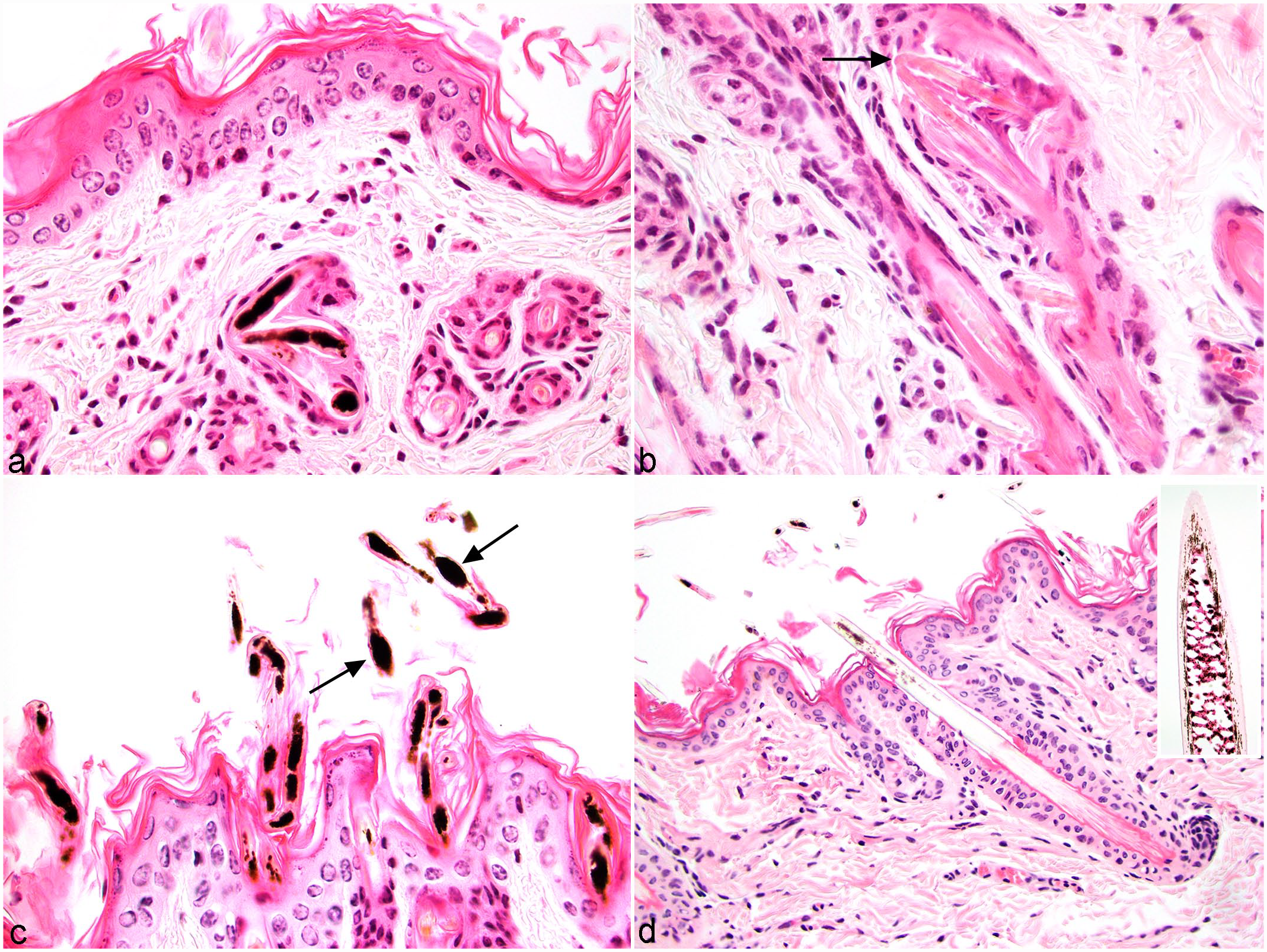
Hair follicle dysplasia, American red squirrel (ARS; Tamiasciurus hudsonicus), skin, squirrel 1. Hematoxylin and eosin. (a) Heavily pigmented coiled hair shaft within a hair follicle lumen. (b) Coiled hair shaft with early perforating folliculitis (arrow). (c) Multiple pigmented and bent hair shafts within the infundibulum of multiple follicles and on the surface. Large melanin clumps distort multiple hair shafts (arrows). (d) Normal haired skin from an unaffected ARS. The inset depicts a higher magnification of a normal hair shaft with small melanin granules within the medulla and cortex.
HFD make up a group of only partially understood genodermatoses with various causes and phenotypes. They have been described in multiple mammalian species including mice, dogs, cats, horses, cattle, and coatimundis.7–12,14,15,18 In dogs, HFDs often spare the head and distal extremities similar to the squirrels reported here. Although some forms of HFD may be congenital, in most cases, the animal is born with hair, but it is lost or partially lost in the first few years of life.9,11,13 Given the unknown history and young age of these animals, it is unclear if they had normal hair coats before presentation. Factors such as alopecia presenting at a young age (6-8 weeks or younger) and the information that 5 of the 8 animals were found with or in close proximity to 2 to 3 other ARS with a similar pattern of alopecia support a genetic etiology.
HFD can be divided into several large categories: color-linked follicular dysplasia, non-color-linked cyclic follicular dysplasia, and non-color-linked, noncyclic follicular dysplasia. The squirrels reported here have some features of all three. Three of the 7 squirrels in which a histopathology was performed had large melanin clumps in hair shafts leading to hair shaft distortion, similar to what is described in color dilution alopecia, black hair follicular dysplasia, and some other forms of HFD in dogs.5,7,8,11,18 Color dilution alopecia is described in color-dilute animals, and the squirrels reported herein are red, a dilute coat color. Most of the squirrels in our study had bent or coiled hair shafts, which is not a histologic feature of canine color dilution alopecia or black hair follicular dysplasia. Coiled hair shafts within the hair canal of nude mice are thought to be a result of abnormal keratinization associated with Foxn1 mutations. 10
It is unknown if there is seasonal or cyclic component to the HFD in these squirrels. Two of the squirrels with documented follicular dysplasia (squirrels 2 and 3) were described to have had complete hair growth 6 months after rescue. A third ARS (squirrel 8) also had almost complete hair growth, with the exception of the tail, 2 months after being found. This squirrel did not have a skin biopsy performed, so we cannot confirm HFD in this case; however, the pattern of alopecia was the same. Because all other animals died or were euthanized, it is unknown if they too would have grown hair. Interestingly, we have had multiple personal communications regarding ARS with a similar alopecic presentations. In our series of cases, all squirrels presented between the months of September and November with the exception of one in April. Squirrels 2 and 3 grew a full coat by March, and squirrel 8 grew hair by June; all three growing a nearly complete hair coat in the spring. There may be a seasonality of the disorder; however, given the relatively small number of cases and the lack of knowledge of recurrence, this is speculation. It is also possible the alopecic syndrome is associated with age as all animals were juveniles. Furthermore, all ARS with hair growth (squirrels 2, 3, and 8) were released into the wild, so it is unknown if they experienced further alopecic events. Because all the animals were juvenile and we did not see a similar pattern of alopecia in any adult squirrels, it is possible this alopecia is a one-time event and does not recur. It is also likely that juvenile ARS with extensive areas of alopecia do not survive their first winter in a harsh cold climate.
Alopecia is permanent in most cases of HFD. However seasonal alopecia, also known as recurrent flank alopecia, occurs in dogs.5,11,12 The onset of alopecia usually occurs between late autumn and early spring, and hair usually regrows spontaneously within 3-4 months.5,11 Clinically the condition presents as noninflammatory, well-demarcated areas of bilateral flank alopecia often with hyperpigmentation. The cause is not known, but there are some breed predispositions in dogs, and thus, genetics likely play an important role. The seasonal nature of the condition during the winter months suggests that photoperiod may play a role in the pathogenesis as well. 3 A study grafting skin from affected dogs onto nude mice demonstrated that on a different host, the skin still has potential to regrow hair. This suggests that the hair follicles are functional, and there is a systemic source for the alopecia. 17 Histologically in dogs, hair follicles are distorted and may have a “witch’s” foot or “jellyfish” configuration that often lacks hair shafts. 5 This distortion of follicles along with follicular keratosis, sebaceous gland melanization, and plugs of melanin in sebaceous ducts are features of recurrent flank alopecia in dogs but were not features noted in this group of ARS. 5
In this report, we describe a unique syndrome of HFD in juvenile ARS that may be seasonal and is self-resolving in at least some cases. Although the pathogenesis of this condition is not understood, there is some evidence that these animals may grow a normal hair coat. Housing with a licensed wildlife rehabilitator may be necessary over the winter months while these animals grow hair during the cold weather. However, this condition likely predisposes young animals to a variety of adverse events including exposure to the elements and predation, which may decrease their chance of survival in the wild. Housing these animals during the winter or until their hair grows may be perpetuating this trait if it is inherited. Research is needed to further characterize the clinical outcome of this disorder and investigate a possible genetic basis.
Footnotes
Acknowledgements
We would like to thank Kate Isaacs for the photographs of squirrel 8, the licensed wildlife rehabilitators that contributed cases, the staff of the Janet L. Swanson Wildlife Hospital for caring for these animals, and the Anatomic Pathology faculty, staff, and residents at Cornell University, College of Veterinary Medicine, Animal Health Diagnostic Center. We would also like to extend a special thank you to Dr. Danny W. Scott for his careful and insightful review of this manuscript.
Declaration of Conflicting Interests
The author(s) declared no potential conflicts of interest with respect to the research, authorship, and/or publication of this article.
Funding
The author(s) received no financial support for the research, authorship, and/or publication of this article.