
Abstract
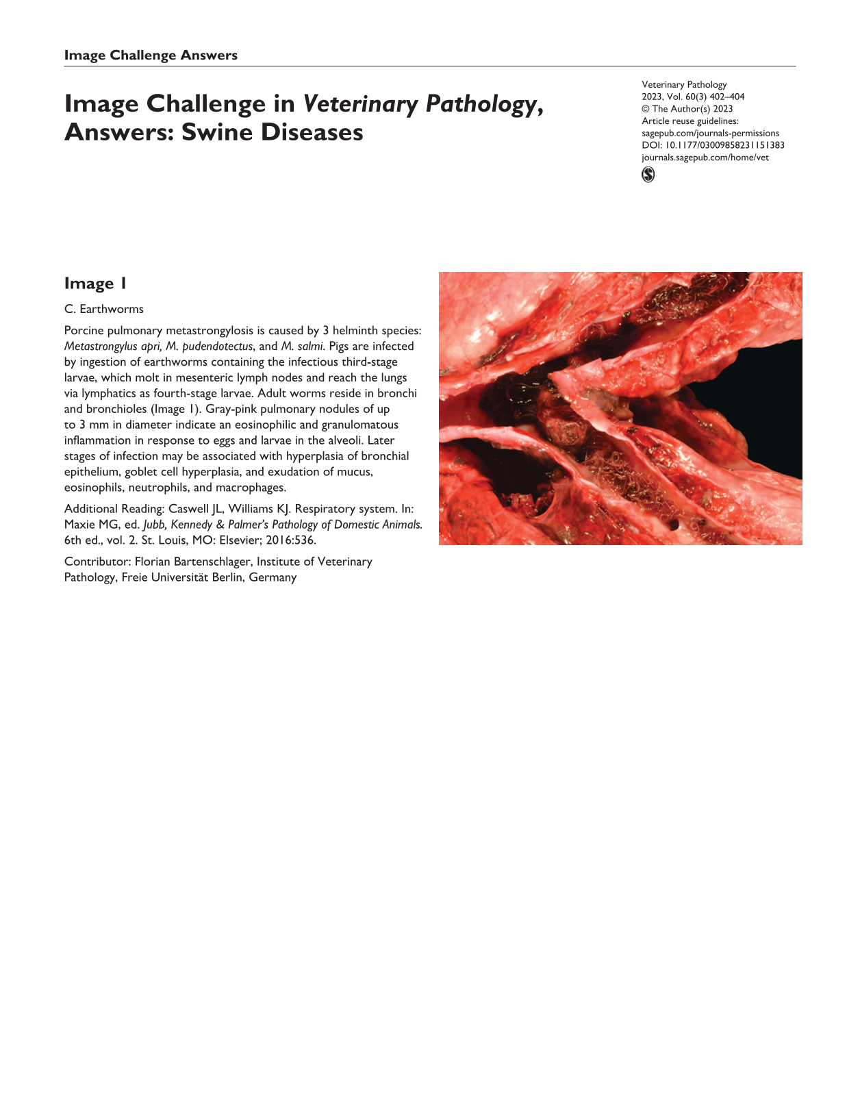
Image 1
C. Earthworms
Porcine pulmonary metastrongylosis is caused by 3 helminth species: Metastrongylus apri, M. pudendotectus, and M. salmi. Pigs are infected by ingestion of earthworms containing the infectious third-stage larvae, which molt in mesenteric lymph nodes and reach the lungs via lymphatics as fourth-stage larvae. Adult worms reside in bronchi and bronchioles (Image 1). Gray-pink pulmonary nodules of up to 3 mm in diameter indicate an eosinophilic and granulomatous inflammation in response to eggs and larvae in the alveoli. Later stages of infection may be associated with hyperplasia of bronchial epithelium, goblet cell hyperplasia, and exudation of mucus, eosinophils, neutrophils, and macrophages.
Additional Reading: Caswell JL, Williams KJ. Respiratory system. In: Maxie MG, ed. Jubb, Kennedy & Palmer’s Pathology of Domestic Animals. 6th ed., vol. 2. St. Louis, MO: Elsevier; 2016:536.
Contributor: Florian Bartenschlager, Institute of Veterinary Pathology, Freie Universität Berlin, Germany
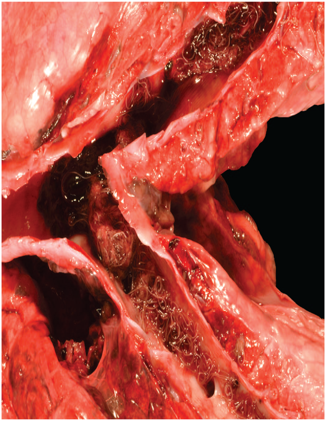
Image 2
A. Influenza A virus
Twenty 80-day-old pigs from a 50-pig lot presented with acute respiratory distress, nasal discharge, and lethargy. One pig was submitted for postmortem examination. Grossly, the lung had dark red firm areas (Image 2a). Histologically, there was attenuation and loss of the bronchiolar epithelium, accompanied by intraluminal neutrophils. Intraepithelial swine influenza virus antigen was detected by immunohistochemistry (Image 2b). Secondary bacterial infection was suspected due to the infiltration of neutrophils. As illustrated by this case, swine influenza is characterized by rapidly spreading respiratory disease with high morbidity, low mortality, and the hallmark histologic lesion of necrosis of the respiratory epithelium.
Additional Reading: Caswell JL, Williams KJ. Respiratory system. In: Maxie MG, ed. Jubb, Kennedy & Palmer’s Pathology of Domestic Animals. 6th ed., vol. 2. St. Louis, MO: Elsevier; 2016:526–527.
Contributor: Bianca Santana de Cecco, Setor de Patologia Veterinária, Universidade Federal do Rio Grande do Sul (UFRGS), Brazil
Photo credit: Francieli Adriane Molossi, UFRGS, Brazil
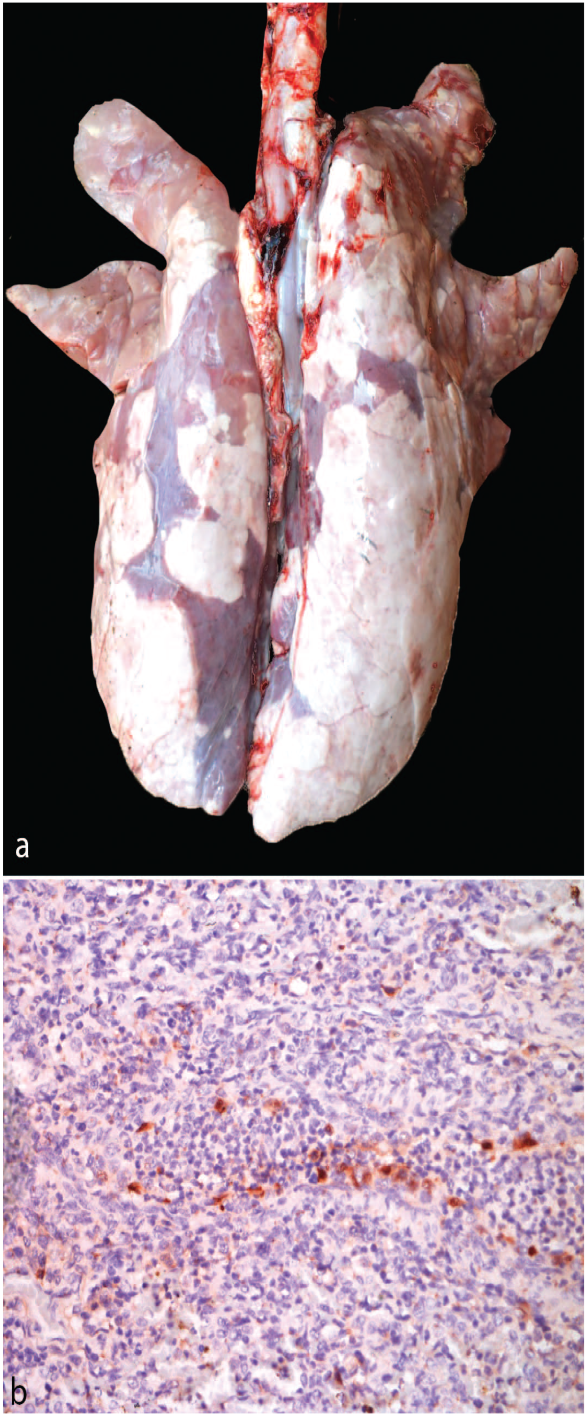
Image 3
A. Ascaris suum
Milk spotted liver is caused by the globally distributed and economically important nematode Ascaris suum. The infective eggs are ingested directly from contaminated environments or via paratenic hosts (worms and dung beetles). The larvae migrate from the intestine to the liver through the portal circulation. The white spots on the surface of the liver (Image 3) correspond histologically to migration tracks with fibrosis and eosinophilic inflammation. The larvae proceed to the pulmonary vasculature, infiltrate alveolar spaces, are coughed up, and then swallowed. The adult worms mature in the small intestine. Infected pigs may have slower growth rates, and severe infections can cause pneumonia.
Additional Reading: Thamsborg SM, Nejsum P, Mejer H. Impact of Ascaris suum in livestock. In: Holland C, ed. Ascaris: The Neglected Parasite. Cambridge, MA: Academic Press; 2013:363–381.
Contributor: Cody Rinker, Colorado State University
Photo Credit: Paula Schaffer, Colorado State University
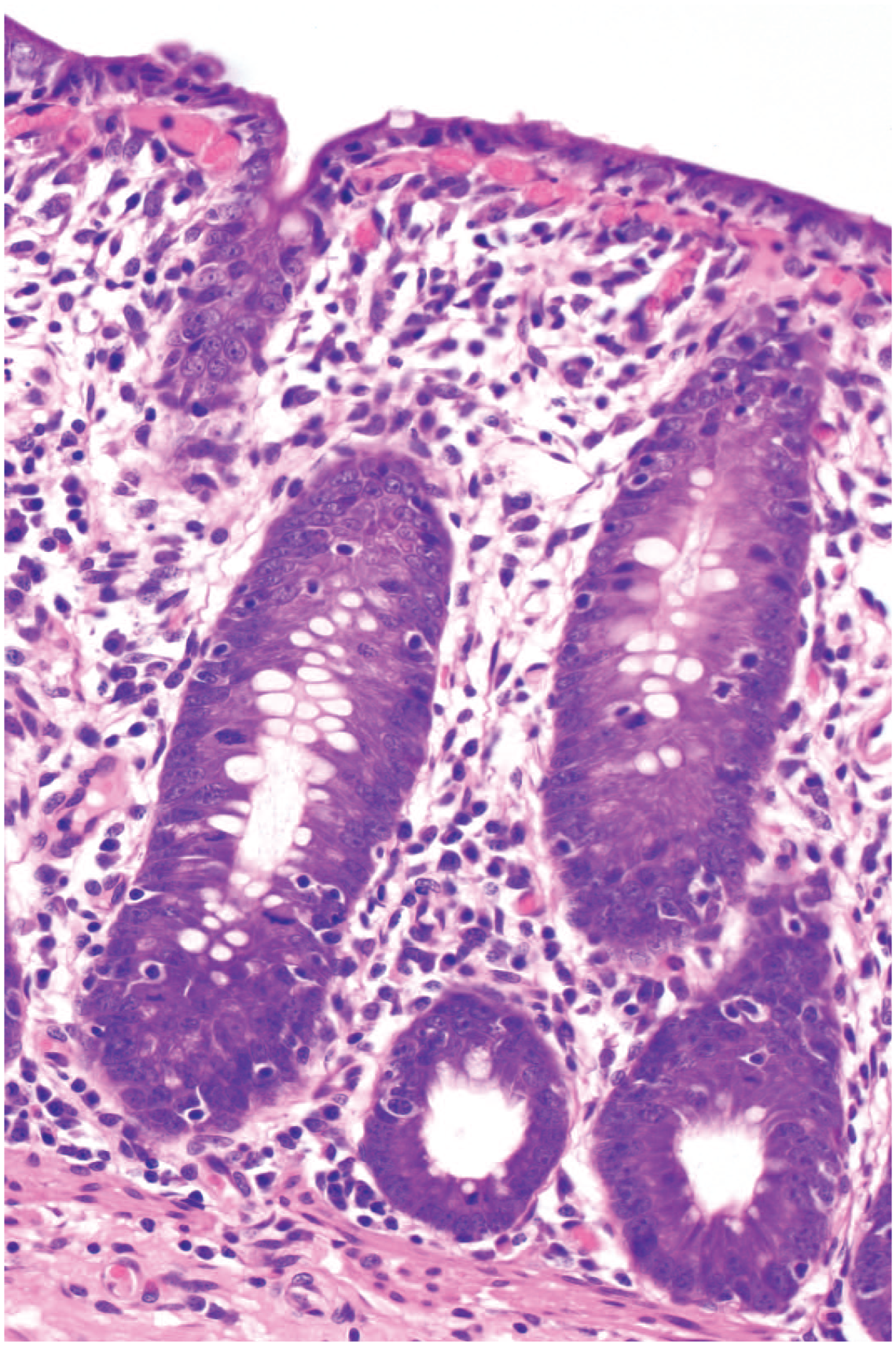
Image 4
B. Brachyspira pilosicoli
Brachyspira pilosicoli causes mucoid to watery diarrhea and colitis (porcine intestinal spirochetosis), which are milder than those caused by Brachyspira hyodysenteriae (swine dysentery). In the routinely stained section, spirochetes are visible in distended and elongated crypts with increased enterocyte mitoses and lymphocytic exocytosis (Image 4). The lamina propria is expanded by edema and increased lymphocytes and plasma cells. The location of the spirochetes within the colonic crypt lumen differentiates them from Lawsonia intracellularis, which are also highlighted by Warthin-Starry stain but are obligate intracellular bacteria. Helicobacter suis is a gastric spirochete. Clostridium perfringens are long thick rods.
Additional Reading: Hampson DJ, Burrough ER. Swine dysentery and brachyspiral colitis. In: Zimmerman JJ, Karriker LA, Ramirez A, et al., eds. Diseases of Swine. 11th ed. Hoboken, NJ: Wiley-Blackwell; 2019:951–970.
Contributor: Chun-Ming Lin, South Dakota State University
Veterinary Pathology invites submission of exceptional gross or microscopic images for consideration as an Image Challenge, along with a multiple-choice question and answer. For details, see the Instructions to Authors on the journal website.