
Abstract
Arteriolar lesions with lipid and/or amyloid deposits are frequently detected in canine gonads by routine histopathologic examination; however, they have never been examined in detail. In the present study, a total of 139 testes/epididymides and 200 ovaries from 72 male (4 months to 14 years old) and 105 female (7 months to 16 years old) dogs were examined for arteriolar lesions. Arteriolar lesions were detected in 21 of 72 male dogs (29%) and 54 of 105 female dogs (51%). These lesions were histologically classified into 4 types: “fibromuscular hypertrophy,” characterized by thickening of the tunica intima; “focal vasculitis,” characterized by mononuclear cell infiltration; “vacuolar change,” consisting of lipid accumulation and infiltration of foamy cells; and “hyalinosis,” characterized by irregular thickening with amyloid deposits. In the lesions of vacuolar change and hyalinosis, lipid deposition and infiltration of α-SMA-positive cells and Iba-1-positive cells were also observed. Foamy cells and amyloid deposits were immunopositive for apolipoproteins and oxidized low-density lipoprotein-related proteins. These results indicate that vacuolar change is possibly an early stage of atherosclerosis, and that amyloid may deposit as a consequence of the microenvironment associated with atherogenesis. Logistic regression analysis revealed that arteriolar lesions with lipid deposits were associated with age and interstitial cell tumors in male dogs, and with age in female dogs. Aging is likely an important risk factor of arteriolar lesions with lipid deposits of the canine gonads.
Atherosclerosis, one of the most common vascular diseases in humans, causes ischemic heart disease, and myocardial and cerebral infarction. It is not commonly seen in domestic animals with the exception of birds, rabbits, and pigs. 5,12,34,43 Spontaneous lesions of atherosclerosis have been reported in dogs with hypothyroidism and are thought to be associated with hypercholesterolemia. 18,24 In dogs with hypothyroidism, atherosclerotic lesions were observed systemically, affecting the heart, spleen, and urogenital organs. 14,21,22 Histopathologically, atherosclerotic lesions involved the tunica intima and media of elastic and muscular arteries and were characterized by deposition of lipid and infiltration of foamy cells. 21,22
Hyalinosis is histopathologically characterized by amorphous eosinophilic (hyaline) deposits that consist predominantly of fibrin or glycosaminoglycans or immunoglobulins, and less commonly amyloid. Hyalinosis has been observed in the intramural coronary arteries and the splenic arteries of dogs. 13,30,33 In hyalinosis of the splenic arteries, deposition of lipid and foamy macrophages comprised of apolipoprotein B are frequently detected, which may indicate that hyalinosis of the splenic arteries is a precursor of atherosclerosis. 33
Dogs are often neutered for population control and prevention or treatment of certain diseases that are associated with reproductive hormones. 38,42 In the testes and ovaries of dogs without hypothyroidism, atherosclerotic lesions are thought to be rare. 14 However, in the authors’ experience, routine histopathological examination of the extracted testes/epididymides and ovaries frequently reveals various types of arteriolar changes and deposition of lipid is occasionally detected in the affected arteriolar wall. Arteriolosclerosis in the testis/epididymis and ovary have been reported in buffalos, 11 pigs, 44 horses, 26 primates, 35 and humans, 35 but have never been reported in dogs. Histologically, the most common finding relevant to arteriolosclerosis in the testis/epididymis and ovary of these animals is fibrous hypertrophy of the tunica intima, which is thought to be related to reproductive activity and aging. 11,26,30,35,44
The aim of the present study was to characterize arteriolar lesions in the testes/epididymides and the ovaries of dogs, and to evaluate the relation between the presence of arteriolar lesions and other pathological changes. The findings suggest that some of the arteriolar lesions are associated with lipid deposition, comparable to atherosclerosis.
Materials and Methods
Sample Collection
One hundred and thirty-nine testes and epididymides and 200 ovaries from 72 male (0.42-14.1 years old) and 105 female (0.58-16.0 years old) dogs were reexamined from the sample archive of the Laboratory of Veterinary Pathology, the University of Tokyo. Samples were submitted for routine histopathological examination. Testes from 46 males and ovaries from 89 females were clinically considered to be normal and were resected for reproduction control and/or disease prevention purposes.
Histopathology
The testes/epididymides and the ovaries were fixed in 10% neutral-buffered formalin and embedded in paraffin. Four-micrometer-thick sections were stained with hematoxylin and eosin (HE) for detection of arteriolar lesions and general pathological examination. Further examinations of arteriolar lesions were performed with Elastica van Gieson, Congo red, and oil red O staining. The resultant slides were reviewed by 3 veterinary pathologists (NU, JKC, and KU).
Antisera for Canine Apolipoprotein B Preparation
Antigenic peptides of amino acid residues 143 to 158 (CRETLGPRGDAGSPRAL) of canine apolipoprotein (apo) B (XP_005630653) were synthesized and conjugated with keyhole limpet hemocyanin. The synthetic peptide was administered 4 times to 2 different conventional rabbits. Forty-nine days after the first administration, whole blood was collected, and the antisera were obtained (Cosmobio).
Immunohistochemistry
Immunohistochemistry (IHC) was performed to examine the distribution of lipid-laden foamy cells and apolipoproteins. After deparaffinization, antigen retrieval procedures were performed as described in Table 1. The primary antibodies for apolipoprotein (apo) A1, apoB, apoE, oxidized phospholipid (oxPL), and lectin-like oxidized LDL receptor 1 (LOX1) demonstrated reactivity with the appropriate molecular weight 7,8,10,15,17,22,39 by Western blot analysis (Supplemental Fig. S1). In order to inactivate endogenous peroxidase, the sections were immersed in 3% hydrogen peroxide in methanol for 5 minutes. Subsequently, the sections were washed with Tris-buffered saline (TBS), and then blocked with 8% skim milk in TBS at 37 °C for 40 minutes. The primary antibodies used in the present study are listed in Table 1. After incubation with a primary antibody at 4 °C overnight, the sections were incubated with the Dako Envision+ system horseradish peroxidase–labeled polymer anti-rabbit/mouse secondary antibody (Dako) or biotinylated rabbit anti-goat IgG antibody (Bethyl Laboratory Inc) at 37 °C for 40 minutes. After incubation with biotinylated rabbit anti-goat IgG antibody, the sections were incubated with streptavidin horseradish peroxidase conjugate (Dako) at 37 °C for 40 minutes. Immunolabeled antigens were visualized using 0.05% 3,3′-diaminobenzidine plus 0.03% hydrogen peroxide in a Tris-hydrochloric acid buffer and then counterstained with hematoxylin. Canine liver tissues with no pathological abnormalities and heart tissues with atherosclerotic lesions were used as positive controls. As negative controls, the primary antibodies were replaced with TBS.
Primary Antibodies Used for Immunohistochemistry.
Abbreviations: Apo, apolipoprotein; OxPL, oxidized phospholipid; LOX1, lectin-like oxidized low-density lipoprotein receptor-1; mAb, monoclonal antibody; pAb, polyclonal antibody.
Statistical Analysis
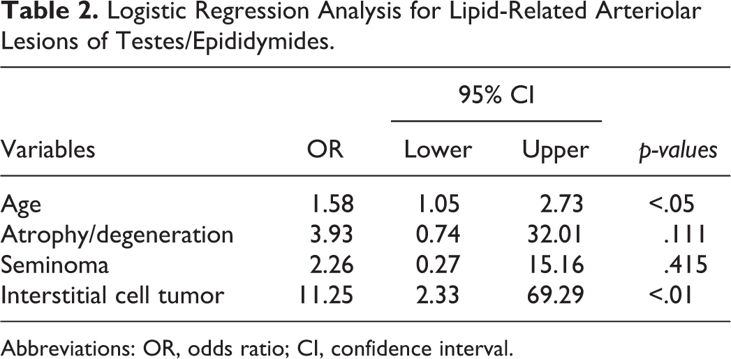
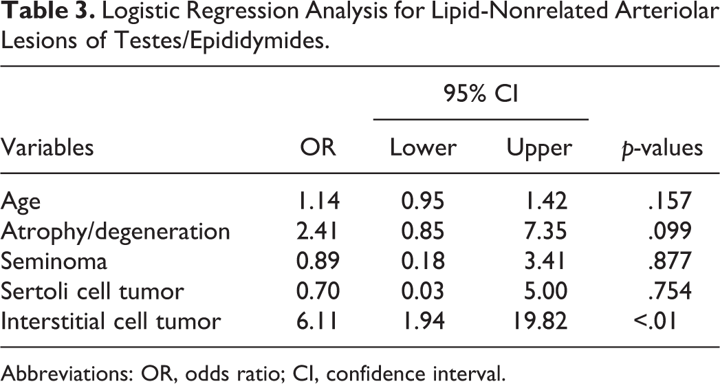
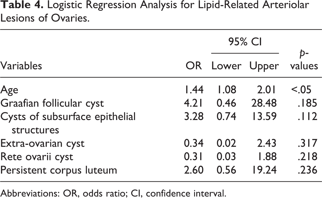
Statistical analysis was performed to assess the relation between the presence of arteriolar lesions with or without lipid deposits and other factors including age and other pathological findings. Arteriolar lesions with lipid deposits included hyalinosis and vacuolar change, whereas arteriolar lesions without lipid deposits included fibromuscular hypertrophy and vasculitis. Odds ratios (ORs) and the 95% profile likelihood-based confidence intervals (CI) were calculated using logistic regression analysis. Other pathological findings included age, seminiferous tubule atrophy and degeneration, seminoma, interstitial cell tumor, Sertoli cell tumor, and mix germ cell-sex cord-stromal tumor in the testes/epididymides; age, Graafian follicular cyst, cysts of subsurface epithelial structures, extra-ovarian cyst, rete ovarii cyst, corpus luteum cyst, persistent corpus luteum, adenoma/adenocarcinoma (epithelial cell tumor), and granulosa cell tumor in the ovaries. Age was included as a control variable in each of the logistic regression analyses. The binary variables were coded as 1 or 0 to represent the presence or absence of pathological findings except for age. Age was treated as a continuous variable. A P value was calculated by likelihood ratio test and that of <.05 was considered statistically significant. All statistical analyses were carried out using R statistical software (version 4.0.2; R Foundation for Statistical Computing).
Results
Histopathological Findings Other Than Arteriolar Lesions
Evaluation of 139 testes and epididymides samples from 72 male dogs revealed diagnoses of seminiferous tubule atrophy and degeneration (n = 68), orchitis and epididymitis (n = 1), necrosis (n = 1), seminoma (n = 20), Sertoli cell tumor (n = 8), interstitial cell tumor (n = 20), and mixed germ cell-sex cord-stromal tumor (n = 2; Supplemental Table S1). Evaluation of 200 ovaries sampled from 105 female dogs revealed diagnoses of Graafian follicular cyst (n = 13), cysts of subsurface epithelial structures (n = 40), extra-ovarian cyst (n = 33), rete ovarii cyst (n = 41), corpus luteum cyst (n = 10), persistent corpus luteum (n = 130), epithelial cell tumor (n = 4), and granulosa cell tumor (n = 7; Supplemental Table S2).
Histopathology of Arteriolar Lesions in Testes/Epididymides of Dogs
Arteriolar lesions in the testis/epididymis were classified into 4 types according to their histopathological features: fibromuscular hypertrophy, vasculitis, vacuolar changes, and hyalinosis (Figs. 1–6). Fibromuscular hypertrophy was characterized by thickening of the tunica intima with collagen fiber and smooth muscle (Figs. 1, 2). Vasculitis was characterized by infiltration of mononuclear cells in the arteriolar wall (Fig. 3). In severe lesions of vasculitis, the arteriolar layers were obscured (Fig. 4). Vacuolar change was characterized by thickening of the tunica intima with infiltration of oil red O-positive lipid-laden foamy cells and deposition of oil red O-positive granules (Figs. 5a, 5b). In some lesions with vacuolar change, the internal elastic lamina was disrupted or duplicated, and the tunica media was severely thin (Fig. 5c). Hyalinosis was characterized by irregular thickening of the tunica intima with Congo red–positive amyloid deposits (Figs. 6a, 6b). Oil red O-positive granules were colocalized with amyloid deposits (Fig. 6c). The internal elastic lamina was disrupted or duplicated in the lesions of hyalinosis (Fig. 6d). Fibromuscular hypertrophy, hyalinosis, and vacuolar change were observed in both the epididymis and testis, but vasculitis was only observed in the epididymis. Fibromuscular hypertrophy was found in 13 of 72 dogs (18%), which was unilateral in all 13 dogs. Vasculitis was found in 10 of 72 dogs (14%), which was unilateral in 8 of the 10 dogs and bilateral in 2 dogs. Vacuolar change was found in 2 of 72 dogs (3%), which was unilateral in 2 dogs. Hyalinosis was found in 5 of 72 dogs (7%), which was unilateral in 3 of the 5 dogs and bilateral in the other 2 dogs.
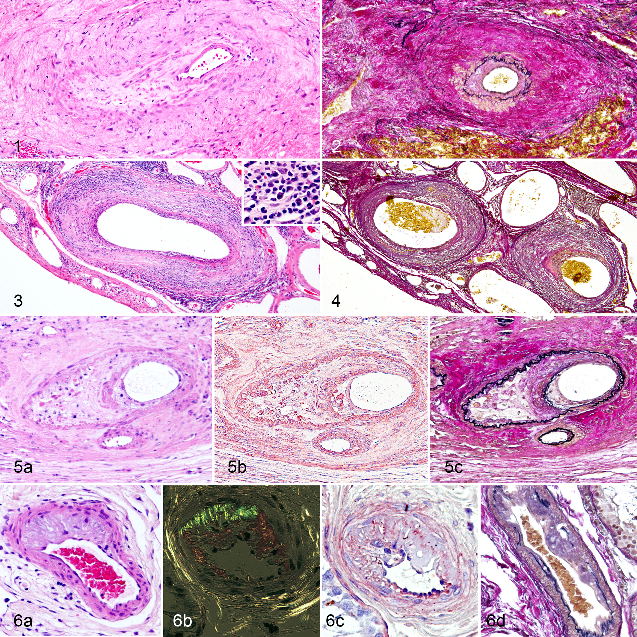
Fibromuscular hypertrophy, testis, dog, case 3.
Histopathology of Arteriolar Lesions in Ovaries of Dogs
Arteriolar lesions in the ovary were classified into 4 types according to their histopathological features: fibromuscular hypertrophy, vasculitis, vacuolar changes, and hyalinosis (Figs. 7–10). Fibromuscular hypertrophy was characterized by irregular thickening of the tunica intima and the internal elastic lamina with loose elastic fiber and collagen fiber (Figs. 7a, 7b). Vasculitis was characterized by infiltration of mononuclear cells within the arteriolar wall (Fig. 8a). In severe lesions of vasculitis, the arteriolar layers were obscured (Fig. 8b). Vacuolar change was characterized by deposits of oil red O–positive granules in the tunica intima and tunica media (Figs. 9a, 9b). The internal elastic lamina was disrupted or duplicated in the lesions of vacuolar change (Fig. 9c). Hyalinosis was characterized by irregular thickening of the tunica intima with Congo red–positive amyloid deposits (Figs. 10a, 10b). Oil red O–positive granules were colocalized with amyloid deposits (Fig. 10c). The internal elastic lamina was disrupted or duplicated in the lesions of hyalinosis (Fig. 10d). Fibromuscular hypertrophy was most frequently observed in the 4 types of arteriolar lesions in 50 of 105 dogs (48%). The lesion was detected unilaterally in 28 dogs (27%) and bilaterally in 22 dogs (21%). Vasculitis was found in 2 of 105 dogs (2%) and was detected unilaterally in both dogs. Vacuolar change was found in 7 of 105 dogs (7%). The lesion was found unilaterally in 5 dogs and bilaterally in 2 dogs. Hyalinosis was found in 3 of 105 dogs (6%) and was detected bilaterally.
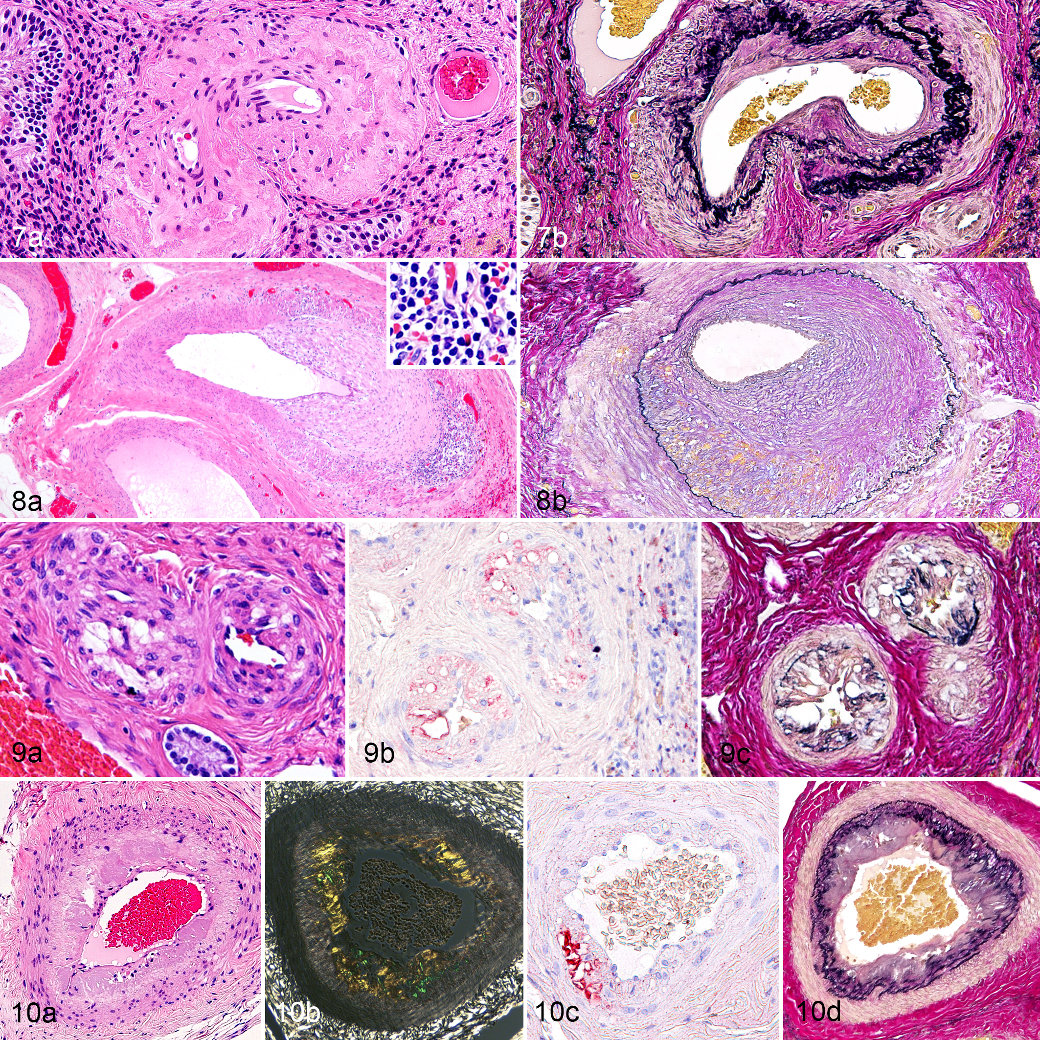
Fibromuscular hypertrophy, ovary dog, case 60. (a) Thickening of tunica intima. Hematoxylin and eosin (HE). (b) Increased collagen, hyperplasia/hypertrophy of smooth muscle cells in the tunica intima, and multiplication of elastic fibers in internal elastic lamina. Elastica van Gieson (EVG).
Immunohistochemical Analysis of Vacuolar Changes and Hyalinosis
Cells infiltrating the tunica intima were immunopositive for α-SMA and Iba-1 (Figs. 11–14) and some of those cells were swollen and vacuolated (foamy cells). The number of Iba-1-positive foamy cells in vacuolar change of the testes was larger than that of α-SMA-positive foamy cells (Fig. 11), whereas the number of α-SMA-positive foamy cells in vacuolar change of the ovaries was larger than that of Iba-1-positive foamy cells (Fig. 13). There were a few swollen and vacuolated cells that were immunopositive for Iba-1 and α-SMA within the lesions of amyloid deposits (Figs. 12, 14). Immunoreactivity for apoA1, apoE, apoB, oxPL, and LOX1 were observed in amyloid deposits and subendothelial space of the tunica intima (Figs. 15–18). Strong cytoplasmic immunoreactivity for apoA1, apoE, apoB, oxPL, and LOX1 were observed in the infiltrated foamy cells of the tunica intima in ovaries, while foamy cells in the testes were positive for apoE and negative for apoA1, apoB, oxPL, and LOX1 (Figs. 15, 17). The staining intensity for apoE and apoB was greater in amyloid deposits than that for apoA1 (Figs. 16a–c, 18a–c).
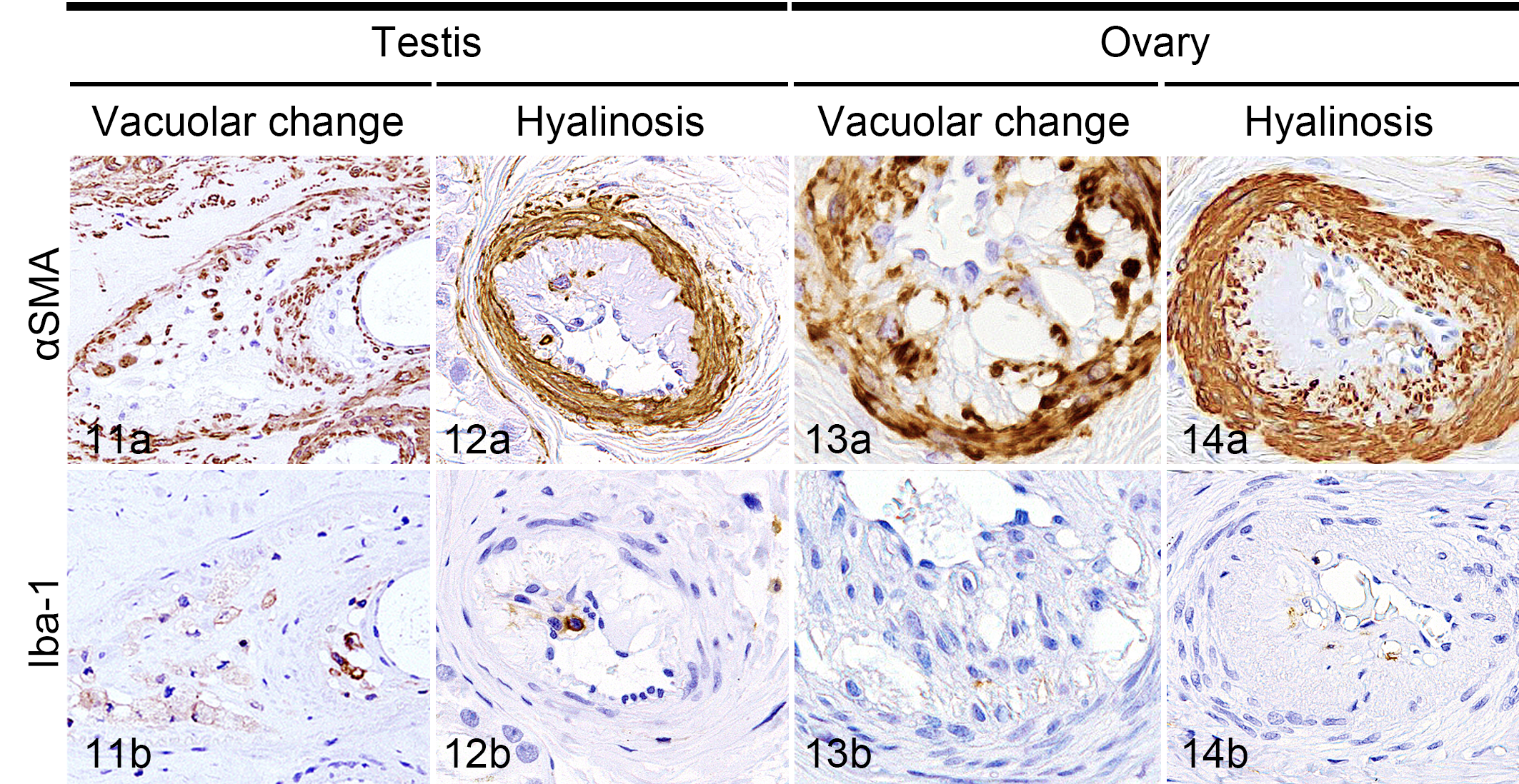
Immunohistochemistry for (a) α-SMA and (b) Iba-1, hematoxylin counterstaining.
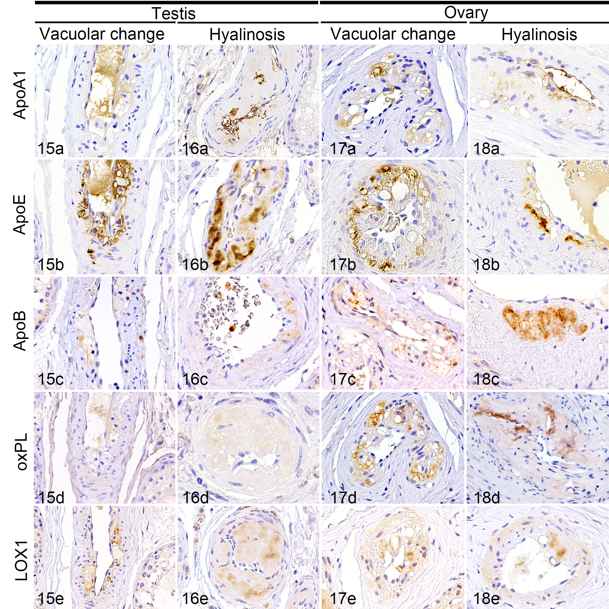
Immunohistochemistry for (a) apolipoprotein (apo) A1, (b) apoE, (c) apoB, (d) oxidized phospholipid (OxPL), and (e) lectin-like oxidized LDL receptor 1 (LOX1), hematoxylin counterstaining.
Statistical Evaluation
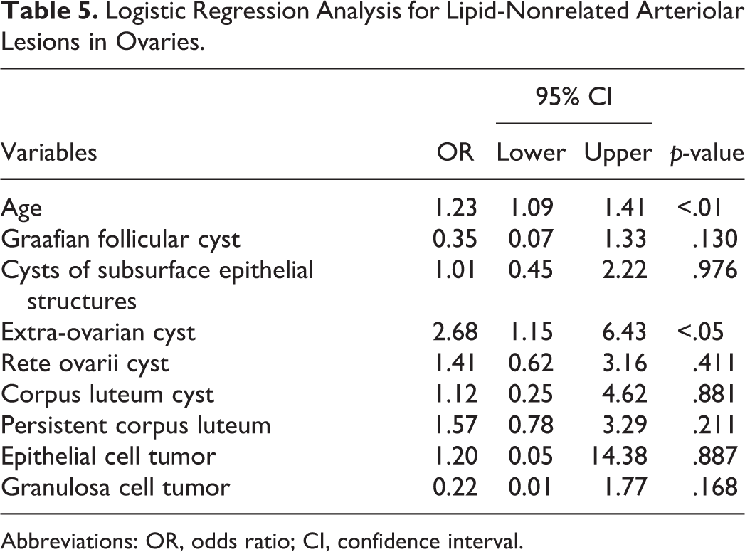
Logistic regression analysis revealed that age (p < .05, OR = 1.58, 95% CI = [1.05, 2.73]) and interstitial cell tumor (p < .01, OR = 11.25, 95% CI = [2.33, 69.29]) were associated with hyalinosis and vacuolar change (Table 2). In addition, interstitial cell tumor (p < .01, OR = 6.11, 95% CI = [1.94, 19.82]) was also associated with fibromuscular hypertrophy and vasculitis (Table 3). As for the ovaries, age (p < .05, OR = 1.44, 95% CI = [1.08, 2.01]) was associated with hyalinosis and vacuolar change (Table 4). Age (p < .01, OR = 1.23, 95% CI = [1.09, 1.41]) and extra-ovarian cyst (p < .05, OR = 2.68, 95% CI = [1.15, 6.43]) were associated with fibromuscular hypertrophy and vasculitis (Table 5).
Logistic Regression Analysis for Lipid-Related Arteriolar Lesions of Testes/Epididymides.
Abbreviations: OR, odds ratio; CI, confidence interval.
Logistic Regression Analysis for Lipid-Nonrelated Arteriolar Lesions of Testes/Epididymides.
Abbreviations: OR, odds ratio; CI, confidence interval.
Logistic Regression Analysis for Lipid-Related Arteriolar Lesions of Ovaries.
Abbreviations: OR, odds ratio; CI, confidence interval.
Logistic Regression Analysis for Lipid-Nonrelated Arteriolar Lesions in Ovaries.
Abbreviations: OR, odds ratio; CI, confidence interval.
Discussion
The present study focused on the histopathological features of arteriolar lesions in the testes/epididymides and the ovaries of dogs and classified the lesions into 4 types: fibromuscular hypertrophy, vasculitis, vacuolar change, and hyalinosis. Fibromuscular hypertrophy was more common in ovaries than in testes/epididymides. Multiplication of the internal elastic lamina was more remarkable in the ovaries than in the testes/epididymides. These features were similar to the arteriolar lesions in the reproductive organs of buffalos, pigs, equines, primates, and humans. 11,26,35,44 Some reports have indicated that arteriolosclerosis in the ovaries and the uteri may be associated with the reproductive activity of the animal. 11,26,30,35,44 In the present study, logistic regression analysis revealed that age and extra-ovarian cyst were significantly related to the presence of arteriolar lesions without lipid deposits in the ovaries. Other hormone-producing lesions (Graafian follicular cyst, corpus luteum cyst, persistent corpus luteum, and granulosa cell tumor) were not related to the presence of arteriolar lesions without lipid deposits. Extra-ovarian cysts usually do not produce sex hormones; 4,25 therefore, this may indicate that sex hormones are not related to fibromuscular hypertrophy and vasculitis of the ovaries.
Vasculitis was more common in the epididymides than in the ovaries. Various degrees of mononuclear cell infiltration were observed in the arteriolar walls of epididymides. In severe lesions, necrosis and fibrosis of the arteriolar wall were found, resulting in loss of the arteriolar layers. Idiopathic canine polyarteritis, or canine juvenile polyarteritis (beagle pain syndrome) often involved the thymus, the heart, and the epididymis in beagles. 16,32,36,37 In the present study, none of the dogs with vasculitis showed any signs related to idiopathic canine polyarteritis, or canine juvenile polyarteritis. It remains unknown whether the vascular inflammation affected other organs of these animals.
Diffuse intimal thickening consisting of the infiltration of activated smooth muscle cells is considered to be a precursor lesion of atherosclerosis in humans. 2,6 During the progression from diffuse intimal thickening to atherosclerosis, vascular smooth muscle cells decreased as macrophage infiltration increased. 27 Oxidized low-density lipoprotein (oxLDL), which contains oxPL, induces endothelial cells, smooth muscle cells, and macrophages to upregulate LOX1, which results in increased uptake of oxLDL and consequently apoptosis. 3,9,45 In the present study, foamy smooth muscle cells in the vacuolar change of ovaries exhibited cytoplasmic immunoreactivity for apoA1, apoE, apoB, oxPL, and LOX1, but foamy macrophages in the testes/epididymides only for apoE. These results might indicate that vacuolar change with intimal foamy smooth muscle cells is a precursor of atherosclerotic plaques similar to humans. Smooth muscle cells may upregulate the expression of LOX1 and induce apoptosis by up-taking of oxLDL. After apoptosis of smooth muscle cells, macrophages increasingly might infiltrate and contribute to form mature atherosclerotic plaques.
In certain types of amyloidosis, including nonhereditary apoA1-associated amyloidosis, amyloid deposits are detected in atherosclerotic plaques. 19,29,31 It is speculated that proteolysis of apoA1-containing HDL by protease secreted by foamy cells is involved in the pathogenesis of apoA1-associated amyloidosis. 31 In the present study, deposition of lipid and infiltration of foamy smooth muscle cells and macrophages were detected in amyloid deposits. Immunohistochemistry revealed that amyloid deposits as well as the foamy smooth muscle cells in the vacuolar change of the ovaries were immunopositive for apoA1, apoE, apoB, oxPL, and LOX1. These results suggest that hyalinosis may be associated with vacuolar change of the ovaries. Some amyloid precursor proteins, such as apoA1, may have undergone proteolysis and formed amyloid fibrils during progression of vacuolar change in the arterioles of the ovary.
Logistic regression analysis revealed that the presence of arteriolar lesions with lipid deposits, hyalinosis, and vacuolar change was associated with age and interstitial cell tumor in the testes/epididymides and with age in the ovaries. In addition, the presence of arteriolar lesions without lipid deposits, fibromuscular hypertrophy, and vasculitis was associated with interstitial cell tumor in the testes/epididymides and with age and extra-ovarian cyst in the ovaries. Aging is one of the risk factors of atherosclerosis and cellular senescence is associated with atherosclerosis. 41 Therefore, it is reasonable to assume that cellular senescence, including increase of reactive oxygen species and decrease of endothelial proliferation, is involved in the pathogenesis of arteriolar lesions with age in the ovaries. Although the relevance of extra-ovarian cysts and arteriolar lesions in the ovary is unknown, dilated cysts may compress the surrounding vessels and alter blood pressure, which may cause hyperplastic change of the arteriolar wall. 4,25
A previous study showed that dogs with interstitial cell tumor had lower serum concentration of testosterone than dogs with normal testes. 28 Testosterone deficiency in men is associated with obesity, metabolic syndrome, type 2 diabetes, and atherosclerosis. 20,23 Although serum testosterone levels were not measured in the present dogs, imbalance of sex hormones may be associated with the development of arteriolar lesions with lipid deposits including vacuolar change and hyalinosis. The presence of interstitial cell tumor was not only associated with the presence of vacuolar change and hyalinosis but also with other lesions such as fibromuscular hypertrophy and vasculitis. Interstitial cell tumors cause hypervascularity combined with testicular feeding vessels and increase peripheral and circumferential blood flow of the testis in humans. 1,40 In dogs, microscopic hypervascularity is commonly observed in testis with interstitial cell tumor. Hypervascularity and increase of blood flow may increase blood pressure and damage endothelial cells. As a consequence, endothelial dysfunction may promote arteriolar lesions with/without lipid deposits.
In conclusion, hyalinosis and vacuolar change in the testes/epididymides and ovaries are characterized by deposition of lipid and proteins related to atherosclerosis. Amyloid deposits were detected in these lesions, possibly due to proteolysis of amyloid precursor proteins in association with aberrant lipid metabolism. These arteriolar lesions with lipid deposits were associated with age and interstitial cell tumor in the dogs.
Supplemental Material
Supplemental Material, sj-pdf-1-vet-10.1177_0300985821996670 - Age-Related Arteriolar Changes With Lipid and Amyloid Deposits in the Gonads of Dogs
Supplemental Material, sj-pdf-1-vet-10.1177_0300985821996670 for Age-Related Arteriolar Changes With Lipid and Amyloid Deposits in the Gonads of Dogs by Nanako Ushio, James K. Chambers, Kenichi Watanabe, Mitsunori Kayano and Kazuyuki Uchida in Veterinary Pathology
Footnotes
Declaration of Conflicting Interests
The author(s) declared no potential conflicts of interest with respect to the research, authorship, and/or publication of this article.
Funding
The author(s) disclosed receipt of the following financial support for the research, authorship, and/or publication of this article: The present study was supported by Grant-in-Aid for JSPS Research Fellow (17J09424).
Supplemental material for this article is available online.
References
Supplementary Material
Please find the following supplemental material available below.
For Open Access articles published under a Creative Commons License, all supplemental material carries the same license as the article it is associated with.
For non-Open Access articles published, all supplemental material carries a non-exclusive license, and permission requests for re-use of supplemental material or any part of supplemental material shall be sent directly to the copyright owner as specified in the copyright notice associated with the article.