Abstract
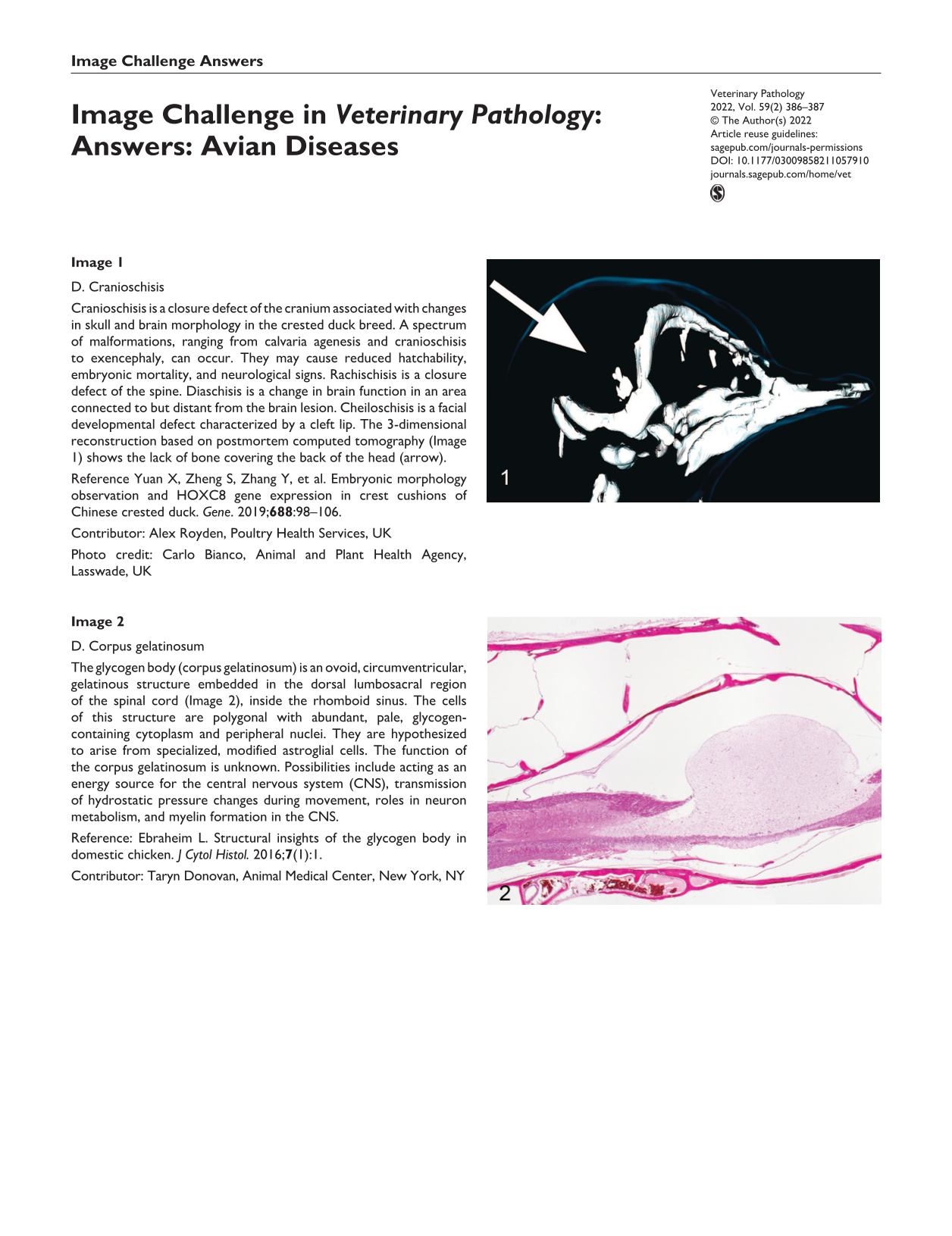
Image 1
D. Cranioschisis
Cranioschisis is a closure defect of the cranium associated with changes in skull and brain morphology in the crested duck breed. A spectrum of malformations, ranging from calvaria agenesis and cranioschisis to exencephaly, can occur. They may cause reduced hatchability, embryonic mortality, and neurological signs. Rachischisis is a closure defect of the spine. Diaschisis is a change in brain function in an area connected to but distant from the brain lesion. Cheiloschisis is a facial developmental defect characterized by a cleft lip. The 3-dimensional reconstruction based on postmortem computed tomography (Image 1) shows the lack of bone covering the back of the head (arrow).
Yuan X, Zheng S, Zhang Y, et al. Embryonic morphology observation and HOXC8 gene expression in crest cushions of Chinese crested duck. Gene. 2019;
Contributor: Alex Royden, Poultry Health Services, UK
Photo credit: Carlo Bianco, Animal and Plant Health Agency, Lasswade, UK

Image 2
D. Corpus gelatinosum
The glycogen body (corpus gelatinosum) is an ovoid, circumventricular, gelatinous structure embedded in the dorsal lumbosacral region of the spinal cord (Image 2), inside the rhomboid sinus. The cells of this structure are polygonal with abundant, pale, glycogen-containing cytoplasm and peripheral nuclei. They are hypothesized to arise from specialized, modified astroglial cells. The function of the corpus gelatinosum is unknown. Possibilities include acting as an energy source for the central nervous system (CNS), transmission of hydrostatic pressure changes during movement, roles in neuron metabolism, and myelin formation in the CNS.
Ebraheim L. Structural insights of the glycogen body in domestic chicken. J Cytol Histol. 2016;
Contributor: Taryn Donovan, Animal Medical Center, New York, NY
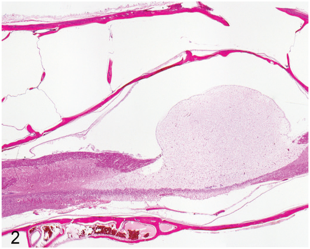
Image 3
A. Bornavirus
This is lymphoplasmacytic ganglioneuritis in the crop caused by avian bornavirus (proventricular dilatation disease). Also known as macaw wasting disease, this virus is neurotropic and affects autonomic ganglia of the upper and middle digestive tract. Captive psittacines are most often affected. Flaccidity and dilation of the proventriculus and/or crop are common gross findings (Image 3). The diagnosis is made by immunohistochemistry for avian bornavirus, which is 100% specific and sensitive in sections of brain, spinal cord, and adrenal glands with lymphoplasmacytic infiltrate similar to that seen in the myenteric ganglia of the digestive tract.
Raghav R, Taylor M, Delay J, et al. Avian bornavirus is present in many tissues of psittacine birds with histopathologic evidence of proventricular dilatation disease. J Vet Diagn Invest. 2010;
Contributor: Ariel Carlson, University of Tennessee
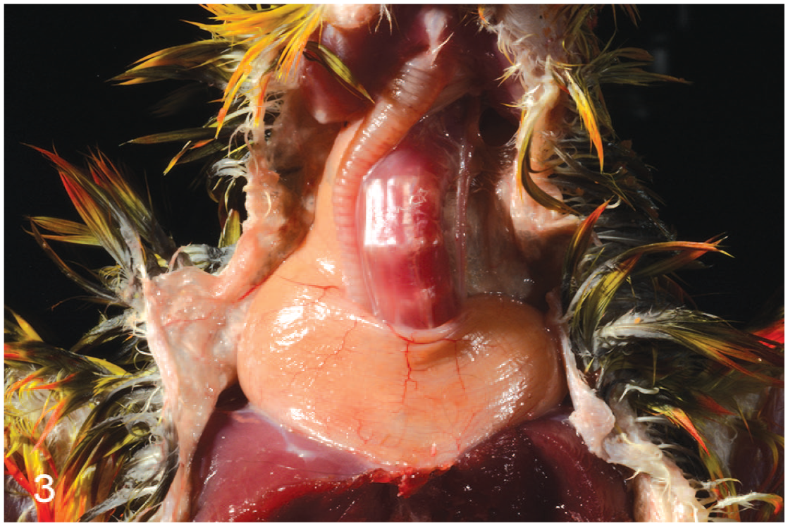
Image 4
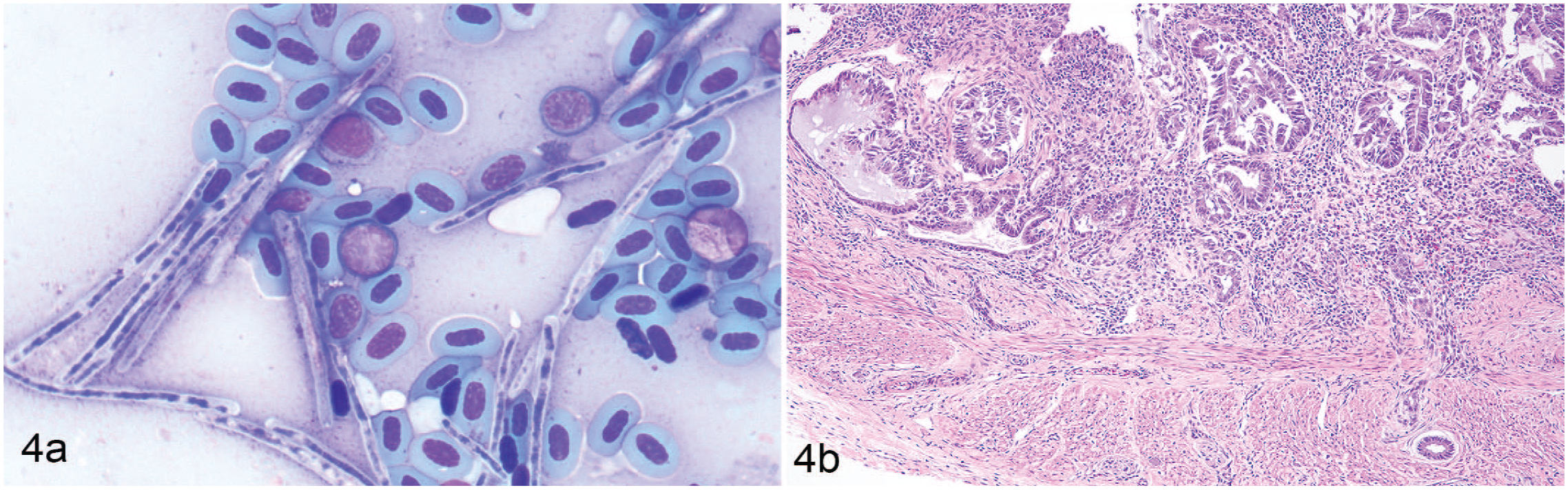
B. Melena
The black intestines and anemic pallor of the lung and liver indicate melena. This budgerigar had Macrorhabdus ornithogaster diagnosed by cytology (Image 4A) and proventricular adenocarcinoma diagnosed by histopathology (Image 4B) leading to gastrointestinal bleeding. M. ornithogaster is an avian gastric yeast affecting the proventricular-ventricular junction of psittacines, and infection has been linked to proventricular adenocarcinoma. The bright yellow fat is due to normal dietary carotenoids. Birds lack biliverdin reductase, the enzyme that converts biliverdin to bilirubin, and therefore are unable to develop icterus. Melanosis and pseudomelanosis would be unlikely to affect only the intestines, and would not explain the anemic pallor.
Powers LV, Mitchell MA, Garner MM. Macrorhabdus ornithogaster infection and spontaneous proventricular adenocarcinoma in budgerigars (Melopsittacus undulatus). Vet Pathol. 2019;
Contributor: Camille Cordero-Aponte, University of Tennessee
Veterinary Pathology invites submission of exceptional gross or microscopic images for consideration as an Image Challenge, along with a multiple choice question and answer. For details, see the Instructions to Authors on the journal website.