
Abstract
Given their genetic and anatomic similarities to humans, nonhuman primates (NHPs) may serve as animal models for urogenital diseases of humans. The purpose of this study was to examine the frequency of spontaneous urogenital lesions occurring over a 30-year period at the Yerkes and Southwest National Primate Research Centers and to compare and contrast lesions occurring in Old World versus New World primates. Lesions occurring in the chimpanzee (Pan troglodytes), baboon (Papio spp.), rhesus macaque (Macaca mulatta), cynomolgus macaque (Macaca fascicularis), pig-tailed macaque (Macaca nemestrina), sooty mangabey (Cercocebus atys), common marmoset (Callithrix jacchus), cotton-top tamarin (Sanguinus oedipus), and squirrel monkey (Saimiri sciureus) are discussed. The most common lesions of the kidney were medullary amyloidosis, renal cysts, renal tubular degeneration, glomerulonephritis or glomerulopathy, nephritis, nephrocalcinosis, pyelonephritis, and hydronephrosis. Specific causes of renal tubular disease included pigmentary nephrosis and tubular lipidosis. Renal tumors, including renal adenoma and carcinoma, lymphoma, and nephroblastoma, were infrequent diagnoses in all species. Endometriosis was the most frequently diagnosed lesion of the female genital tract. Of the animals examined in this study, it was most frequent in Old World primates. Leiomyoma was the most common uterine tumor. Granulosa cell tumor was the most frequently observed neoplasm of the ovaries, followed by teratoma. Of animals included in the study, most ovarian tumors occurred in baboons. Neoplasms of the male reproductive tract included interstitial cell tumor, seminoma, penile squamous cell carcinoma, penile papilloma, and histiocytoma. In New World monkeys, renal lesions were reported more frequently than genital lesions.
Keywords
Nonhuman primates (NHPs) are used as laboratory animal models in biomedical, pharmacological, and toxicological studies. The number of NHPs used in research studies has continued to rise annually since 2014, with rhesus and cynomolgus macaques being the most commonly used species. 24 Diseases of the urinary and reproductive tracts are a significant cause of morbidity and mortality in humans worldwide; urologic diseases and reproductive health are among the top primary research areas for studies involving NHPs. 24,49 In order to effectively utilize these invaluable animals in research, it is critical for veterinary pathologists and researchers involved in NHP study design and interpretation to have a thorough understanding of species-specific, commonly occurring spontaneous diseases.
The microscopic anatomy of the NHP kidney closely resembles that of humans and other laboratory animals. 20 Age-associated structural and functional abnormalities have been characterized in a few NHP species. 43 Among the order Primates, humans and members of the family Atelidae (spider and woolly monkeys) are unique in that they have multipapillate, multipyramidal kidneys, in which urine flows through a system of calyces to reach the renal pelvis and ureter. 20,22 The remaining primate species have unipapillate kidneys. 20 Despite these anatomical differences, similar functional renal capacity has been reported for humans and NHPs, so they purportedly serve as excellent models of renal transplantation. 20
Most male NHPs have an external, fibrovascular penis with a rounded tip (glans), an os penis (baculum), prepuce, and scrotal testes. The male accessory sex glands are located within the pelvic cavity, and typically consist of paired seminal vesicles, a single- or bi-lobed prostate, and paired bulbourethral glands. 20 Testicular size varies with the reproductive season in some species of NHPs. 36
The normal anatomical and histological features of the female genital system are noteworthy. New and Old World monkeys (NWMs and OWMs) have a simple uterus, while prosimians have a bicornuate uterus. 53 Normal ovarian histology of NWMs is notable for prominent interstitial tissue, luteal tissue, and granulosa cell nests or clusters. 21,79 Macaques and baboons have well-developed perineal sex skin that displays marked swelling and erythema during estrus. Old World primates have a similar endometrial physiology to that of humans, with regular, roughly monthly menstrual cycles occurring in great apes and macaques; they also exhibit prominent keratinization of the vaginal mucosa under the influence of estrogen, and the vaginal mucosa may become permanently thickened with age. 21,61 The cervix of rhesus and cynomolgus macaques is sigmoid with blind folds, making transcervical uterine access extremely difficult, while baboons and marmosets have a straight cervix. 4,31,84 This anatomical difference is of particular importance for insemination procedures and gives the baboon an advantage as a model for reproductive research. Unlike OWMs, the majority of NWMs do not exhibit menses. 41 The reproductive cycles of rhesus macaques and marmosets are closely linked to social structure, frequently resulting in ovarian cycle suppression of subordinate females in group settings. 1,19
The purpose of this study is to provide veterinary pathologists and researchers with an overview of spontaneous urogenital lesions occurring in NHPs during a defined period (30 years) and to compare urogenital lesions occurring in OWMs versus NWMs. Spontaneous lesions are defined as those occurring naturally (ie, not experimentally induced). To this end, we conducted a 30-year retrospective study of the Yerkes National Primate Research Center (YNPRC) and Southwest National Primate Research Center (SNPRC) pathology records from January 1989 to February 2019. For the purposes of this review, the term “study period” will refer to the >30-year period (January 1989 to February 2019). Data from both Primate Centers were pooled.
Methods
Pathology records from the YNPRC and SNPRC were reviewed to identify all spontaneous lesions occurring in the male and female reproductive and urinary tissues of NHPs. A combination of manual and automated data mining techniques was employed using keywords relating to the urinary system (kidney, ureter, urinary bladder, and urethra) and male and female reproductive systems (testis, epididymis, prostate, seminal vesicles, penis, prepuce, scrotum, ovary, oviduct, uterus, cervix, vagina, and vulva) of NHPs. All spontaneous lesions were included, including those interpreted to be clinically significant and those deemed to be insignificant or background lesions. Lesions in the tissues of interest that have a known association with experimental manipulation (ie, nephritis or glomerulonephritis associated with simian immunodeficiency virus [SIV] infection in macaques) and experimental NHP renal transplant cases were excluded from the data analysis. 27 Spontaneously occurring cases of SIV infection were also excluded.
Many animals had lesions affecting more than one tissue; if these lesions were considered separate processes, they were counted individually. However, if the same process was present in multiple tissues within the same animal (ie, endometriosis or metastatic/multifocal neoplasms), this lesion was counted once. If the same, nonmetastatic primary neoplasm was diagnosed in paired organs in the same animal (ie, bilateral ovarian or testicular neoplasms) it was considered as a separate lesion in each paired organ. If a tumor had multiple sites within an organ, it was counted as one neoplasm (ie, multiple uterine leiomyomas). If an animal had 2 identical diagnoses, one from a previous biopsy and one from necropsy (SNPRC data only), only the necropsy diagnosis was counted. Procedures involving animals at each institution were conducted in compliance with applicable regulations, including oversight by the YNPRC and SNPRC Institutional Animal Care and Use Committees.
When reviewing the data from female NHPs, we focused on lesions of the non-gravid uterus, oviducts, cervix, ovaries, vagina, and vulva. Diseases of the postpartum uterus either resulting from normal parturition, abortion, or stillbirth were excluded. The spectrum of normal uterine changes across the menstrual cycle were not described in this study. 19,20
For each necropsy case included in the analyses, standard necropsy procedures were followed at each institution. A full postmortem exam was completed for all necropsy cases, including gross examination of vital organs. Histopathology was performed for further characterization of most of gross lesions.
Results
The most frequent nonneoplastic and neoplastic lesions are summarized in Tables 1 and 2. Species-specific data for the macaque and New World monkey species are included in Supplemental Tables S1 and S2.
Nonneoplastic Lesions of the Urogenital Tract in Nonhuman Primates Observed Over a >30-Year Period (1989–2019) at 2 Primate Centers.
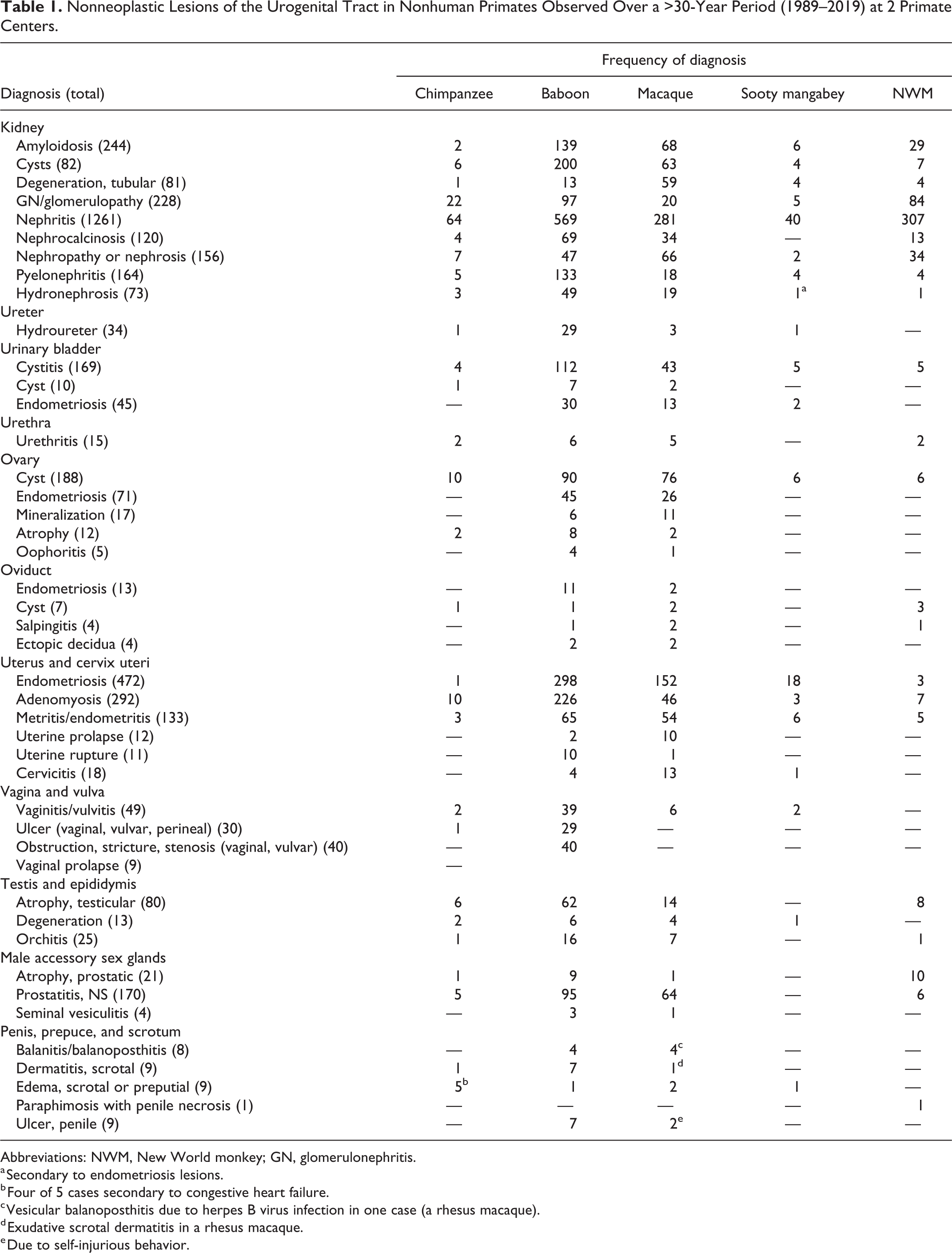
Abbreviations: NWM, New World monkey; GN, glomerulonephritis.
a Secondary to endometriosis lesions.
b Four of 5 cases secondary to congestive heart failure.
c Vesicular balanoposthitis due to herpes B virus infection in one case (a rhesus macaque).
d Exudative scrotal dermatitis in a rhesus macaque.
e Due to self-injurious behavior.
Hyperplastic and Neoplastic Lesions of the Urogenital Tracts in Nonhuman Primates Observed Over a >30-Year Period (1989–2019) at 2 Primate Centers.
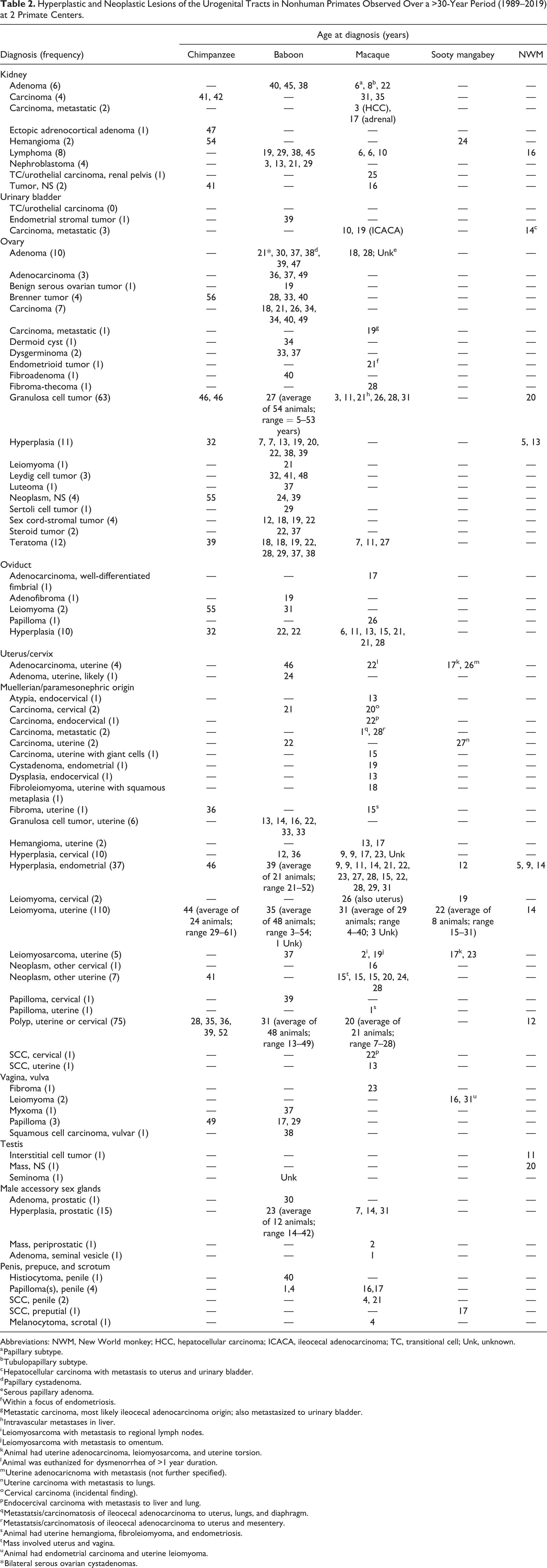
Abbreviations: NWM, New World monkey; HCC, hepatocellular carcinoma; ICACA, ileocecal adenocarcinoma; TC, transitional cell; Unk, unknown.
a Papillary subtype.
b Tubulopapillary subtype.
c Hepatocellular carcinoma with metastasis to uterus and urinary bladder.
d Papillary cystadenoma.
e Serous papillary adenoma.
f Within a focus of endometriosis.
g Metastatic carcinoma, most likely ileocecal adenocarcinoma origin; also metastasized to urinary bladder.
h Intravascular metastases in liver.
i Leiomyosarcoma with metastasis to regional lymph nodes.
j Leiomyosarcoma with metastasis to omentum.
k Animal had uterine adenocarcinoma, leiomyosarcoma, and uterine torsion.
l Animal was euthanized for dysmenorrhea of >1 year duration.
m Uterine adenocaricnoma with metastasis (not further specified).
n Uterine carcinoma with metastasis to lungs.
o Cervical carcinoma (incidental finding).
p Endocercival carcinoma with metastasis to liver and lung.
q Metastatsis/carcinomatosis of ileocecal adenocarcinoma to uterus, lungs, and diaphragm.
r Metastatsis/carcinomatosis of ileocecal adenocarcinoma to uterus and mesentery.
s Animal had uterine hemangioma, fibroleiomyoma, and endometriosis.
t Mass involved uterus and vagina.
u Animal had endometrial carcinoma and uterine leiomyoma.
* Bilateral serous ovarian cystadenomas.
Urinary Tract (Kidneys, Ureters, Urinary Bladder, and Urethra)
The most common lesions of the kidney were amyloidosis, cysts, tubular degeneration, glomerulonephritis or glomerulopathy, nephritis (including chronic renal disease), nephrocalcinosis, nephropathy or nephrosis, pyelonephritis, and hydronephrosis (Fig. 1). Animals with chronic renal disease resulting in renal failure were included in the diagnostic category of nephritis (21 cases; Fig. 2). Ureteral lesions were rare; the most frequent diagnosis was hydroureter (34 cases; Fig. 1), and this was observed more frequently in females compared to males and was occasionally attributed to endometriosis-related stricture of the ureter.
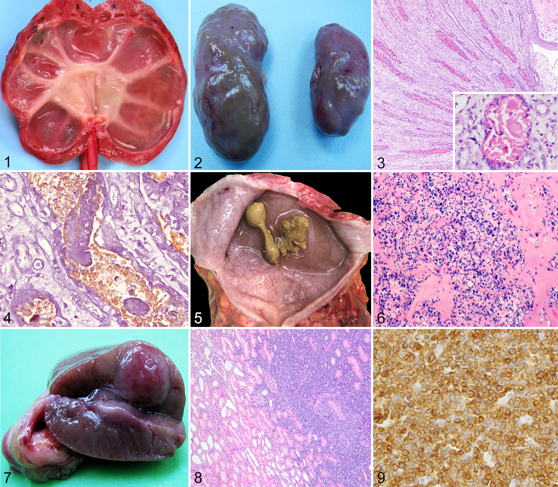
Hydronephrosis and hydroureter with secondary renal atrophy, kidney and ureter, chimpanzee. Figure 2. Chronic renal disease, kidneys, rhesus macaque. Parenchymal loss, fibrosis, and severe unilateral renal atrophy are evident.
There were 16 cases of rhabdomyolysis-associated pigmentary nephrosis (15 rhesus macaques and 1 sooty mangabey; Figs. 3, 4). Many additional animals had nephrosis or nephropathy of undetermined cause. We identified one rhesus macaque with severe renal and hepatic lipidosis at necropsy, suggestive of fatal fasting syndrome; this animal had a history of obesity, rapid significant weight loss, and severe azotemia. There were 41 additional cases of renal tubular lipidosis or steatosis, the cause of which was not further specified.
Glomerular disease was uncommon in NHPs during the study period. Of the animals with glomerulopathy or glomerulonephritis, baboons and common marmosets were highly represented. One female chimpanzee had both glomerular and medullary renal amyloidosis, along with extensive hepatic and splenic amyloidosis. This animal had hypoproteinemia (total serum protein 2.4 g/dL), and ascites and hydropericardium at necropsy. Protein loss was largely attributed to the glomerular amyloidosis, but urine protein was not measured, so hypoproteinemia due to decreased liver function as a result of hepatic amyloidosis, or a combination of the two, could not be ruled out.
Congenital renal lesions observed during the study period included polycystic kidneys (3 adult rhesus macaques and 1 baboon), ectopic adrenal tissue (3 rhesus macaques and 1 chimp), renal ectopia (2 rhesus macaques), adrenal-renal fusion (1 baboon), and unilateral renal aplasia (1 cynomolgus and 2 rhesus macaques).
The most frequent lesions of the urinary bladder were cystitis, endometriosis, and urinary bladder cysts. The cause of cystitis was most often undetermined, and the character of inflammation was not specified for most cases. In a minority of cases (16 rhesus macaques), the inflammation was specified as lymphocytic, lymphoplasmacytic, lymphofollicular, eosinophilic, or suppurative. Suppurative bacterial cystitis was associated with Escherichia coli, Klebsiella pneumonia, and Proteus mirabilis infections. Other findings in urinary bladders included polyarteritis nodosa (1 baboon), mineralization (1 baboon), and cystolithiasis (7 cases). In rhesus macaques, 6 cases of cystolithiasis were associated with retrograde ejaculation (Figs. 5, 6), while the seventh case was of unknown mineral composition in a pig-tailed macaque.
Lesions of the urethra were rare. Specific causes of ulcerative urethritis included retrograde ejaculation (4 rhesus macaques), bacterial infections (2 male baboons and a female chimpanzee), and trauma (1 male chimpanzee and 1 female rhesus macaque).
Thirty neoplasms involving the kidney were diagnosed during the study period (Table 2). These included renal lymphoma (Figs. 7–9), carcinoma, adenoma, nephroblastoma, urothelial (transitional cell) carcinoma of the renal pelvis, renal hemangioma, ectopic adrenocortical carcinoma, and renal sarcoma. Renal lymphoma was the most frequent tumor. One rhesus macaque had T-cell lymphoma affecting the kidney, liver, tonsils, thymus, and lymph nodes; SIV and Epstein-Barr virus infections were ruled out in that case. Two additional SIV-negative macaques had multicentric lymphoma affecting the kidneys; their simian T-lymphotropic virus (STLV) and lymphocryptovirus (LCV) serologic statuses are unknown. Four baboons and one common marmoset were diagnosed with renal lymphoma; serologic information for those animals was not available. Metastatic carcinomas involved the kidneys of 2 rhesus macaques (ileocecal adenocarcinoma) and one common marmoset (hepatocellular carcinoma).
Primary neoplasia of the urinary bladder and urethra was not diagnosed during the study period. Nonprimary tumors of the urinary bladder included an endometrial stromal tumor in a baboon, metastatic ileocecal adenocarcinoma in 2 rhesus macaques, and metastatic hepatocellular carcinoma in a common marmoset.
Female Reproductive Tract
Endometriosis was by far the most common diagnosis of the female genital tract (Figs. 10, 11). Adenomyosis, defined as the presence of endometrial glands within the myometrium, and endometrial or cervical polyps (Fig. 12) were frequent lesions. Other uterine lesions included endometrial hyperplasia (Figs. 13, 14), metritis or endometritis, uterine prolapse, uterine rupture, and cervicitis (Table 1). Ovarian cysts were the most frequent nonneoplastic lesion of the ovary (Table 1). Ovarian atrophy, aplasia (Fig. 15), hypoplasia, mineralization, oophoritis, ectopic adrenal tissue, ectopic decidua, and ovotestes were occasionally diagnosed.
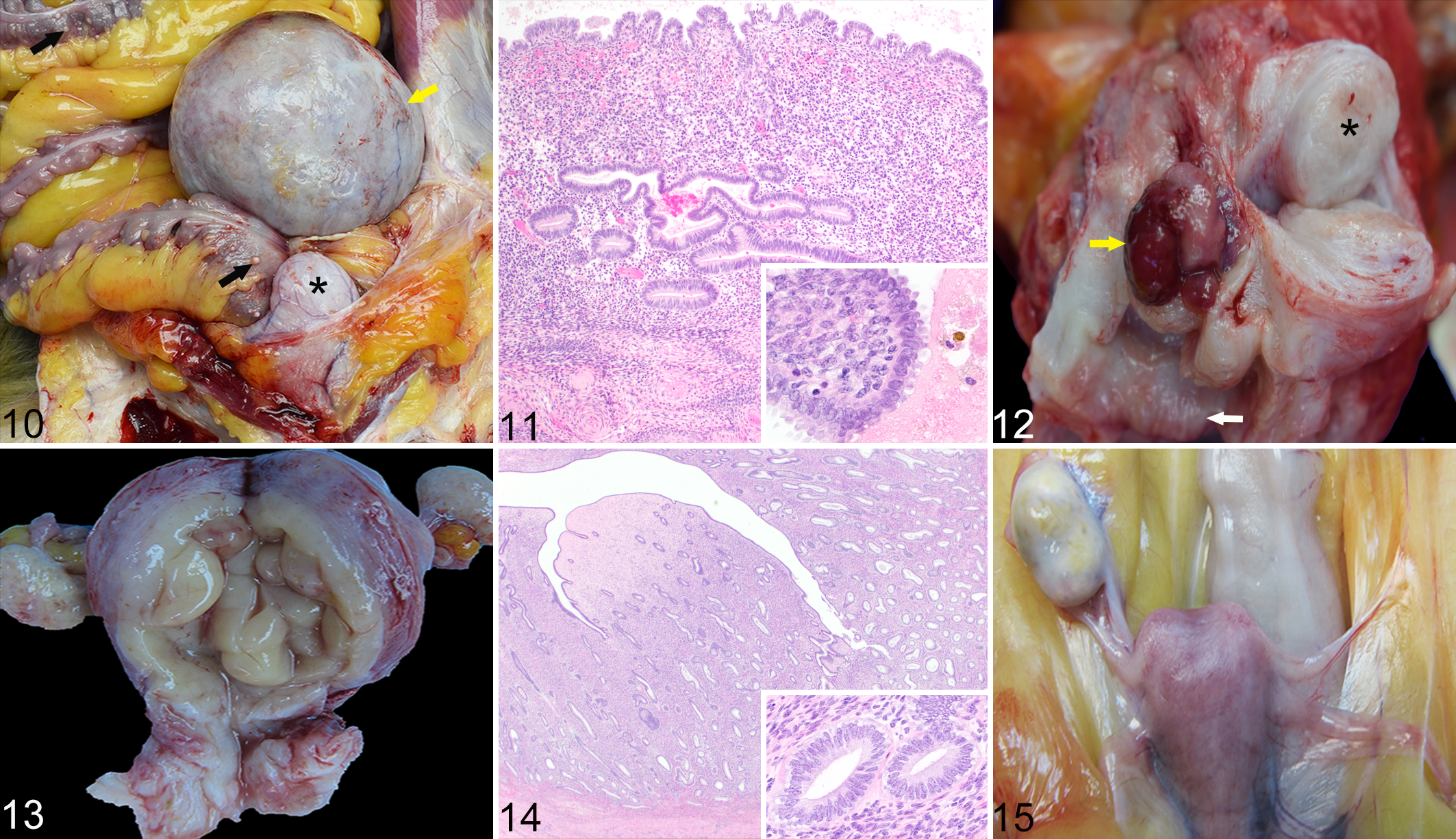
Endometriosis; ovary, uterus and colon; 18-year-old female rhesus macaque.
In baboons, vaginal, vulvar or perineal ulceration, vaginal obstruction, stenosis or stricture, and vulvitis was often attributed to herpesvirus papio 2 infection. Mild vaginitis or vulvitis were the most frequent diagnoses of the lower female genital tract. Chronic vaginal prolapse was seen in 3 baboons and 6 rhesus macaques; at least one of these macaques was a breeding female.
Leiomyoma was the most frequently diagnosed uterine neoplasm during the study period (Fig. 12; Table 2); this was most often an incidental finding. An unusual presentation of malignant uterine neoplasia was a leiomyosarcoma in a 2-year-old rhesus macaque with extension to the cervix and vagina.
Granulosa cell tumors (GCTs; Figs. 16, 17) accounted for 63 of 123 (51%) ovarian neoplasms diagnosed during the study period. The second most common ovarian tumor was teratoma (1 chimpanzee, 8 baboons, and 3 macaques; Figs. 18, 19). Of animals included in the study, most ovarian tumors occurred in baboons (108/134; 80%).
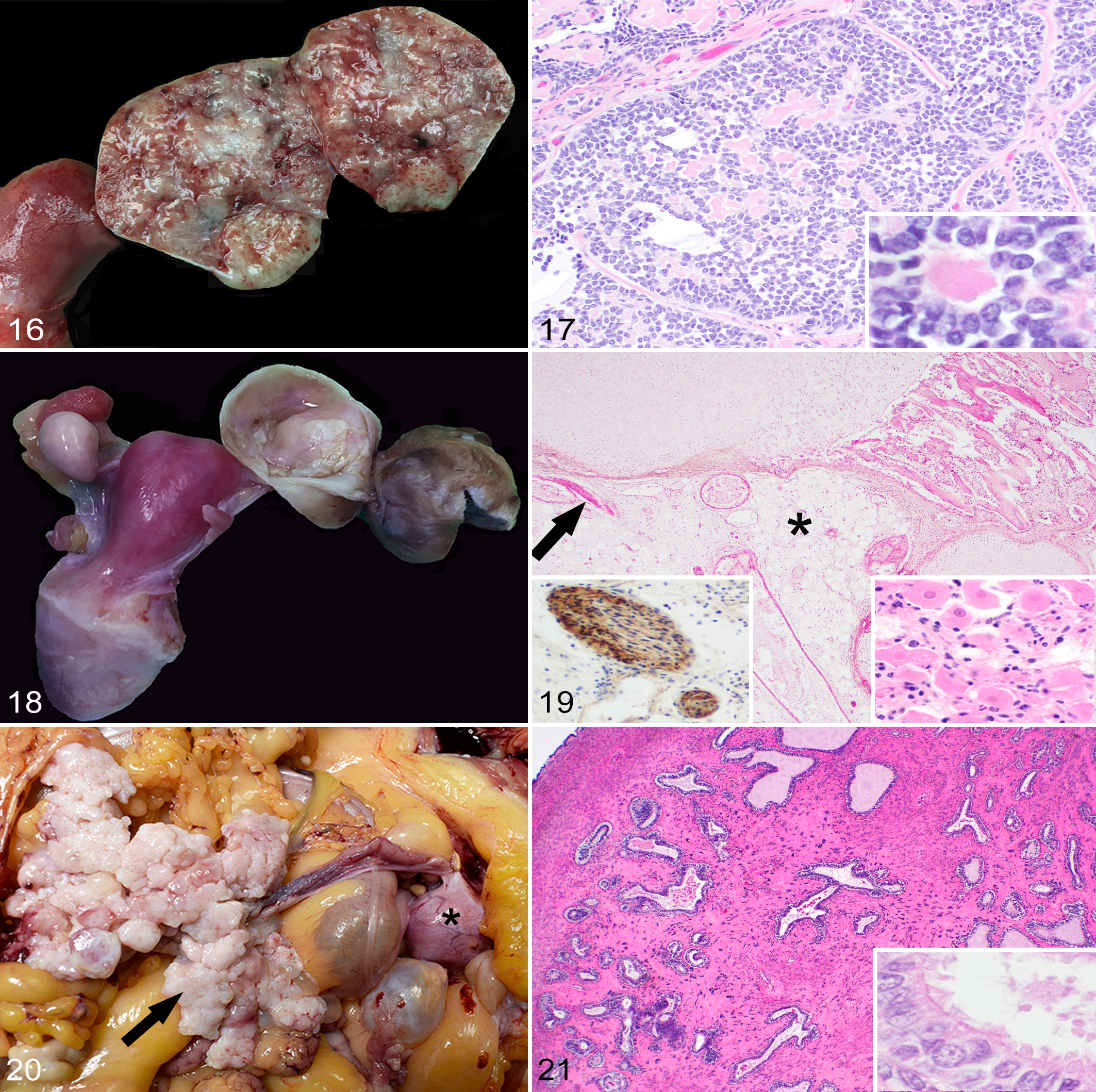
Granulosa cell tumor, left ovary, 21-year-old female rhesus macaque.
During the study period, there were 4 benign and one malignant oviductal tumor, including 1 fimbrial adenocarcinoma in a rhesus macaque (Figs. 20, 21). Ten cases of oviductal hyperplasia were observed during the study period in OWMs. The hyperplastic change was further specified as fimbrial in 2 cases (1 rhesus macaque and 1 baboon).
Six benign vaginal and vulvar neoplasms were diagnosed, including a fibroma in a rhesus macaque, 2 leiomyomas in cynomolgus macaques, a papilloma in a chimp, and a myxoma and 2 papillomas in baboons (Table 2). The PV status of the chimpanzee and 2 baboons with vaginal papillomas is unknown.
Developmental anomalies of the female genital tract included uterus didelphys (1 rhesus macaque), uterine hamartoma (1 rhesus macaque and 1 common marmoset), ectopic ovarian tissue (1 rhesus and 1 baboon) and an imperforate vulva in a stump-tailed macaque fetus, who also had atresia coli, imperforate anus, pulmonary hypoplasia and right-sided hydronephrosis.
Male Genital Tract
Testicular atrophy (80 cases) and minimal to mild prostatitis (170 cases) were the most frequently diagnosed lesions of the male genital tract (Table 1). These were almost always incidental findings. Herpesvirus papio 2 was the single most frequent confirmed or suspected cause of infectious posthitis or balanoposthitis in baboons during the study period (10 cases). A single case of B virus-associated ulcerative balanitis associated with macacine alphaherpesvirus 1 (herpes B virus) infection was diagnosed in a 7-year-old rhesus macaque.
Neoplasia of the testes, epididymis, accessory sex glands, and external genitalia was rare during the 30-year period, with a total of 15 tumors. One interstitial cell tumor was diagnosed in a squirrel monkey (Figs. 22–24). Malignant prostatic neoplasia and neoplasia of the seminal vesicles were not observed during the study period. Prostatic hyperplasia was a rare diagnosis across all NHP species (12 baboons and 3 rhesus macaques). The most common penile neoplasm during the study period was squamous cell carcinoma (3 cases; Figs. 25–27); the papillomavirus (PV) status of these cases is undetermined.
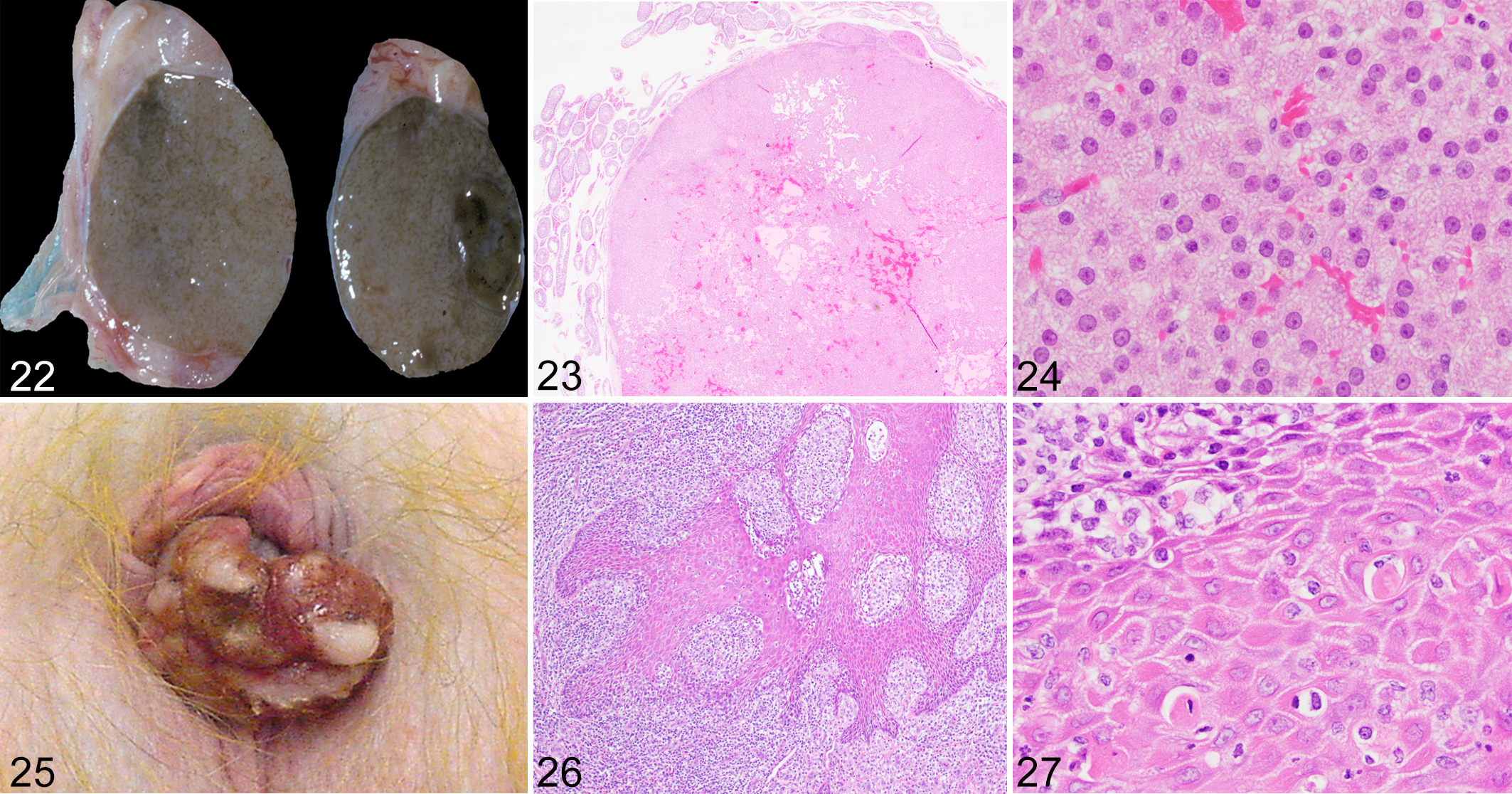
Interstitial (Leydig) cell tumor, testis, 20-year-old male squirrel monkey.
Cryptorchidism (16 cases) was the most frequent congenital lesion of the male reproductive tract. Hypospadias (2 rhesus macaques), preputial orifice hypoplasia, and penile aplasia (1 baboon) were also observed.
Discussion
We conducted a comprehensive review of spontaneous genitourinary lesions occurring in NHPs over a 30-year period at 2 primate centers to serve as a resource for veterinarians, pathologists, and researchers working with these species. Background histopathologic findings in kidneys and in male and female reproductive tracts of NHPs have been previously described in the literature. 16,46,63,64,70,73,88 Data gathered from this study highlighted some differences between OWMs and NWMs. A possible limitation of this study is bias toward more florid, grossly apparent lesions such as endometriosis and lesions of the vital organs (ie, kidney) versus other subtle microscopic lesions of the male and female reproductive tracts.
Amyloidosis was the most common renal lesion. In NHPs, amyloid often accumulates within the renal interstitium (especially in the medulla) as a result of chronic inflammation (enterocolitis, rheumatoid arthritis, chronic catheterization, viral infection, and parasitism in macaques). This finding is most often incidental, but amyloid can compress and replace tubules in some advanced cases. 38,52 Compared to medullary amyloidosis, glomerular amyloidosis was rare across all NHP species. Few cases of clinically significant glomerular amyloid deposition have been described in macaques and marmosets. 20,38,48
The cause of nephrosis or nephropathy was infrequently identified during the study period. Reported causes of renal tubular disease in NHPs include acute rhabdomyolysis, fatal fasting syndrome, aminoglycoside antibiotics, and nonsteroidal anti-inflammatory drugs. 2,9,20,28,42,68,78 Fatal fasting syndrome is a disease entity of obese macaques following anorexia and weight loss characterized by moderate to severe hepatic and proximal renal tubular epithelial cell lipidosis; focal pancreatic necrosis and pancreatitis have also been reported. 9 Azotemia is the most common laboratory abnormality. 9,20 Antemortem clinicopathologic data are necessary to make a diagnosis of fatal fasting syndrome. Correlative data was not available for all cases during the study period; thus, fatal fasting syndrome may be underestimated in our data set.
Renal lesions (particularly nephritis, glomerulonephritis, and glomerulonephropathy) were frequent diagnoses in NWMs (primarily marmosets and tamarins), while genital lesions were infrequent in those species. Glomerulonephritis and glomerulopathy were more frequently diagnosed in baboons and common marmosets compared to macaques. The occurrence of spontaneous glomerulopathy in young common marmosets and additional lesions in aging marmosets suggests that this species may be a useful animal model for studying spontaneous renal diseases. 43,87 In laboratory chimpanzees, cardiac disease may lead to progressive renal dysfunction (ie, “cardiorenal syndrome”), with the most common associated lesions being glomerulosclerosis and interstitial nephritis. 17
Lesions of the urinary bladder were rare during the study period. Cystolithiasis was rare, with most cases attributed to retrograde ejaculation in male rhesus macaques. 29 Tumors of the kidney and urinary bladder were uncommon, with 30 and 4 cases diagnosed, respectively. Lymphoma was the most common renal tumor. Lymphomas in NHPs are often associated with oncogenic viruses such as rhesus lymphocryptovirus and simian T-cell leukemia virus; oncogenic viral infection could not be ruled out in several of our cases. 12,57,66 Only a single neoplasm of the urinary tract (renal hemangioma) was observed in sooty mangabeys.
Female reproductive tract lesions were more frequently encountered than male reproductive tract lesions; this observation may be related to a number of potential factors, including more limited postmortem examination of male reproductive tissues, variable demographics of the NHP colonies, and differences in age at study assignment and experimental endpoint for males versus females. These factors are potential limitations of this study.
Endometriosis was the most frequent lesion of the female reproductive tract in NHPs. Of the animals examined in this study, it was most frequent in OWMs (baboons > macaques > mangabeys), and rarely in chimpanzees and common marmosets (Supplementary Table 1), making OWMs attractive animal models for the corresponding human condition. Endometriosis, defined as the presence of ectopic endometrial tissue outside the uterus, is the leading cause of presenescent reproductive failure in rhesus and cynomolgus macaques. 26,46,76 In baboons, the prevalence of spontaneous endometriosis has been shown to increase with the duration of captivity. 23 Endometriosis may be encountered as an incidental lesion or as a clinically significant finding in NHPs. Secondary lesions of the urinary tract were occasionally observed, such as adhesions resulting in hydroureter or hydronephrosis, and are also seen in women as sequelae of endometriosis. 80 Endometrial hyperplasia and uterine or cervical polyps were also diagnosed more frequently in OWMs compared to NWMs.
Leiomyoma was the most frequent neoplasm of the female genital tract in NHPs, and granulosa cell tumor was the most frequent ovarian tumor; these findings are consistent with previous reports. 7,10,13,18,19,25,39,44,45,47,50,54,60,67 Uterine leiomyomas are generally considered benign tumors in NHPs, but complications may arise when the tumors become excessively large, leading to uterine torsion or compression of surrounding structures such as the ureters, aorta or vena cava. 33 Leiomyoma is the most common indication for hysterectomy in women and several research groups are currently working to establish a NHP model of uterine transplantation. 40,56,77,82 Neoplasia of the urinary and genital tracts was rare in the NWMs examined in this study.
Dysplastic and neoplastic lesions of the cervix are associated with species-specific papillomavirus infections in baboons and macaques making these species attractive animal models for studies of human papillomavirus oncogenesis. 6,35,58,83 –85 As in humans, benign and malignant squamous genital proliferations in NHPs are likely caused by separate viruses. 19,58 In women, the target for oncogenic papillomaviruses is the squamo-columnar junction of the cervix uteri. 58 High-grade cervical intraepithelial neoplasia is also reported at the squamo-columnar junction in cynomolgus macaques. 84 There were a small number of cervical lesions in macaques and baboons during the study period; the papillomavirus status of these animals remains undetermined.
Papillomavirus-associated vaginal papillomas and carcinomas of the vulva and sex skin have previously been reported in NHPs. 5,30,84 Squamous cell carcinoma of the vulva, perineum, and/or sex skin has been observed in baboons and chimpanzees; potential risk factors include prolonged sunlight exposure and repetitive trauma to the area. 5,30 Chronic herpesvirus papio 2 and Histoplasma capsulatum var duboisii infections may confer additional risk in baboons. 11,30,51
There are rare reports of papillomavirus infection leading to penile carcinoma in rhesus macaques as well as spontaneous squamous cell carcinoma at the mucocutaneous junction of the penis and prepuce. 37 Penile squamous cell carcinoma in men is associated with human papillomavirus (predominantly human papillomavirus 16) infection. 62 Chronic preputial inflammation due to phimosis or lichen sclerosis is also associated with penile squamous cell carcinoma in men. 32 Recently, genital condyloma-like lesions have been described in cynomolgus monkeys originating from the island of Mauritius; using PCR, macaque lymphocryptovirus, but not papillomavirus or poxvirus, was identified in those lesions. 34
Malignant ovarian epithelial tumors comprise more than 90% of ovarian neoplasms in women over 40 years of age, so there is considerable interest in developing an NHP model of ovarian adenocarcinoma. 81 However, spontaneous ovarian tumors are rarely reported in NHPs, a trend that our data further supports (Table 2). 19 Rhesus macaques have been evaluated as a model for the study of ovarian cancer chemopreventive drugs, but the low incidence of spontaneous ovarian neoplasia in this species may limit the utility of this model. 8 Other research groups have studied hormonal regulation and epitheliectomy of the ovarian surface epithelium in macaques as means of reducing ovarian cancer risk. 69,86
Oviductal neoplasia is rare in NHPs, with leiomyoma, atypical polypoid adenomyofibroma, and adenomyofibroma being previously reported. 10,71,74 We identified a single malignant oviductal neoplasm during the 30-year study period. Given the minimal anisokaryosis and high degree of differentiation, the differential diagnosis was fimbrial adenomyofibroma, which has previously been reported in a cynomolgus macaque. 74 However, the tumor was unencapsulated, expansile, and multifocally incited a prominent scirrhous response, which favored a diagnosis of adenocarcinoma. This tumor is of particular interest because a high proportion of high-grade serous ovarian cancers in women are now thought to be of oviductal origin. 81 In contrast to benign prostatic hyperplasia (BPH), a frequent diagnosis of aging men, prostatic hyperplasia was rarely observed in all NHPs during the study period. 59 In 2 retrospective studies of aging (25- to 29-year-old) male chimpanzees, spontaneous BPH was reported in one group but not in the other. 13,75 Hyperplastic and neoplastic lesions originating from the prostatic basal cell population, histologically similar to those reported in men, were previously reported as common findings in a group of 19 aged, male macaques. 55 These lesions likely have little, if any, clinical consequence in macaques, as they are found in the middle to outer regions of the prostate and the macaque prostate gland does not completely encircle the urethra as it does in man. 55 Malignant prostatic tumors were not observed during the study period.
A variety of congenital or developmental lesions were diagnosed at low frequency during the study period. Polycystic kidney disease is the most commonly reported renal anomaly of NHPs, and is common among slender lorises (Loris lydekkerianus). 20,65 Infantile polycystic kidney disease has been reported in rhesus macaques although this condition was not observed during the study period. 3 Other developmental renal anomalies previously reported in NHPs but not represented in our data set include renal fusion and renal ectopia. 14,15,72 Uterus didelphys was diagnosed in a 23-year-old rhesus macaque as an incidental finding. A single case of uterus didelphys has previously been reported in a neonatal tamarin (Sanguinus fuscicollis). 15
Nonhuman primates are among the most commonly used large preclinical animal models in drug development, primarily for safety assessment and pharmacokinetics studies. Although spontaneous clinically significant urogenital disease is uncommon in NHPs, diseases of the urinary and genital systems are common in humans and several NHP species are used as animal models of these conditions. 24 Furthermore, as gene modification technologies emerge, the epidemiology and/or phenotype of these disease entities in NHPs may change over time. Knowledge of the background lesions occurring in NHP species may assist pathologists and investigators in the evaluation of new animal models as they are developed.
Supplemental Material
Combined_supplemental_materials-Kirejczyk_et_al - Urogenital Lesions in Nonhuman Primates at 2 National Primate Research Centers
Combined_supplemental_materials-Kirejczyk_et_al for Urogenital Lesions in Nonhuman Primates at 2 National Primate Research Centers by Shannon Kirejczyk, Christopher Pinelli, Olga Gonzalez, Shyamesh Kumar, Edward Dick and Sanjeev Gumber in Veterinary Pathology
Footnotes
Acknowledgements
The authors would like to thank the veterinary pathologists and necropsy staff, past and present, of the Yerkes National Primate Research Center.
Declaration of Conflicting Interests
The author(s) declared no potential conflicts of interest with respect to the research, authorship, and/or publication of this article.
Funding
The author(s) disclosed receipt of the following financial support for the research, authorship, and/or publication of this article: This investigation used resources that were supported by the Yerkes Base Grant P51OD11132, and Southwest National Primate Research Center Grant P51OD011133 from the Office of Research Infrastructure Programs, National Institutes of Health. This investigation was conducted in facilities constructed with support from the Office of Research Infrastructure Programs (ORIP) of the National Institutes of Health through Grant Numbers C06 RR014578, C06 RR016228, C06 RR015456, and C06 RR017332.
Ethical Approval
All applicable international, national, and/or institutional guidelines for the care and use of animals were followed.
Supplemental Material
Supplemental material for this article is available online.
References
Supplementary Material
Please find the following supplemental material available below.
For Open Access articles published under a Creative Commons License, all supplemental material carries the same license as the article it is associated with.
For non-Open Access articles published, all supplemental material carries a non-exclusive license, and permission requests for re-use of supplemental material or any part of supplemental material shall be sent directly to the copyright owner as specified in the copyright notice associated with the article.