
Abstract
Odontogenic lesions are well described in domestic cats, but published literature describing these lesions in nondomestic felids is limited. This study reports oral lesions in 109 captive, non-domestic felids. Ten cases of odontogenic lesions were diagnosed, including 9 with fibromatous epulis of periodontal ligament origin (FEPLO) and one odontogenic cyst in a cougar. FEPLO was common in lions. FEPLO did not recur after surgical removal in any of the 3 cases for which follow-up information was available. Increased occurrences of oral papillomas in snow leopards and eosinophilic granulomas in tigers were identified, which is consistent with the reported literature. With the exception of oral papillomas in snow leopards and FEPLO in lions, the spectrum of oral lesions in nondomestic felids was similar to what is reported in domestic cats, with squamous cell carcinoma being the most common oral malignancy, and stomatitis/gingivitis/glossitis accounting for approximately one third of all cases. Rare diagnoses with one case each included hemangioma, fibrosarcoma, melanoma, cleft palate, and glossal amyloidosis.
Keywords
Odontogenic lesions are well described in animals. Although classification schemes in veterinary oral pathology generally closely follow those in human oral pathology, there are notable exceptions of lesions that may be unique to animals, such as canine acanthomatous ameloblastoma and canine odontogenic parakeratinized cyst. 1,5,7,13 While recent advances have been made in the nomenclature and classification of odontogenic tumors in animals, reports on the incidence of odontogenic lesions among various species are generally sparse, particularly for nondomestic species. A recent review of reported odontogenic tumors in domestic animals is available. 1 Reports of odontogenic lesions in nondomestic species, including nondomestic felids, are usually single cases, and are generally uncommon.
The purpose of this study is to report oral lesions diagnosed in nondomestic felids in human care and compare findings to those in domestic cats and current literature. This study focused on odontogenic lesions, which are not commonly reported in these animals.
Materials and Methods
For this retrospective study, the Northwest ZooPath (Monroe, WA) archives were searched for all diagnoses in felids. All diagnoses of oral lesions were summarized in table format. Diagnoses were grouped into the following categories: non-odontogenic neoplasia included benign and malignant neoplasms; inflammation included all lesions that were composed primarily of inflammation; odontogenic lesions included all proliferative odontogenic lesions and odontogenic cysts; other diagnoses included non-odontogenic developmental and deposition disorders. Hematoxylin and eosin (HE)-stained histologic sections from formalin-fixed paraffin-embedded tissue blocks were reviewed and the diagnoses made by one pathologist (MMG). Cases of odontogenic lesions were reviewed histologically by 2 additional pathologists (BGM and EEBL). For odontogenic lesions, additional case information, including clinical history, outcome, radiographs, and gross photographs, was requested from clinicians, and collated when available.
Results
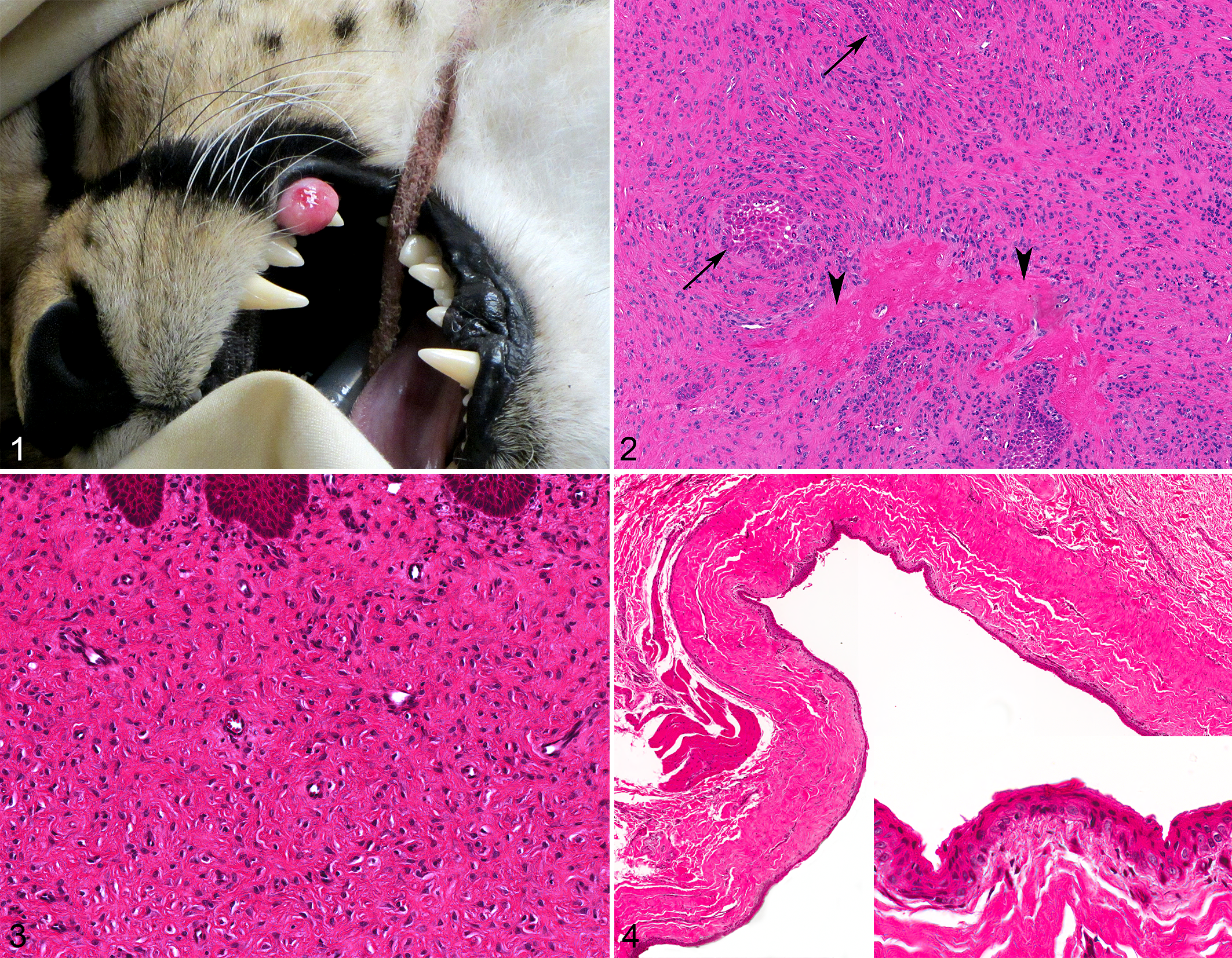
Oral lesions were diagnosed in 109 captive felids, including 36 snow leopards (Panthera uncia), 25 tigers (Panthera tigris), 13 lions (Panthera leo), 12 cheetahs (Acinonyx jubatus), 5 cougars (Puma concolor), 5 leopards (Panthera pardus), 3 jaguars (Panthera onca), 3 ocelots (Leopardus pardalis), 2 lynx (Lynx sp.), 2 servals (Leptailurus serval), 1 Geoffrey’s cat (Leopardus geoffroyi), 1 bobcat (Lynx rufus), and 1 fishing cat (Prionailurus viverrinus; Table 1). Each animal was diagnosed with only one oral lesion. The most common diagnostic category was neoplasia (56/109; 51%). Papillomas were the most common neoplasm diagnosed with 39 cases, and were most common in snow leopards (30/39 cases; 77%). At least 19 of the 39 papilloma cases had evidence of viral cytopathic effects consistent with viral etiology (ie, large cytoplasmic keratohyalin granules, koilocytes, and/or intracytoplasmic viral inclusions); further diagnostics to confirm papillomavirus infection were not available. Squamous cell carcinoma was the second most common neoplasm diagnosed with 14 cases. The second most common diagnostic category was inflammation (41/109; 38%) with predominantly lymphoplasmacytic (non-eosinophilic) inflammation in 33 cases and predominantly eosinophilic inflammation in 8 cases, 7 of which were tigers. Odontogenic lesions were the third most common diagnostic category (10/109; 9%; see below). Other diagnoses were rare (2/109; 2%).
Oral Lesions in 109 Captive Nondomestic Felidsa.
Abbreviations: SCC, squamous cell carcinoma; FEPLO, fibromatous epulis of periodontal ligament origin.
a Data in parentheses are number of cases.
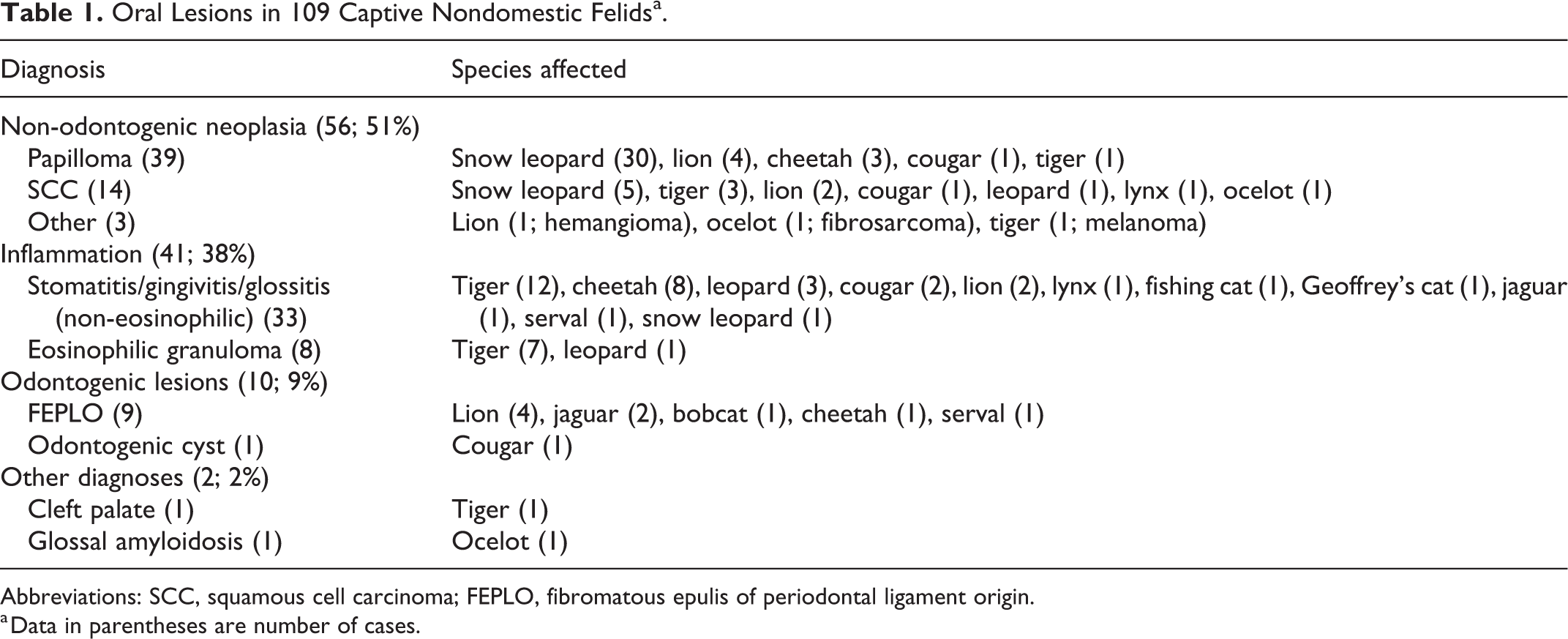
The most common odontogenic lesion that was diagnosed was fibromatous epulis of periodontal ligament origin (FEPLO), which was diagnosed in 4 lions, including 3 females and 1 male ranging in age from 13 to 21 years; age for 1 “adult” was not specified. FEPLO was also diagnosed in 2 jaguars, 1 bobcat, 1 cheetah, and 1 serval that included 3 females and 2 males ranging in age from 3 to 12 years. FEPLOs were reported to arise from the maxillary or mandibular gingiva around the canine, premolar, or molar teeth. Gross photographs of FEPLO were available in 1 cheetah (case 6; Fig. 1) and revealed an exophytic mass arising from the tooth-associated gingiva that partially entrapped the adjacent tooth. Microscopically, in all of the FEPLO cases, the majority of the lesion was composed of proliferative periodontal ligament-like stroma with a moderate number of spindle to stellate mesenchymal cells embedded in fibrous tissue composed of finely bundled collagen with regularly spaced, dilated, small-caliber blood vessels (Figs. 2, 3). These lesions often had variable amounts of interspersed odontogenic epithelium, characterized by small to moderately sized rests or branching plexiform ribbons of epithelial cells with a peripheral palisade of columnar cells in which the nuclei were frequently antibasilar, with rare cytoplasmic basilar clearing. Islands of cemento-osseous matrix (COM) were variably present, and occasionally abundant. COM demonstrated histologic features consistent with both osteoid and cementoid matrices, and was sometimes mineralized and had embedded cells within lacunae. Additional clinical history and follow-up information was available in 3 cases (cases 1, 6, and 7). In cases 1 and 7, an excisional biopsy of the FEPLOs was performed, but surgical margins were incomplete and the FEPLOs extended histologically to the deep surgical margins. Despite incomplete excision, surgical removal/debulking was presumably curative in both cases as there was no regrowth at annual examinations for 7 years in case 1, at which point the animal was euthanized for unrelated disease, and for 2 years in case 7, at which point the animal was lost to follow-up. In case 6, an excisional biopsy of the FEPLO was performed, and surgical margins were narrow with cells of the FEPLO extending <100 µm from the surgical margin. Despite narrow excision, there was no regrowth of the FEPLO at annual examinations for 3 years, at which point the animal died of unrelated disease.
Fibromatous epulis of periodontal ligament origin, gingiva.
A single odontogenic cyst was identified in a 20-year-old, female cougar who was euthanized for degenerative joint disease and urinary tract disease. Perimortem dental radiographs were performed due to a history of periodontal disease and revealed a periapical lucency associated with the root of one of the mandibular canine teeth. A necropsy sample taken from the cyst wall was submitted for histology. Histologically, a cyst expanded the gingival subepithelial stroma (Fig. 4). The lining epithelium of the cyst was non-keratinizing stratified squamous epithelium, the basal layer of which was palisading and rarely exhibited features of odontogenic epithelium (antibasilar nuclei and basilar clearing). Epithelial thickness varied from 2 to 10 layers. Lymphocytic and plasmacytic infiltration was mild in the stroma around the cyst and within the epithelium.
Discussion
Odontogenic lesions comprised only 9% of the oral lesions diagnosed in this group of nondomestic felids. Of the odontogenic lesions identified in this population, FEPLO was diagnosed in 9/10 cases. FEPLO is also called peripheral odontogenic fibroma; the authors prefer the former term, which indicates that the lesion originates from the periodontium, as opposed to the latter term, which implies a neoplasm. 5 While common in domestic dogs, FEPLO are less common in domestic cats, and the number of cases diagnosed in nondomestic felids was surprising. 5,12 FEPLO was diagnosed in 5 different species, and lions were the most commonly affected species. There is only one previous report of any proliferative odontogenic lesion in any nondomestic felid, and it was a FEPLO in a lion. 3 Lions may be predisposed to this condition, similar to domestic dogs. Recurrence of FEPLO was not noted in any of the animals for which follow-up information was available. This is similar to the biologic behavior of FEPLO in domestic animals, where regrowth is rare even in cases of incomplete excision. 5
The only other odontogenic proliferative lesion diagnosed in nondomestic felids was a single odontogenic cyst diagnosed in a cougar. Odontogenic cysts have been rarely reported in domestic cats and dentigerous cysts are the most common-type reported. 5 The odontogenic cyst in this case was not anatomically consistent with a dentigerous cyst as it was associated with an erupted tooth. The cougar had a history of severe dental disease, and the cyst was radiologically associated an inflamed tooth root. These findings are consistent with a radicular cyst, which is uncommon in domestic animals, but occurs secondary to tooth-associated periapical inflammation. 5
Non-odontogenic neoplasia and oral inflammatory lesions collectively comprised 89% of oral lesions of nondomestic felids. Feline papillomavirus-associated oral papillomas have been reported in a variety of nondomestic felids and have been well documented as a relatively common finding in captive snow leopards. 8,9,11 The histologic characteristics of oral papillomas in captive snow leopards include viral cytopathic changes within the granular cell layer such as large cytoplasmic keratohyalin granules, koilocytes, and rare intracytoplasmic viral inclusions. 9,11 This report is consistent with previous literature as oral papillomas were diagnosed in 30/36 of snow leopards with oral lesions. The occurrence of oral papillomas in snow leopards was more frequent than in all other nondomestic felids combined. With the exception of snow leopards, oral papillomas were relatively uncommon in nondomestic felids and accounted for 9/26 of all other non-odontogenic neoplasms identified. This is similar to domestic cats, in which oral papillomas are infrequently reported. 5
Squamous cell carcinoma (SCC) is the most common oral malignancy in domestic cats, and accounts for over 60% of all feline oral tumors. 5 Results of this study are consistent with this finding as 14/17 of the oral malignancies diagnosed in nondomestic felids were SCC. The incidence of SCC was higher in snow leopards compared with other nondomestic felids in this study and has been previously reported in this species. 11 Given the high incidence of papillomas in snow leopards, a relationship between SCC and papillomavirus-induced neoplasia has been suggested. 11 However, the role of papilloma virus in the pathogenesis of oral SCC in domestic cats is controversial as the incidence of oral SCC in domestic cats is much greater than the incidence of oral papillomas. 5
Inflammatory lesions are the most common oral lesions in domestic cats and chronic gingivostomatitis is the most commonly diagnosed of these lesions. 12 The cause of gingivitis is multifactorial and often associated with bacterial plaques/calculi. 5 With the exception of snow leopards, inflammatory lesions were the most common oral lesion in all species of nondomestic felids in this study; lesions of stomatitis, gingivitis, or glossitis were diagnosed in 33/41 cases of oral inflammatory lesions. The incidence of oral inflammatory lesions was highest in tigers, which accounted for 12/33 of stomatitis/gingivitis/glossitis cases and 7/8 of oral eosinophilic granuloma cases. The incidence of oral eosinophilic granulomas in tigers reported here is consistent with previous reports suggesting oral eosinophilic granulomas are an underreported and possibly common lesion of tigers. 4,10 Oral eosinophilic granuloma complex lesions are fairly common in domestic cats, with which tigers share 95.6% similarity in their genome sequence. 2 The genetic relatedness of domestic cats and tigers may make these species similarly predisposed to oral eosinophilic granulomas. Further research is needed to elucidate what role (if any) genetics plays in the development of oral inflammatory diseases. Cheetahs accounted for 8/33 cases of stomatitis, gingivitis, or glossitis. Captive cheetahs have been reported to have a higher incidence of focal palatine erosions compared to other nondomestic felids. 8 Specific location of biopsy specimens was not always provided in biopsy submission forms in this study, and as such incidence of focal palatine erosions could not be determined. One of the cases of stomatitis in a cheetah was associated with intranuclear inclusion bodies; this animal also had rhinotracheitis and was confirmed to have feline herpesvirus-1, to which cheetahs may be predisposed. 6 There were multiple limitations of the interpretation of inflammatory lesions in this series. Anatomic location of the lesions within the oral cavity, and/or any associated periodontal disease (eg, dental calculi) were often not specified. Additionally, polymerase chain reaction or cultures for infectious agents were either not performed or not available to the pathologist.
Conclusion
With the exception of papillomas in snow leopards and FEPLO in lions, the spectrum of oral lesions in nondomestic felids is similar to what is reported in domestic cats. Inflammatory lesions are the most common oral lesions, SCC is the most common oral malignancy, and odontogenic lesions comprised a minority of the lesions. The results of this study largely agree with the previously reported species-specific data regarding well-documented oral lesions in nondomestic felids, specifically papillomas and SCC in snow leopards and eosinophilic granulomas in tigers. As follow-up information was not consistently available in the cases presented here, additional data are needed to understand the clinical relevance and prognoses of oral lesions in nondomestic felids.
Footnotes
Authors’ Note
The views expressed in this article are those of the authors and do not reflect the official policy of the Department of the Army/Navy/Air Force, Department of Defense, or the US government.
Acknowledgements
The authors thank Cathy Minogue and Christie Buie of Northwest ZooPath for collection and processing of materials. We also thank Leroy Brown of Histology Consultation Services for specimen preparation. The authors are also grateful to the institutions and individuals who contributed case material and follow-up information, including Fort Worth Zoo (especially Kim Rainwater), Fossil Rim Wildlife Center (especially Holly Haefele), Hendricks County Animal Hospital, Indianapolis Zoo (especially Jeff Proudfoot and Abigail Rosenblum), Jacksonville Zoo (especially Tiffany Martin), San Diego Zoo (especially Bruce Rideout), and Zoo Miami (especially Gwen Myers).
Declaration of Conflicting Interests
The author(s) declared no potential conflicts of interest with respect to the research, authorship, and/or publication of this article.
Funding
The author(s) received no financial support for the research, authorship, and/or publication of this article.