
Abstract
Biopsy samples of colorectal polyps were collected and examined from 67 Miniature Dachshund dogs (including 35 cases with an additional biopsy). Histopathologic diagnoses of the initial biopsy samples were “inflammatory polyp” in 52 cases (78%), “adenoma” in 10 cases (15%), and “adenocarcinoma” in 5 cases (8%). Eight of 10 cases (80%) diagnosed as adenoma also had inflammatory polyp lesions in the same specimen. A second biopsy was performed in 25 cases (48%) initially diagnosed with inflammatory polyp. Pathologic diagnoses for the second biopsy were inflammatory polyp in 11 cases (44%), adenoma in 9 cases (36%), and adenocarcinoma in 5 cases (20%). The number of beta-catenin-positive nuclei in epithelial cells was significantly higher in adenoma (46%) and adenocarcinoma (75%) as compared with inflammatory polyp (6%). Normal epithelial cells and hyperplastic goblet cells in inflammatory polyps showed homogeneous positive cytoplasmic immunoreactivity for adenomatous polyposis coli (APC) antigen. However, APC expression was decreased in areas of intense nuclear beta-catenin expression in adenoma and adenocarcinoma lesions. Foci of cytokeratin 5/6–positive squamous cell-like neoplastic cells showed intense beta-catenin nuclear expression that was similar to squamous morules described in human colorectal tumors. The results of the present study suggest that the inflammatory polyp in Miniature Dachshunds is a progressive disease that may develop into adenoma and/or adenocarcinoma. In addition, immunohistochemical findings suggest that aberrations of APC and beta-catenin expression may be involved in tumor development within the inflammatory polyp lesions.
Keywords
Inflammatory polyps are uncommon in the canine colorectum 13 but are often seen in Miniature Dachshunds in Japan. 25 The inflammatory polyp of Miniature Dachshunds (IPMD) shows unique histologic features that differ from so-called inflammatory polyps, which are characterized by inflammatory cell infiltration and epithelial hyperplasia with lamina propria fibromuscular hyperplasia. IPMD is confined to the colorectum, and it consists of a stroma resembling granulation tissue, which is composed of hyperplastic goblet cells with increased mucus production and infiltration of inflammatory cells, predominantly neutrophils. Several inflammatory factors—including cyclooxygenase 2, fibroblast growth factor 2, 36 interleukins 8 and 17, 26,33 and pattern recognition receptors 15,16 —are considered to be involved in the pathogenesis of IPMD. Treatments for IPMD include surgical resection, endoscopic polypectomy, immunosuppression, and argon plasma coagulation. 25,35
Interestingly, Miniature Dachshunds in Japan are predisposed to develop not only inflammatory polyps but also adenomas in the colorectum. 36 In addition, several confirmed cases developed adenoma secondary to inflammatory polyp, both in the same location of the colorectum. 17 The purpose of this study was to investigate the possible progression of inflammatory polyps to neoplastic lesions and the factors that may promote such progression to neoplastic lesions.
To investigate the potential mechanisms of the progression of IPMD to neoplasia, it may be relevant to study the Wnt/beta catenin pathway, which plays an essential role in gastrointestinal tumorigenesis in humans and experimental animals. 19 In the normal intestine, beta-catenin is expressed on the epithelial cell membrane and forms adherens junctions with cadherin and alpha-catenin. 28 Adenomatous polyposis coli (APC) is a tumor suppressor gene, and its mutation was discovered in patients with human familial adenomatous polyposis. 18,24 Mutational inactivation of APC causes a dysfunction of ubiquitin-mediated degradation of beta-catenin and results in its cytoplasmic accumulation. 32 Subsequently, beta-catenin translocates into the nucleus, binds to transcription factors, and promotes the transcription of Wnt target genes. 3,6,23,38 In human and experimental animals, intestinal tumors that are associated with the APC mutation, beta-catenin, and transcription factor 4 expression form a complex that suppresses cell differentiation and promotes progenitor cell proliferation. 39
A prospective study was performed with sequential biopsy sampling of colorectal polyps in Miniature Dachshunds. The results show that beta-catenin dysregulation is likely to be involved in the progression of IPMD into adenoma and adenocarcinoma.
Materials and Methods
Tissue Samples and Histopathology
Endoscopic examination was performed on Miniature Dachshunds that presented at the Veterinary Medical Center, the University of Tokyo, or at the Japan Small Animal Medical Center between 2013 and 2016 and had suspected bowel disorders with clinical signs such as bloody stools, reluctance, and rectal prolapse. The Miniature Dachshunds that had polypoid lesions in the colorectum within the clinical manifestations were used in this study. Polypoid lesions were extirpated by biopsy forceps polypectomy under endoscopic examination. A total of 108 colorectal polyp lesions collected from 67 Miniature Dachshunds were examined histologically.
All tissues were fixed in 10% neutral buffered formalin. Paraffin sections (4 μm thick) were stained with hematoxylin and eosin, and immunohistochemistry was performed. Hematoxylin and eosin–stained sections were evaluated by 2 experienced veterinary pathologists (J.K.C. and K.U.) accredited by the Japanese College of Veterinary Pathology.
The diagnostic criteria for tumors and IPMDs were based on World Health Organization classification 11 and a previous paper, 36 respectively. Diagnostic criteria are described briefly here. For IPMD, polyps consisted of infiltrating neutrophils and macrophages, hemorrhage, overproduction of mucin, angiogenesis, proliferation of fibroblasts, and osteoid formation. Occasionally, cuboidal epithelial cells with polyps of granulation tissue were observed. For adenoma, the polyps showed focal proliferating epithelial cells arranged in tubular/papillary structures, and cuboidal epithelial cells occupied large regions of the polyps. Adenocarcinoma was defined by the presence of epithelial cells infiltrating through the basement membrane or stromal invasion. Neoplastic epithelial cells showed highly irregular glands, complete loss of cell polarity, irregular nuclear size, large numbers of mitoses, and penetration of the basement membrane. Invasion to the submucosal layer was occasionally observed.
Immunohistochemistry
Histologic sections were deparaffinized and treated with 3% hydrogen peroxidase in methanol at room temperature for 5 minutes and then incubated with 8% skimmed milk in Tris-buffered saline at 37°C for 40 minutes to block nonspecific reactions. The primary antibodies used were as follows: rabbit anti-APC polyclonal antibody (1:100; Thermo Fisher Scientific), mouse anti-beta-catenin monoclonal antibody (1:1000, clone 14/beta-catenin; BD Transduction Laboratories), mouse anti-Ki-67 monoclonal antibody (ready to use, clone MIB-1; Dako), and mouse anti–cytokeratin 5/6 monoclonal antibody (1:50, clone D5/16 B4; Dako). Antigen retrieval was performed by heating the sections with an autoclave at 121°C for 10 minutes in pH 6.0 citrate buffer for APC, beta-catenin, and Ki-67 and in Dako high-pH antigen retrieval solution (Dako) for cytokeratin 5/6 (CK5/6). The sections were reacted with each primary antibody in a moist chamber at 4°C overnight. After washing, the sections were treated with the Dako Envision Plus Kit (Dako). To visualize the immunoreactions, a 3-3′-diaminobenzidine (Dojindo) solution containing 0.03% H2O2 was used. All slides were counterstained with Mayer’s hematoxylin. For negative controls, primary antibody was omitted from the reaction.
Double-Labeling Immunofluorescence
Double-labeling immunofluorescence staining was performed with anti-APC antibody and anti-beta-catenin antibody with the same procedures. After the primary antibody reaction, the slides were incubated with Alexa Fluor 488–conjugated goat anti-rabbit IgG antibody (1:200; Thermo Fisher Scientific) and Alexa Fluor 594–conjugated goat anti-mouse IgG antibody (1:200; Thermo Fisher Scientific) at 37°C for 40 minutes. The specimens were observed with the LSM 700 Laser Scanning Microscope (Carl Zeiss).
Quantitative and Statistical Analyses
Samples with decalcification were excluded from the evaluation. The number of beta-catenin-positive nuclei, at least 1000 cells, was counted in 3 randomly selected high-power fields (×40 objective). In specimens that were too small to count, as many cells as possible were counted. Statistical analysis was performed with SPSS 22 (IBM). Comparisons of the means of different groups were performed with 1-way analysis of variance, followed by the Tukey honestly significant difference test. P values <.05 were considered statistically significant.
Results
Clinical Course and Histopathologic Diagnosis of Colorectal Polyps
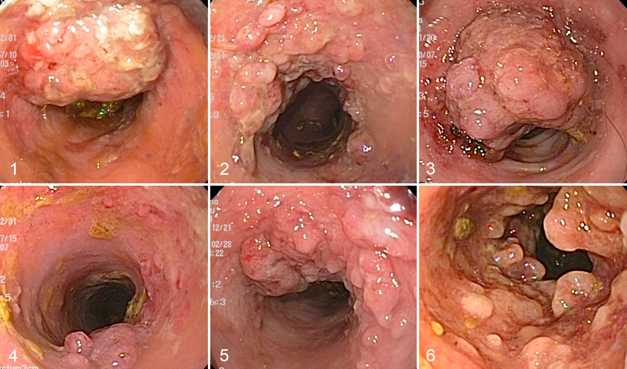
Among the 67 cases of colorectal polyps, 52 (78%) were initially diagnosed as IPMDs, 10 (15%) as adenoma, and 5 (8%) as adenocarcinoma by histopathologic examination of initial biopsy samples (Table 1, Suppl. Table S1). After initial treatment, 35 of 67 cases had polyps collected more than once, whereas 32 did not show any further clinical signs. The polyps were distributed within the colon and rectum, and lesions were not present elsewhere in the gastrointestinal tract. The lesions included single or multiple polyps that were diagnosed as IPMDs at the primary examination (Figs. 1–3). During treatment, secondary colorectal polyps that were diagnosed as not only IPMDs but also neoplastic polyps occurred in some cases (Figs. 4–6). The macroscopic morphology of the polyps was variable, whereby it was difficult to distinguish IPMDs and neoplastic polyps.
Histopathologic Diagnoses for 67 Colorectal Polyp Lesions in Miniature Dachshund Dogs.
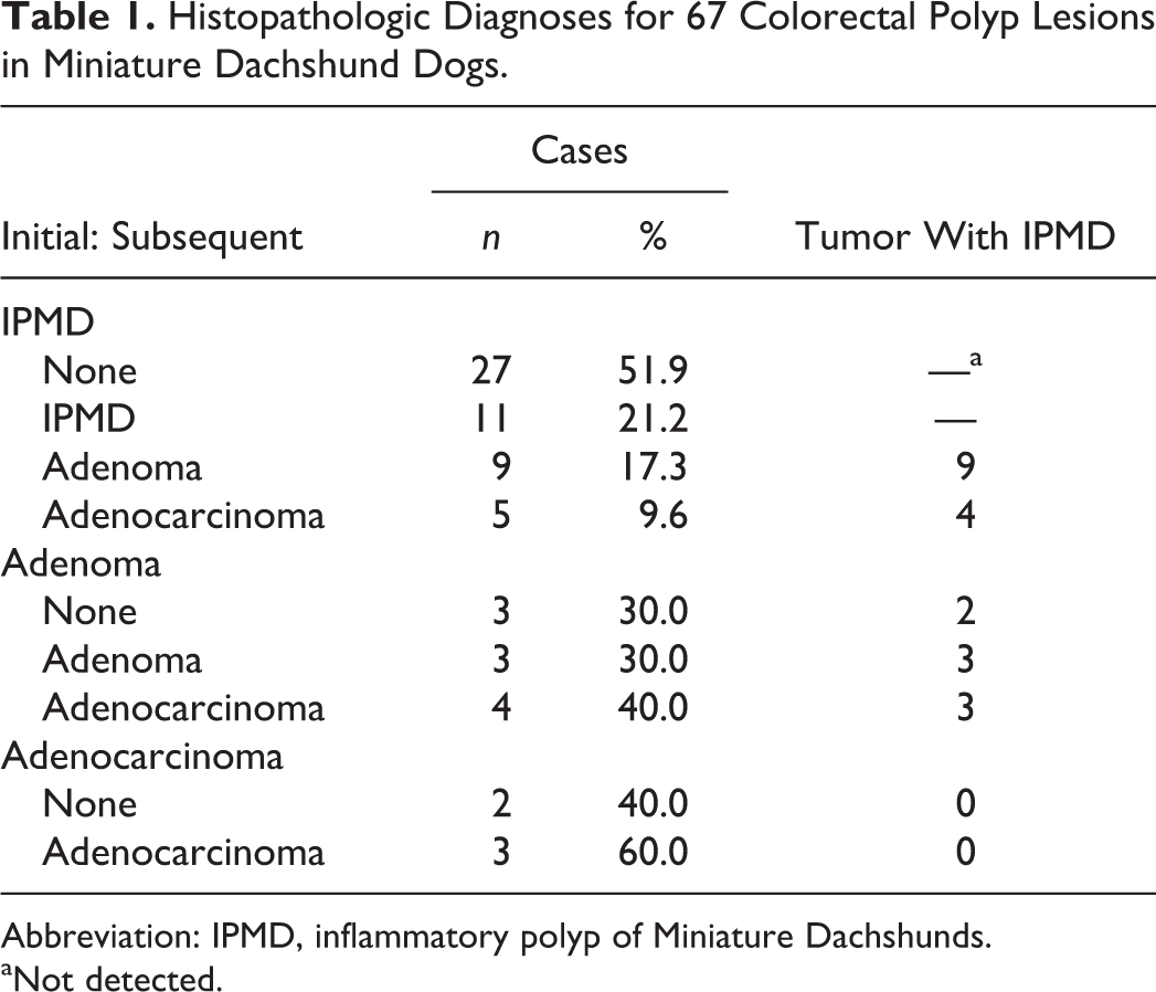
Abbreviation: IPMD, inflammatory polyp of Miniature Dachshunds.
aNot detected.
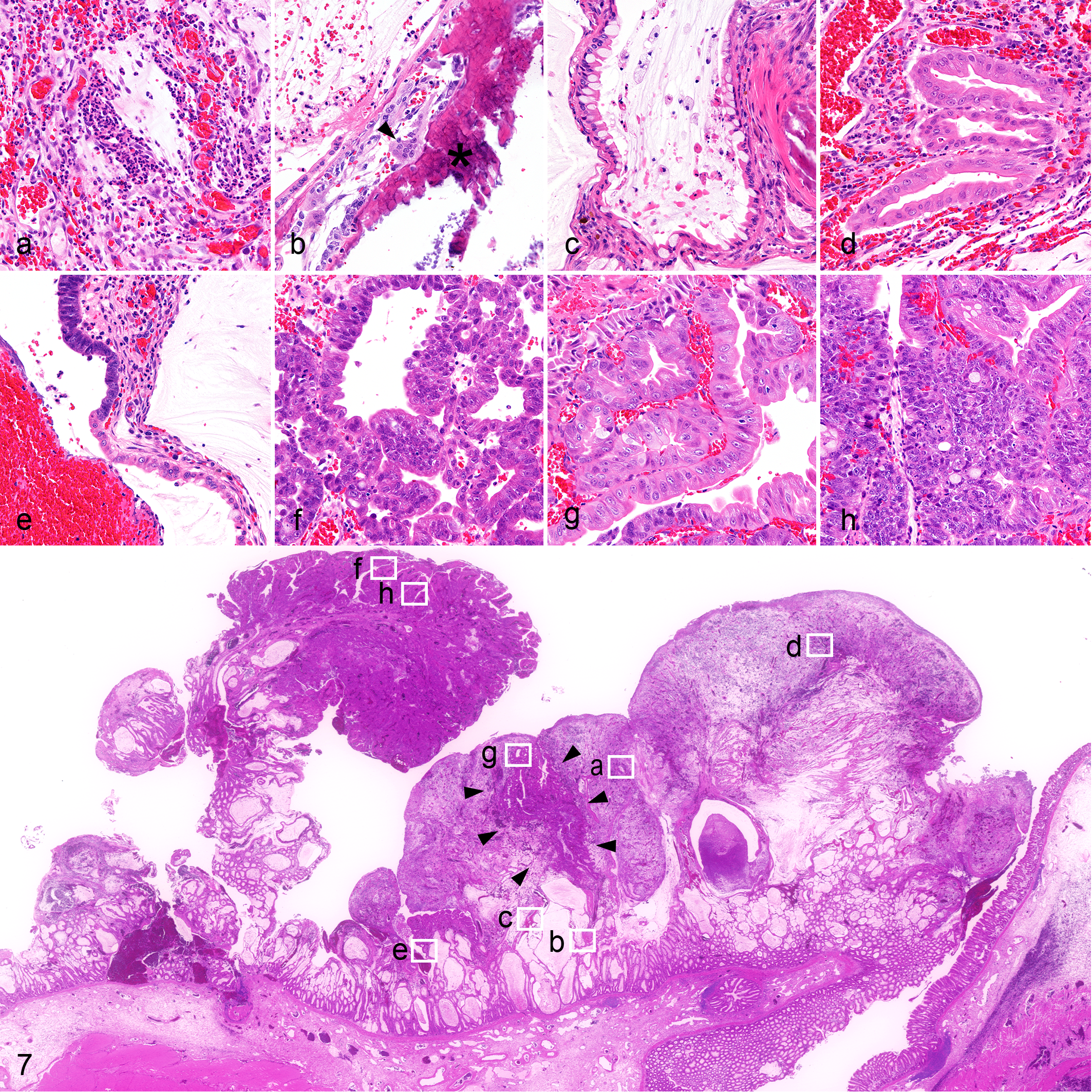
Histopathologically, IPMD consisted of granulation-like tissue that formed polypoid protrusions from the tip of the mucosa to the lumen; occasionally, the base of the lamina propria occasionally, the base of the lamina propria
Subsequent follow-up biopsies were performed in 25 cases that developed polyp lesions (48%) initially diagnosed as IPMD. Histopathologic diagnoses of the second to fourth biopsy specimens were IPMD in 11 cases (21%), adenoma in 9 (17%), and adenocarcinoma in 5 (10%; Table 1, Suppl. Table 1).
Adenoma was diagnosed on the initial biopsy for 10 cases. These consisted of columnar epithelial cells forming tubular structures without goblet cell differentiation. In many cases, the neoplastic epithelium developed within the granulation tissue of IPMDs (Figs. 8, 9). Subsequent follow-up biopsies were performed in 7 cases that developed polyp lesions initially diagnosed as adenoma. Histopathologic diagnoses of the secondary specimens were adenoma in 3 cases and adenocarcinoma in 4 cases (Table 1, Suppl. Table 1). Most cases (6 of 7) had the typical histopathologic features of IPMD within the same specimens. Overall, 18 of 20 cases (90%) that developed adenoma during the study had a previous diagnosis of IPMD or histopathologic features considered more typical for IPMD.
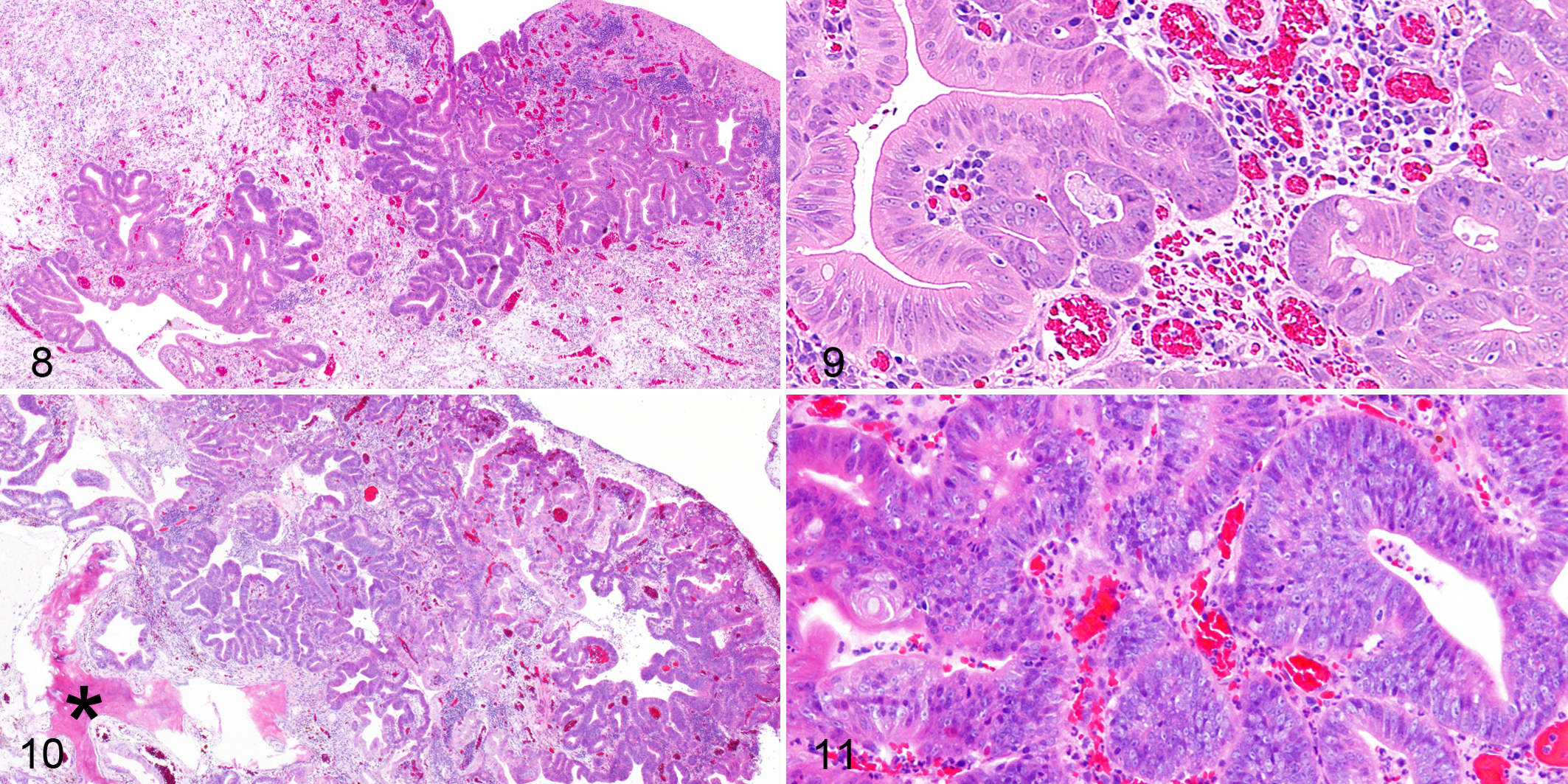
In the adenocarcinoma lesions, atypical epithelial cells showing anisocytosis and anisokaryosis formed disordered tubular structures (Figs. 10, 11). Subsequent second biopsies were performed in 3 cases, and histopathologic diagnoses were adenocarcinoma in all 3 (Table 1, Suppl. Table 1). Overall, 8 of 14 cases (57%) that developed adenocarcinoma during the study had a medical history of an IPMD or a pathologic finding of an IPMD lesion within the same section as the adenocarcinoma. However, the 5 cases that were diagnosed as adenocarcinoma at the initial examination did not show any histopathologic features of IPMD.
Positive Immunostaining of Nuclear Beta-catenin and APC
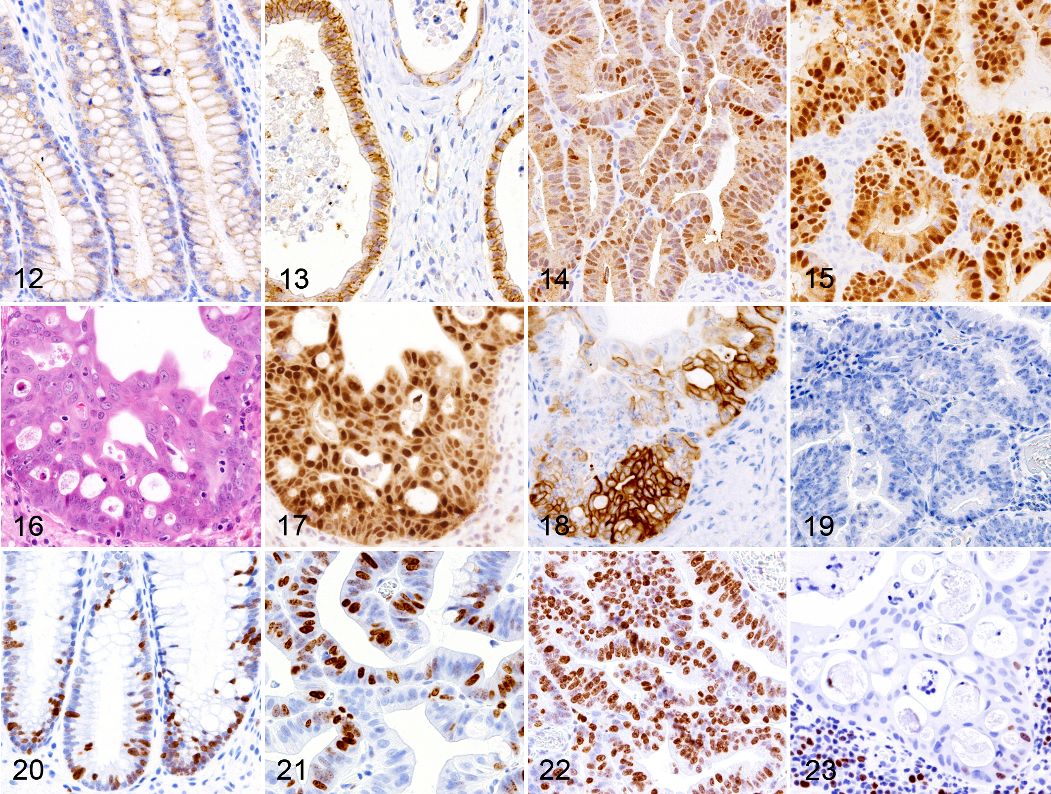
Immunohistochemical examinations were performed to assess the patterns of beta-catenin and APC immunopositivity in colorectal polypoid lesions of Miniature Dachshunds, including IPMD and neoplasia. Normal epithelial cells of the large intestine and hyperplastic goblet cells of IPMD were weakly positive for beta-catenin along the cell membrane (Fig. 12). In the dysplastic epithelial cells within the lesion of IPMD, the membranous immunopositivity for beta-catenin was either intense or weak as compared with normal epithelial cells, and a cytoplasmic positive reaction was also observed (Fig. 13). In adenomas and adenocarcinomas, the cytoplasm and nucleus of neoplastic cells were positive for beta-catenin, but the cell membrane was negative (Figs. 14, 15). Nuclear immunostaining was stronger in adenocarcinoma cells versus adenoma cells. Foci of epithelial cells with abundant eosinophilic cytoplasm and large nuclei showing stratified and/or cribriform patterns were frequently observed in IPMD lesions and occasionally in adenomas and adenocarcinomas (Fig. 16). These cells were intensely positive for nuclear beta-catenin (Fig. 17). Also, cells in these foci were strongly positive for CK5/6, a squamous epithelial cell marker (Fig. 18), although typical features of squamous epithelial cells, such as keratin formation and intercellular bridges, were absent. Columnar epithelial cells in the adenocarcinoma lesions, however, were negative for CK5/6 (Fig. 19). Epithelial cell nuclei in the lesions of IPMD as well as in the deeper crypts of the normal colorectal mucosa were positive for Ki-67 (Fig. 20), whereas those in adenoma and adenocarcinoma lesions were diffusely positive (Figs. 21, 22). The foci of squamous-like cells were mostly negative for Ki-67 (Fig. 23). CK5/6-positive cells were excluded from the quantitative analysis for beta-catenin because epithelial cells in the area should be considered intestinal epithelium rather than squamous morules (see Discussion section).
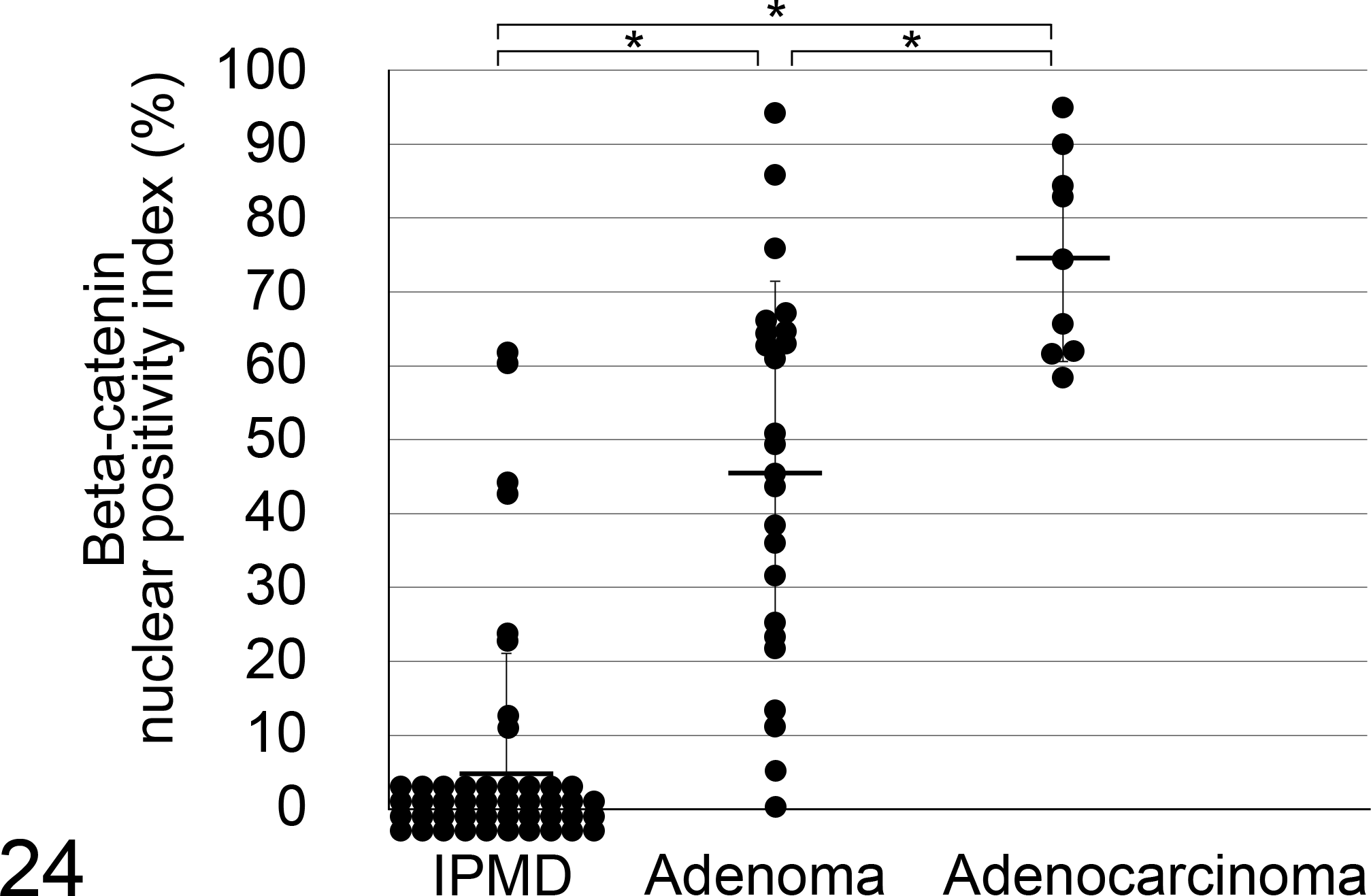
Percentages of beta-catenin-positive nuclei were 6.4%, 46.4%, and 75.4% in IPMD, adenoma, and adenocarcinoma cases, respectively (Fig. 24). The ratios were significantly higher in adenoma and adenocarcinoma versus IPMD (for the results of the statistical tests, see Suppl. Table 2).
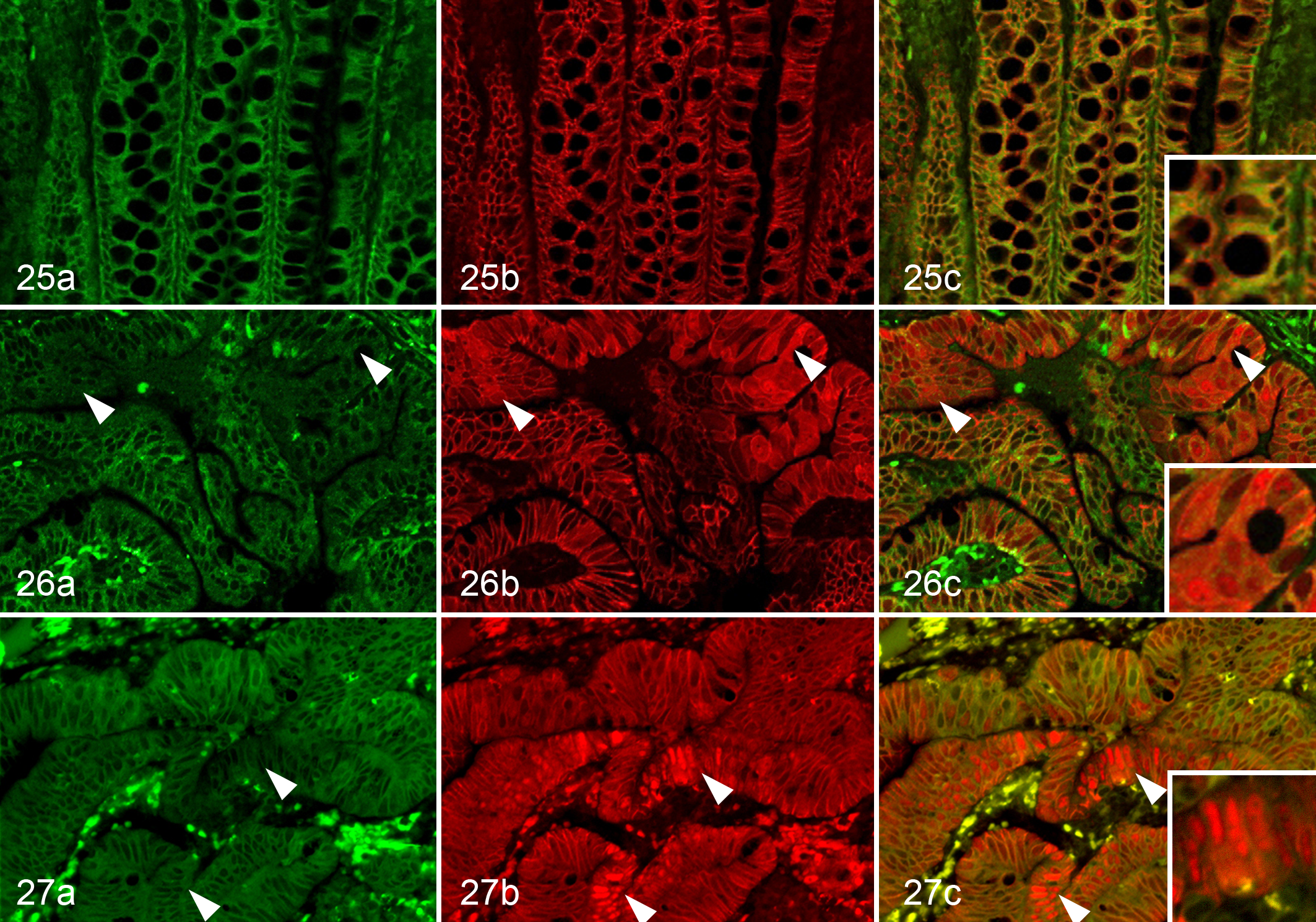
Normal epithelial cells of the intestine and hyperplastic goblet cells of IPMD showed homogeneous cytoplasmic immunostaining for APC (Fig. 25). In contrast, in adenomas and adenocarcinomas, the APC immunostaining pattern was heterogeneous, and there were both intensely and faintly stained areas (Figs. 26, 27). Interestingly, in cells strongly positive for beta-catenin, APC immunostaining was weaker and vice versa.
Discussion
Intestinal epithelial tumors are rare in dogs. 8,12,13,37 Although nonneoplastic polyps in the canine colorectum are also uncommon, Miniature Dachshunds tend to develop a nonneoplastic polyp in the colorectum, termed IPMD. 25 In the present study, we were able to examine sequential biopsy samples of colorectal polyp lesions in Miniature Dachshunds. In sum, 48% (25 of 52) of the IPMD cases were subsequently biopsied, at which point 56% (14 of 25) developed adenoma and/or adenocarcinoma. Moreover, in 22 of 29 (76%) cases diagnosed as either adenoma or adenocarcinoma during the study, the dog had been previously diagnosed with IPMD, or some adenomas and adenocarcinoma had features that were considered more typical for IPMD. These results suggest that IPMD may consist of preneoplastic lesions that can progress into adenoma and adenocarcinoma. However, cases diagnosed as adenocarcinoma at the first biopsy did not show any histopathologic features of IPMD. In these cases, adenocarcinoma may have developed independently from IPMD, or possibly the precedent IPMD lesion was completely replaced by adenocarcinoma. There is no direct evidence that it is the same polyp that recurred; it is also possible that a new lesion developed at a similar site, or lesions were not completely removed. However, the colorectal site was confined to a small region because Miniature Dachshunds are small-breed dogs. Although the occurrence of canine intestinal tumors is rare, 8,12,13,37 of the 52 dogs that developed inflammatory polyps, 14 (27%) developed neoplastic polyps during treatment. In addition, the occurrence of neoplastic epithelium and IPMD within the same specimen suggests that IPMD is potentially the origin of the tumors.
In humans, it has been suggested that colorectal polyps are caused by abnormalities of beta-catenin and APC. In this study, to clarify the contribution of beta-catenin to the development of colorectal tumors in dogs, we performed a quantitative analysis of beta-catenin-positive nuclei, which demonstrated special cells showing highly positive intensity of beta-catenin. At first glance, these cells had malignant features, such as abundant eosinophilic cytoplasm and large nuclei, and showed stratified and/or cribriform patterns. To investigate proliferative activity of the atypical cells, immunostaining of the polyps was performed for Ki-67. A cell proliferation marker, Ki-67 was expressed in the IPMD lesion as well as in the crypts of normal colorectal mucosa, whereas it was expressed diffusely in the lesions of adenomas and adenocarcinomas with disorganized cell proliferation. Interestingly, foci of atypical cells did not show a positive reaction for Ki-67, whereas the atypical cells were positive for CK5/6, although these foci lacked the typical morphologic features of squamous cells.
In humans, squamous metaplasia has been reported in various epithelial tumors, including colorectal adenoma and adenocarcinoma. 1,2,5,10,20 Besides the typical squamous metaplastic lesions, foci of squamous-like cells are occasionally found in tumors of the endometrium, 14,40 stomach 29 and colorectum, 22,27 and these foci are called squamous morules. Cells in such squamous morules are reported to be positive for CK5/6, although they lack typical morphologic features of squamous epithelium, such as keratin formation, intercellular bridges, and/or prominent cell membranes, 14 as observed in the present canine intestinal lesions. Thus, such canine and human morules are considered to exhibit incomplete or immature squamous differentiation. 4 The foci of CK5/6-positive squamous-like cells were also strongly positive for nuclear beta-catenin in the present study. Despite a broad distribution of nuclear beta-catenin, Ki-67- and p53-positive cells are rarely observed in the squamous morules in human colorectal and gastric tumors. 22,29 Thus, there is still an ongoing debate on whether squamous morules are inert malignant or benign elements of a tumor. With the morphologic features and immunohistochemical properties, the foci of CK5/6-positive squamous-like cells in the IPMD, adenoma, and adenocarcinoma are considered to be similar to the squamous morules described in human tumors. Therefore, CK5/6-positive cells were excluded from the quantitative analysis because they deviated from the objective to evaluate enterocyte involvement with tumorigenesis.
The number of nuclear beta-catenin-positive cells was significantly increased in adenoma and adenocarcinoma versus IPMD. As previously reported for canine intestinal tumors, 21,31 the APC positivity observed in the present study was decreased in the areas of intense nuclear beta-catenin-positive reaction. In human patients with familial adenomatous polyposis, benign polyp lesions often undergo malignant transformation in association with mutational inactivation of the APC gene. 9 Moreover, APC is the most commonly affected tumor suppressor gene in sporadic colorectal tumors of dogs 41 as well as humans. 7,30,34 From the results of the present study, the decreased intensity of APC immunolabeling is likely related to beta-catenin nuclear translocation in IPMD, which can cause dedifferentiation and proliferation of intestinal epithelial cells. These results are consistent with a previous report that evaluated the expression of beta-catenin and APC protein in canine colorectal tumors. 31
In conclusion, the present study revealed that IPMD in Japan is a progressive disorder that may subsequently develop adenoma and adenocarcinoma. In such cases, aberrant APC and beta-catenin expression may be involved in tumor progression.
Supplemental Material
Supplemental Material, DS1_VET_10.1177_0300985818777798 - Histopathologic Features of Colorectal Adenoma and Adenocarcinoma Developing Within Inflammatory Polyps in Miniature Dachshunds
Supplemental Material, DS1_VET_10.1177_0300985818777798 for Histopathologic Features of Colorectal Adenoma and Adenocarcinoma Developing Within Inflammatory Polyps in Miniature Dachshunds by Tsubasa Saito, James K. Chambers, Ko Nakashima, Eri Uchida, Koichi Ohno, Hajime Tsujimoto, Kazuyuki Uchida, and Hiroyuki Nakayama in Veterinary Pathology
Supplemental Material
Supplemental Material, DS2_VET_10.1177_0300985818777798 - Histopathologic Features of Colorectal Adenoma and Adenocarcinoma Developing Within Inflammatory Polyps in Miniature Dachshunds
Supplemental Material, DS2_VET_10.1177_0300985818777798 for Histopathologic Features of Colorectal Adenoma and Adenocarcinoma Developing Within Inflammatory Polyps in Miniature Dachshunds by Tsubasa Saito, James K. Chambers, Ko Nakashima, Eri Uchida, Koichi Ohno, Hajime Tsujimoto, Kazuyuki Uchida, and Hiroyuki Nakayama in Veterinary Pathology
Footnotes
Acknowledgements
We are grateful to Pete Aughton, William Ruddock, and Dr Abbas Fotovati, ITR Laboratories Canada Inc, for carefully proofreading the manuscript.
Declaration of Conflicting Interests
The author(s) declared no potential conflicts of interest with respect to the research, authorship, and/or publication of this article.
Funding
The author(s) disclosed receipt of the following financial support for the research, authorship, and/or publication of this article: This study was supported in part by the Japan Society for the Promotion of Science (grant 15K14863).
Supplementary material for this article is available online.
References
Supplementary Material
Please find the following supplemental material available below.
For Open Access articles published under a Creative Commons License, all supplemental material carries the same license as the article it is associated with.
For non-Open Access articles published, all supplemental material carries a non-exclusive license, and permission requests for re-use of supplemental material or any part of supplemental material shall be sent directly to the copyright owner as specified in the copyright notice associated with the article.