
Abstract
Oral and cutaneous tissues are the most frequent origin in canine squamous cell carcinoma (SSC). In SCC, changes in adhesion molecule expression and transition from epithelial to mesenchymal phenotype are thought to be important in development of invasive behavior of neoplastic cells at the leading front of the tumor. We therefore investigated histological invasive front grading and epithelial-mesenchymal transition (EMT) in both oral SCCs and cutaneous SCCs. EMT was assessed by evaluating immunohistochemical expression of E-cadherin, β-catenin, desmoglein, vimentin, and N-cadherin. Regardless of the anatomic location, invasive front grading resulted in higher histological grades than grading of the surface. Most oral SCCs were of significantly higher histologic grade than cutaneous SCCs (P < .01). Expression of E-cadherin, β-catenin, and desmoglein was significantly lower in oral SCC compared with cutaneous SCC (P < .01). A significant association was found between invasive front grading and loss of E-cadherin, β-catenin, and desmoglein (P < .01). Also, vimentin-positive neoplastic cells had low immunoreactivity of these adhesion molecules, and a few of these neoplastic cells were positive for N-cadherin. These results suggest not only E-cadherin and β-catenin but also desmoglein as markers for predicting biological behavior of canine SCC. Depending on their primary sites, EMT correlates with biological behavior and therefore histological grade of canine SCC. We suggest that combining invasive front grading with assessment of immunohistochemical expression of E-cadherin, β-catenin, and desmoglein may allow more accurate prediction of biological behavior of canine SCCs.
Keywords
Squamous cell carcinoma (SCC) is a malignant epithelial tumor with keratinocyte differentiation. 8,9 SCC is one of the most common neoplasms in the oral cavity of dogs, and the frequency distribution indicates that the gingiva is more often affected than other sites such as the lips, tongue, palate, tonsils, and pharynx. Oral SCC in the gingiva invades the bone of the maxilla and mandible and destroys periodontal structures. Although oral SCC does not commonly metastasize to distant sites, it occasionally metastasizes to regional lymph nodes. 13,14 Cutaneous SCC in dogs can occur anywhere on the skin including the head and neck, abdomen, forelimbs, hindlimbs, perineum, and digits, but it shows slow growth and usually does not metastasize, even to regional lymph nodes. 8,9
This malignant neoplasm is comprised of heterogeneous cells with various phenotypes. Centrally located tumor cells are more differentiated compared with those invading the stromal tissue. The latter probably represent the true biological behavior of SCC. 5,6 In the human oral SCC, an improved prognostic value has been reported, a multifactorial malignancy grading system that evaluates only neoplastic cells of the deep invasive front of the neoplasm. 1,5,6,31
The loss of intercellular adhesion molecules such as E-cadherin, β-catenin, and desmoglein is associated with development of invasiveness of tumor cells in carcinoma progression, and the adhesion molecules are considered useful biological markers for evaluation of prognosis in human carcinoma including oral SCC. 2,3,15,16,23,24,30,32,38 –40 Similarly, epithelial-mesenchymal transition (EMT) plays a key role in carcinoma progression and is necessary for invasion and metastasis. 11,17,35,36,38 Defining characteristics of EMT are loss of the epithelial phenotype and acquisition of a mesenchymal phenotype, including attenuation of E-cadherin and acquisition of vimentin and N-cadherin expression. 11,18,26,29,38 E-cadherin is a major cadherin known as a typical cell-cell adhesion molecule in epithelial cells, and the intracytoplasmic domain of its terminal tail is linked to actin filaments by cytoplasmic proteins called catenins. 21,34 Catenin molecules have 3 types (α-, β-, and γ-catenin), of which β-catenin has a cell-cell adhesion function to bind directly to E-cadherin and form the E-cadherin/β-catenin complex. 21 Many reports have indicated that the loss of E-cadherin and β-catenin plays an important role in the progression and metastasis of human SCC. 2,3,23,24,29,38 –40 Desmoglein is a transmembrane glycoprotein component of desmosome (a cell-cell adhesive structure prominent in epithelial cells), and the expression of desmoglein has been reported to be associated with tumor progression. 15,22,41 N-cadherin is a classic cadherin located mainly in neural tissue and striated muscle. 33 In carcinomas, EMT is characterized immunohistochemically by the expression of N-cadherin and vimentin. 11,18,26,29,38
The aim of this study was to evaluate the expression of adhesion molecules E-cadherin, β-catenin, and desmoglein in canine SCC of the oral cavity and skin, based on invasive front grading. Furthermore, we investigated the expression of vimentin and N-cadherin to evaluate the association between histological grade and EMT. We hypothesized that in neoplastic cells at the invasive front of the tumor, higher grade neoplasms would have loss of E-cadherin, β-catenin, and desmoglein but greater expression of vimentin and N-cadherin.
Material and Methods
Samples
Biopsy samples, 55 of oral SCC and 58 of cutaneous SCC, were collected from 113 dogs. The sites of oral SCC were the mucosa of gingiva (n = 40), tongue (n = 13), larynx (n = 1), and cheek (n = 1); the sites of cutaneous SCC were the epidermis of head (n = 17), neck (n = 7), thorax (n = 3), abdomen (n = 9), dorsal region (n = 1), inguinal (n = 6), forelimb (n = 7), hindlimb (n = 5), digit (n = 1), tail (n = 1), and preputium (n = 1).
Biopsy samples were obtained by mandibulectomy, maxillectomy, incisional biopsy, excisional biopsy, or needle biopsy, fixed in 10% phosphate-buffered formalin, embedded in paraffin, and cut at 4-μm thickness. The sections were stained with hematoxylin and eosin.
Histological Grading and Immunohistochemistry
Histological grading of the deep invasive front of tumors is considered to provide the most useful prognostic information, 1,5,6,23,27,28,31 and Bryne et al 5,6 developed a histological grading system that evaluated only the invasive front of the tumors. This system was used to classify tumors in 3 grades based in a total score obtained from morphological features described in Table 1. In addition, intravasation of tumor cells (the invasion of neoplastic cells into blood or lymphatic vessels) was investigated in each neoplasm.
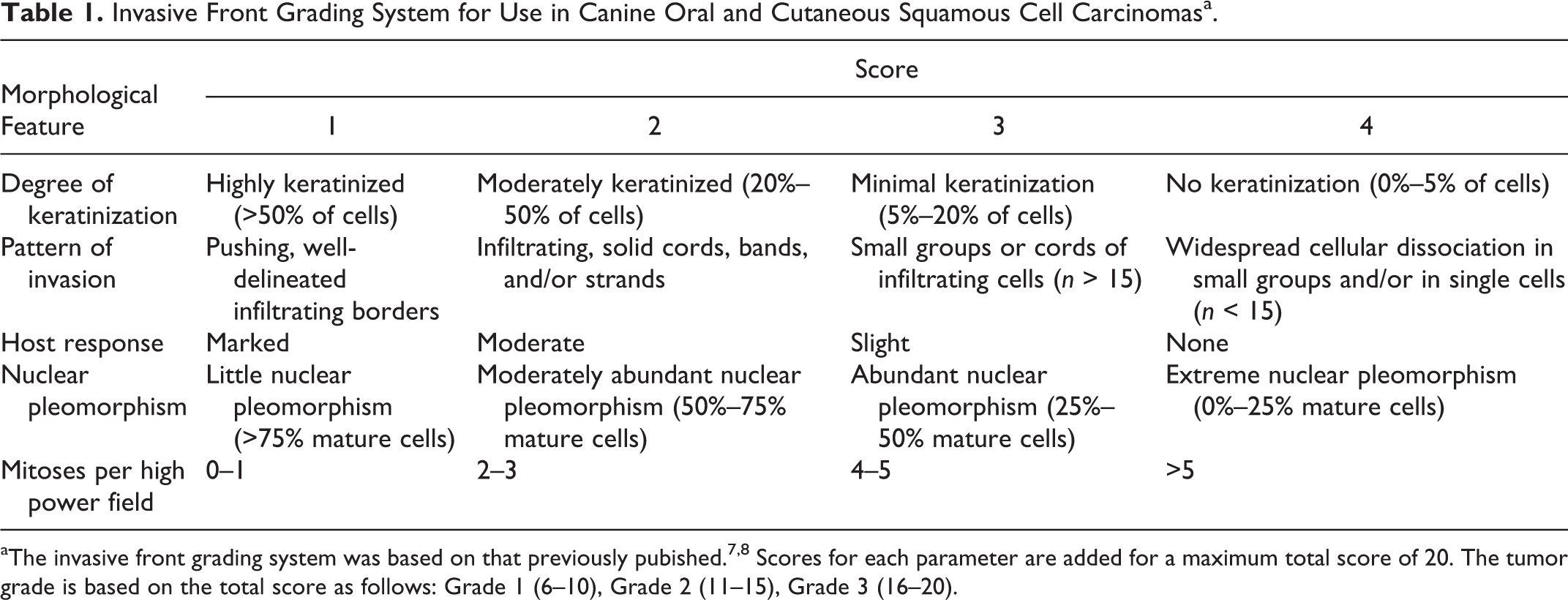
Invasive Front Grading System for Use in Canine Oral and Cutaneous Squamous Cell Carcinomasa.
Histologic sections were examined immunohistochemically using the avidin–biotin–peroxidase complex procedure (Vectastain Elite ABC Kit; Vector Laboratories, Burlingame, California). Details of the primary antibodies used are listed in Supplemental Table S1. To block endogenous peroxidase, all sections were immersed in 0.5% periodic acid solution at room temperature for 10 minutes. All sections were incubated with primary antibody at 4°C for 16 hours, with biotinylated secondary antibody for 30 minutes at room temperature, and with ABC for an additional 30 minutes. Visualization was accomplished using 3,3′-diaminobenzidine. The pattern of immunoreactivity for adhesion molecules (E-cadherin, β-catenin, and desmoglein) was described as normal when homogeneous staining was detected. Abnormal staining included heterogeneous or focal staining. For the adhesion molecules, the cases were scored according to whether 0%–10%, 11%–50%, 51%–75%, or 76%–100% of cells showed a normal pattern of membranous staining and were allocated scores of 0, 1, 2, or 3, respectively. 23 For vimentin, the neoplasms were evaluated according to whether 0, 1%–25%, 26%–50%, or 51%–100% of tumor cells showed cytoplasmic staining and were allocated scores of 0, 1, 2, or 3 respectively. For N-cadherin, the presence or absence of immunolabeling was investigated in the neoplasms (14 cases of oral SCC and 9 cases of cutaneous SCC) that showed attenuation of E-cadherin immunolabeling.
The individual animal raw data, the histologic scores, presence of intravasation, and immunohistochemical scores in oral and cutaneous SCC are summarized in Supplemental Tables S2 and S3.
Statistical Analysis
The relationships of each morphological score, the total score, the appearance of tumor cell intravasation, and the immunohistochemical score for each adhesion molecule and vimentin between oral and cutaneous SCC were compared by use of the Mann-Whitney U test. Furthermore, Spearman’s rank correlation coefficient by rank test was used to investigate the correlations among each morphological score and the total score, along with the immunoreactivity of each adhesion molecule and vimentin. P < .05 was considered statistically significant.
Results
Histologic Grading
The neoplastic cells were evaluated for each morphological feature compared with squamous cells of normal epidermis or mucous epithelium. The neoplastic cells at the invasive front tended to show poorer differentiation and more invasive growth pattern cells at than the surface or center of the neoplasm in both oral and cutaneous SCC. Results of the histologic grading of oral and cutaneous SCC are summarized in Table 2.
Histologic Grading of Oral and Cutaneous Squamous Cell Carcinoma (SCC) in Dogsa.
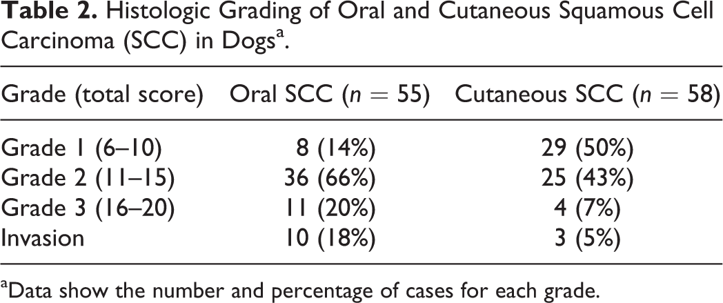
aData show the number and percentage of cases for each grade.
Of the 55 cases of oral SCC, 8 (14%) were classified as Grade 1, forming large nests with differentiation to form a squamous cell layer (Fig. 1). In the 36 (66%) cases classified as Grade 2, dendrites (branching finger-like arrangements of neoplastic cells) infiltrated from the mucosa and formed small nests with moderate lymphocyte infiltration (Fig. 2). In the 11 (20%) cases classified as Grade 3, clumps of a few tumor cells with marked nuclear pleomorphism were sporadically observed (Fig. 3), and undifferentiated tumor cells were isolated as single cells separate from the tumor nests. Intravasation (invasion of neoplastic cells into vessels) was observed in 10 (18%) cases of oral SCC, including 1 of 8 (13%) Grade 1 neoplasms, 6 of 36 (17%) Grade 2 neoplasms, and 3 of 11 (27%) Grade 3 neoplasms.
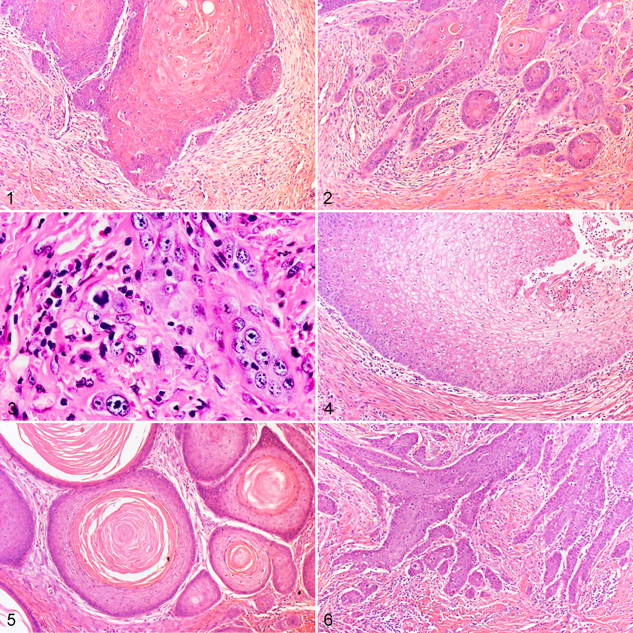
Of the 58 cases of cutaneous SCC, 29 (50%) classified as Grade 1; there were expansive and formed large masses with differentiation similar to the stratum spinosum of the epidermis (Fig. 4). The 25 (43%) neoplasms classified as Grade 2 grew in thick cords from the epidermis, with moderate to slight infiltration of lymphocytes (Fig. 5). In the 4 (7%) neoplasms classified as Grade 3, the tumor cells formed infiltrating dendritic arrangements and isolated single cells with nuclear pleomorphism, and no keratinization of tumor cells was observed (Fig. 6). Intravasation (invasion of neoplastic cells into vessels) was observed in 3 (5%) cases of cutaneous SCC including 0 of 29 Grade 1 neoplasms, 2 of 25 (8%) Grade 2 neoplasms, and 1 of 4 (25%) Grade 3 neoplasms.
Immunohistochemical Findings
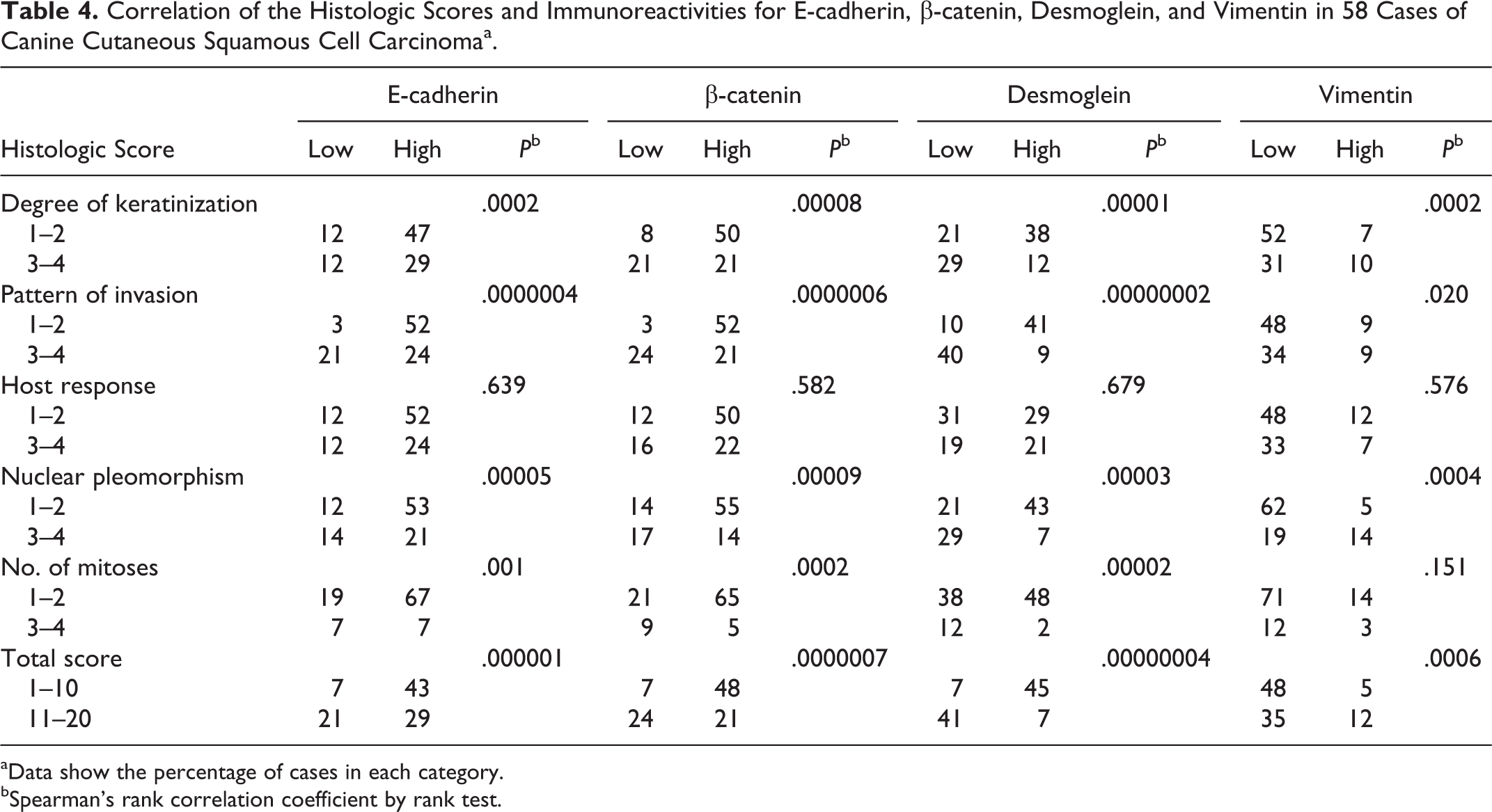
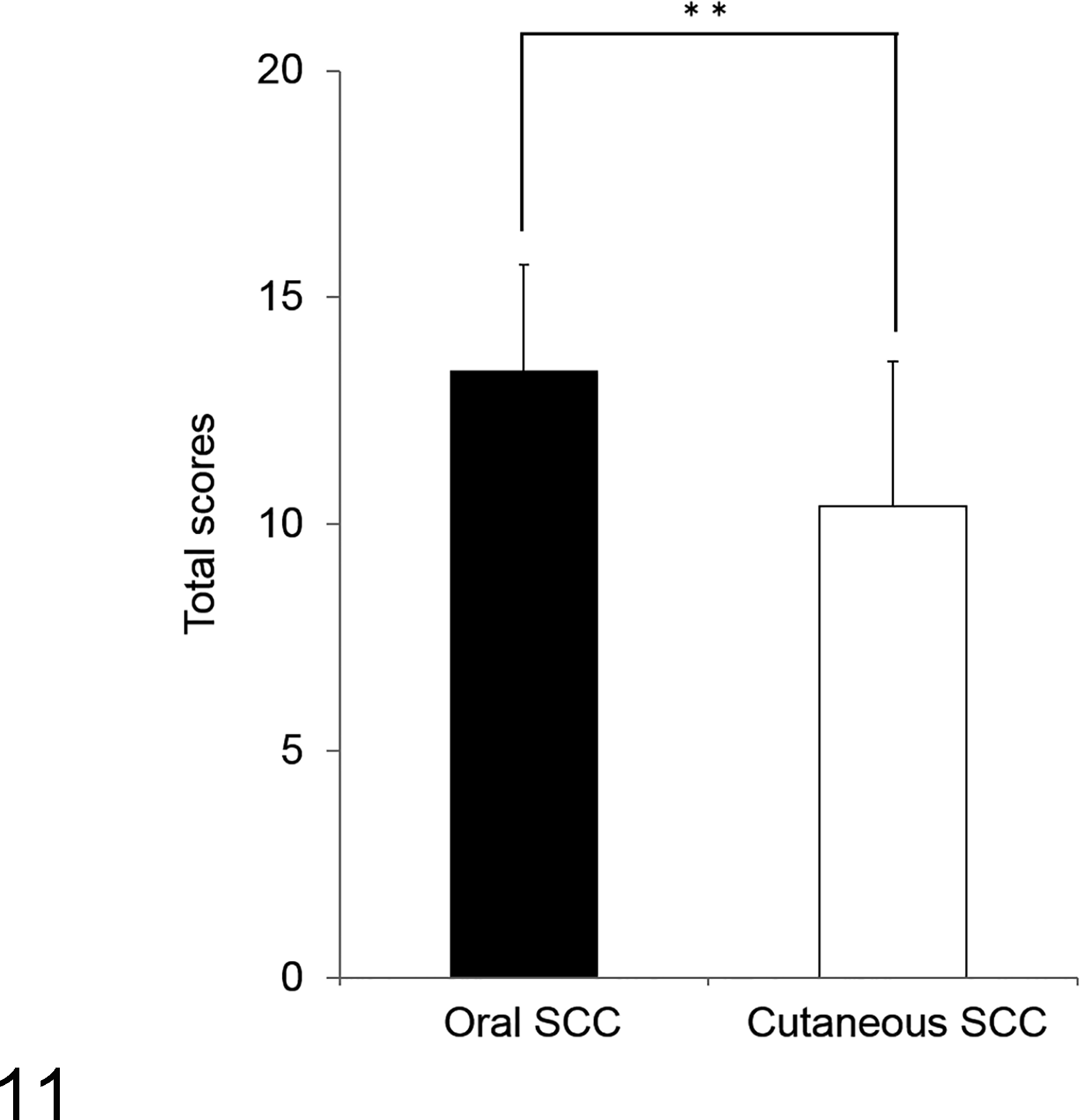
In the normal oral mucosa and cutaneous epidermis, the epithelial cells in the basal and squamous layers showed high membranous immunoreactivity for E-cadherin, β-catenin, and desmoglein but were negative for vimentin and N-cadherin (Suppl. Figs. S1 and S2). Immunohistochemical results for oral and cutaneous SCC are summarized in Tables 3 and 4.
Correlation of the Histologic Scores and Immunoreactivities for E-cadherin, β-catenin, Desmoglein, and Vimentin in 55 Cases of Canine Oral Squamous Cell Carcinomaa.
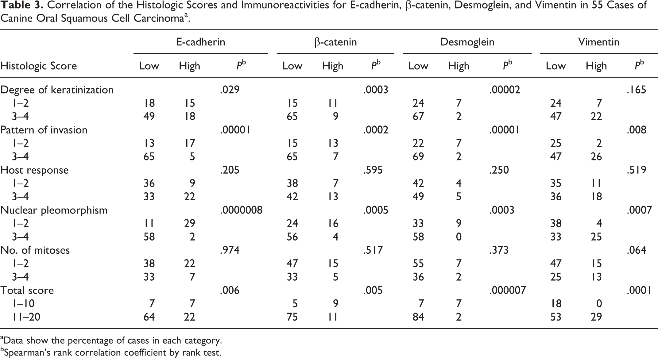
aData show the percentage of cases in each category.
bSpearman’s rank correlation coefficient by rank test.
Correlation of the Histologic Scores and Immunoreactivities for E-cadherin, β-catenin, Desmoglein, and Vimentin in 58 Cases of Canine Cutaneous Squamous Cell Carcinomaa.
aData show the percentage of cases in each category.
bSpearman’s rank correlation coefficient by rank test.
In oral and cutaneous SCC, the tumor cells that formed large nests with differentiation to the squamous cell layer showed high immunoreactivity for E-cadherin, with membranous labeling in a circumferential or honeycomb-like arrangement in the basal cell layer and squamous cell layer (Fig. 7a). In the same region, the tumor cells showed high homogeneous immunoreactivity for β-catenin and desmoglein but were negative for vimentin (Fig. 7b-d). In contrast, the tumor cells that formed small nests or were isolated as single cells showed low immunoreactivities for E-cadherin, β-catenin, and desmoglein (Fig. 8a-c) and high immunoreactivity for vimentin (Fig. 8d). In 2 neoplasms, a few invading tumor cells showed low membranous immunoreactivity for N-cadherin (Fig. 9a and 9d). These N-cadherin-positive tumor cells had low immunoreactivity for E-cadherin and high immunoreactivity for vimentin (9b and 9c).
In oral and cutaneous SCC, intravasated tumor cells exhibited various expressions of each adhesion molecule and vimentin. However, some tumor cells showed high immunoreactivity for vimentin and low immunoreactivity for each adhesion molecule.
Quantitative Analysis of Histologic and Immunohistochemistry Findings
Correlations of histological scores and immunoreactivities for adhesion molecules and vimentin in oral and cutaneous SCC are summarized in Tables 3 and 4. A significant association was found between the total score (leading front grade) and lower expression of all adhesion molecules in oral and cutaneous SCC (P < .01). Also, lower expression of all adhesion molecules was associated with the individual morphological scores including the degree of keratinization, pattern of invasion, and nuclear polymorphism in oral SCC (P < .01–.05) and the degree of keratinization, pattern of invasion, nuclear polymorphism, and number of mitoses in cutaneous SCC (P < .01). Particularly, the pattern of invasion was intimately related to decreased expression of the adhesion molecules. The expression of vimentin was significantly associated with the total score, the pattern of invasion, nuclear polymorphism, and decreased expression of all adhesion molecules in oral SCC (P < .01–.05) and was associated with the total score, the degree of keratinization, pattern of invasion, nuclear polymorphism, and decreased expression of all adhesion molecules in cutaneous SCC (P < .01–.05).
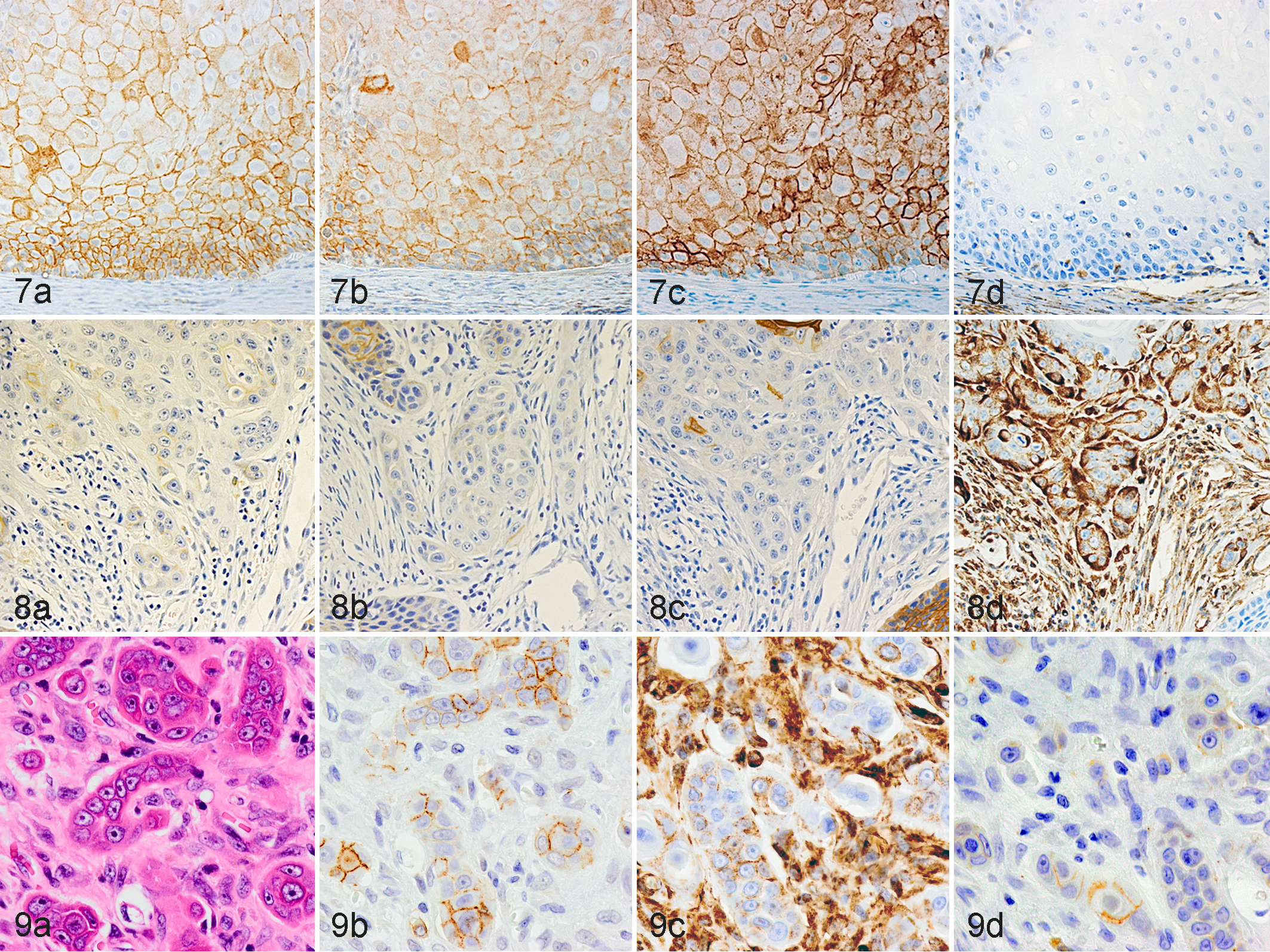
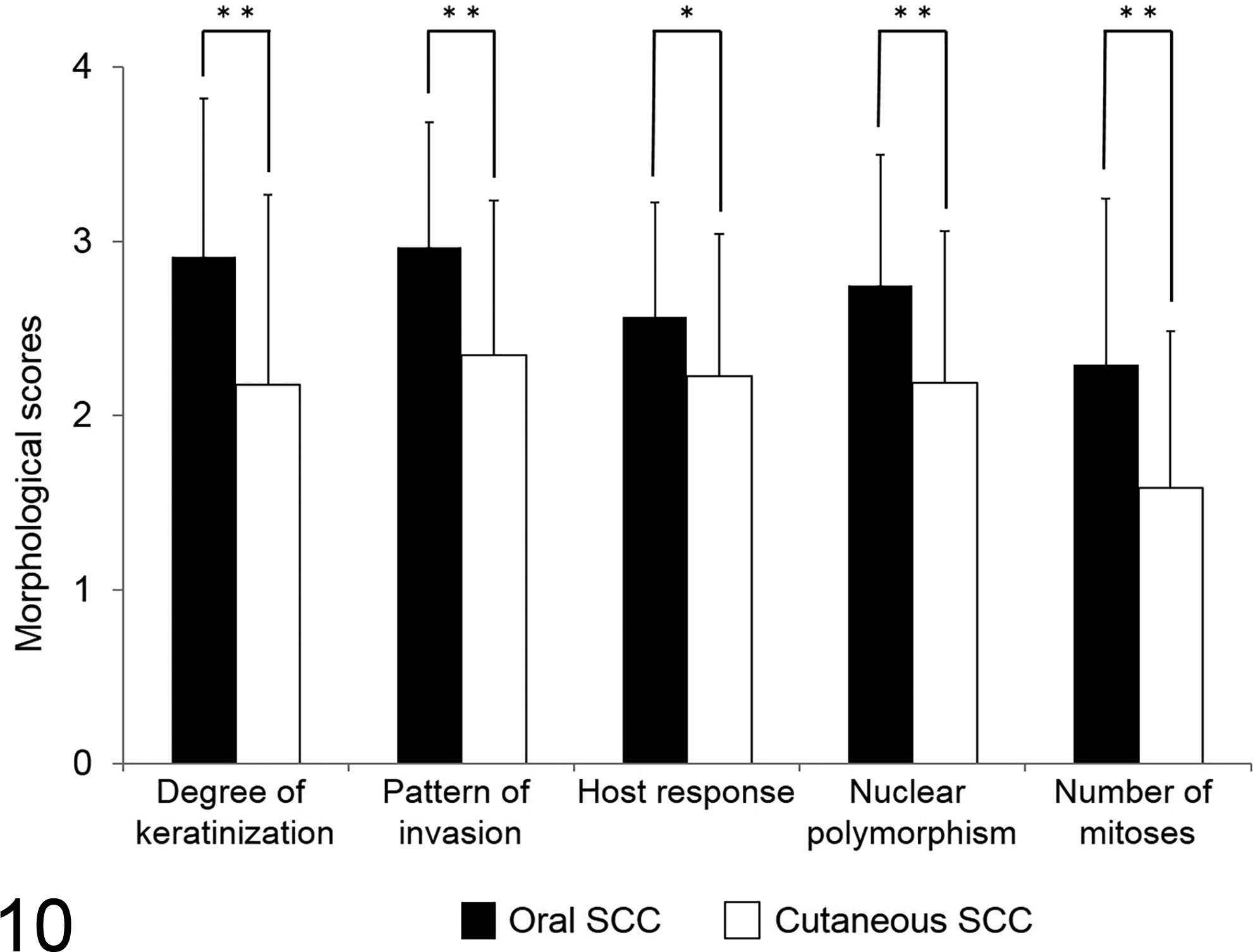
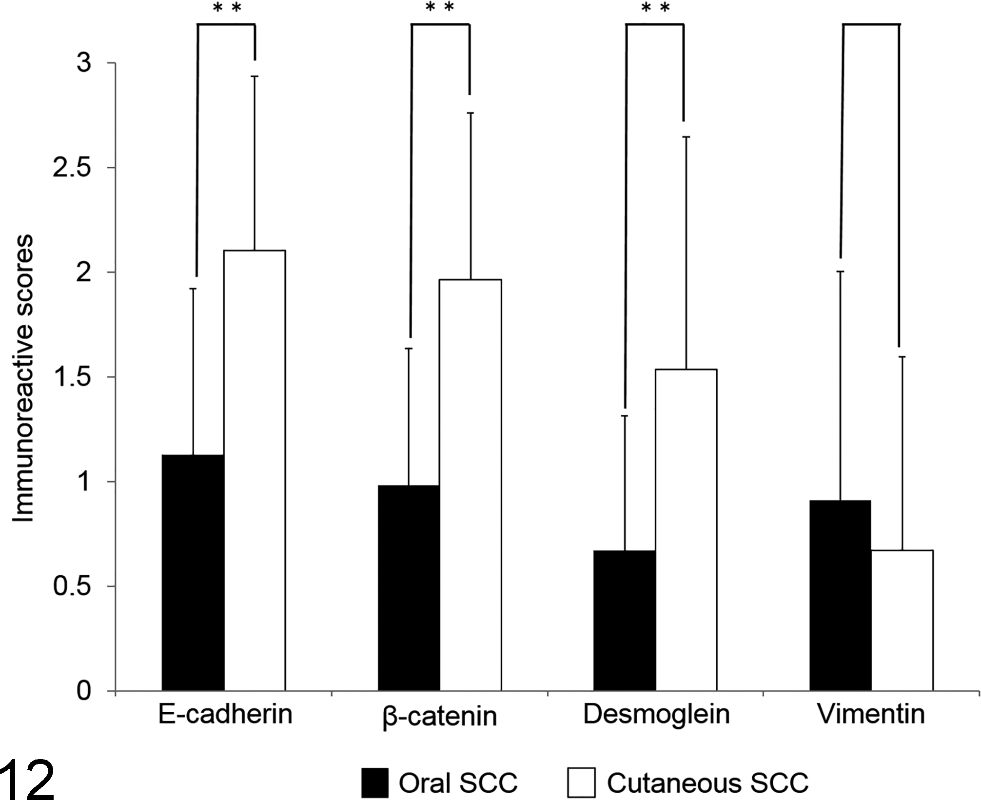
The total score and all individual morphological scores were significantly higher in oral SCC compared with cutaneous SCC (P < .01–.05, Figs. 10 and 11). Also, there was more intravasation in oral SCC than in cutaneous SCC (Table 2). Expression of all adhesion molecules was significantly lower in oral SCC than cutaneous SCC (P < .01, Fig. 12), whereas a significant difference was not observed between oral and cutaneous SCC in their expression of vimentin (P > .05, Fig. 12).
Discussion
Our objective was to evaluate the expression of E-cadherin, β-catenin, and desmoglein based on invasive front grading and to investigate the expression of vimentin and N-cadherin to evaluate the association between histological grade and EMT in canine oral and cutaneous SCC. The results of this study showed that the higher grade neoplasms had loss of these adhesion molecules but gained expression of vimentin and N-cadherin.
In the cases of oral and cutaneous SCC examined in this study, the deep invasive fronts on the neoplasms exhibited higher histological grade than the surface or center of the neoplasm, and the invasive front of oral SCC showed a significantly higher histological grade compared with cutaneous SCC. A number of studies have demonstrated that the invasive front grading system is a significantly better prognostic predictor in human oral SCC. 1,5,6,38,40 Canine oral SCCs have reportedly showed invasiveness, such as bone involvement, as well as frequent local recurrence and low rates of metastasis to regional lymph nodes. Cutaneous SCC, in contrast, showed slow growth. 8,9,13,14 These studies support the hypothesis that canine oral SCC has greater invasiveness than cutaneous SCC.
The decrease of E-cadherin and β-catenin expression was significantly associated with the total score and morphological scores, especially in the pattern of invasion, in both oral and cutaneous SCC. This result suggests that E-cadherin and β-catenin may be useful markers for the evaluation of histological grade. Mestrinho et al 25 reported an association between decreased membranous expression of E-cadherin and tumor grade in the invasive front of canine oral SCC. Also, E-cadherin-negative cells in the invasive front often expressed vimentin, and vimentin expression was significantly associated with the decrease of E-cadherin expression and the total score in both oral and cutaneous SCC. Although the expression of E-cadherin in oral SCC was significantly lower than that of oral SCC, the vimentin immunoreactivity was not statistically different between oral and cutaneous SCC. Similarly, others have reported that the less differentiated tumor cells, mostly at the periphery of the neoplasms on the invading edge, were vimentin positive and E-cadherin negative in canine digital SCC. 4 These findings indicate that EMT is involved with histological grade in the invasive front, and we consider that the loss of E-cadherin rather than the acquisition of vimentin may contribute to the higher histologic grade of oral SCC compared with cutaneous SCC. Recently, several investigations addressing the involvement of EMT with tumor progression have been reported in human SCC. 18 –20,38,39 These studies support the theory that EMT is correlated with tumor progression in the invasive front of canine oral and cutaneous SCC.
Loss of E-cadherin did not necessarily correspond to the expression of vimentin, and tumor cells negative for both E-cadherin and vimentin were also observed in oral and cutaneous SCC. Although the loss of E-cadherin and the acquisition of vimentin are important mechanisms in the EMT process, a detailed cause-and-effect relationship has not been revealed. In other studies, up-regulation of vimentin was not necessarily associated with loss of E-cadherin expression, and the activation of a mesenchymal switch of tumor cells may be at least in part uncoupled from transcriptional down-regulation of E-cadherin in human head and neck SCC. 7 Although the additional investigations are necessary, we consider that loss of E-cadherin precedes the acquisition of vimentin in early-stage EMT in canine oral and cutaneous SCC.
N-cadherin expression was observed in a few tumor cells of oral SCC that showed strong invasiveness but was not observed with any neoplastic cells of cutaneous SCC. N-cadherin-positive cells showed high immunoreactivity for vimentin and low immunoreactivity for E-cadherin. In aggressive carcinomas, the neoplastic cells express N-cadherin with decreased E-cadherin expression due to an EMT phenomenon called the “cadherin switch,” which increases the motility and invasiveness of the neoplastic cells. 12,29 The cadherin switch has been reported in human head and neck and oral SCC. 42 The obtained findings in this study may indicate that the strong invasiveness of oral SCC is attributable to the class switch of cadherin during process of EMT.
Loss of desmoglein expression was associated with higher histological grade in both oral and cutaneous SCC, and desmoglein expression of oral SCC was significantly lower than that of cutaneous SCC. This indicates that expression of desmoglein could be one of the factors behind the difference in histological grade between oral and cutaneous SCC. The loss of desmoglein is reportedly correlated with the degree of differentiation, pattern of invasion, and metastasis into regional lymph nodes in human oral SCC. 15 The results of the present study also led us to conclude that the loss of desmoglein may be one of the important mechanisms of EMT, as with E-cadherin in canine SCC. Although desmoglein and E-cadherin are different components of the cell-cell adherence junction, there may be a common mechanism regulating expression of these adhesion molecules.
Tumor metastasis is a complex multistep process involving invasion in the primary lesion, intravasation into the blood vessels, transport through the circulation, extravasation into the distant organs, formation of micrometastases, and colonization. 36,37 Histopathologically, intravasation is considered to be an important predictor of tumor metastasis and a valuable prognostic factor in human oral and cutaneous SCC. 10 In the present study, intravasation was observed more frequently in oral SCC than cutaneous SCC. This result might suggest that canine oral SCC has a higher metastasis rate and poorer prognosis than cutaneous SCC, although we did not test these outcomes in this study. Also, tumor cells in the vessel showed weak expression for E-cadherin and desmoglein or did not express either, but some of them expressed vimentin. Recently, EMT has been considered to play a critical role in promoting metastasis and encouraging tumor intravasation. 17,35 –37,39 This has suggested that EMT is involved in tumor intravasation in canine SCC.
In conclusion, we demonstrated that the adhesion molecules including E-cadherin, β-catenin, and desmoglein were markers for evaluating the histological grade of canine SCC. Also, the histological grade of canine SCCs differs depending on the primary site, and EMT including the loss of E-cadherin is considered to be one of its determinants. Furthermore, EMT appears to be involved in the acquisition of invasive behavior in canine SCC, and we consider that the loss of E-cadherin and desmoglein may be important mechanisms of EMT in these neoplasms.
Footnotes
Declaration of Conflicting Interests
The author(s) declared no potential conflicts of interest with respect to the research, authorship, and/or publication of this article.
Funding
The author(s) received no financial support for the research, authorship, and/or publication of this article.
References
Supplementary Material
Please find the following supplemental material available below.
For Open Access articles published under a Creative Commons License, all supplemental material carries the same license as the article it is associated with.
For non-Open Access articles published, all supplemental material carries a non-exclusive license, and permission requests for re-use of supplemental material or any part of supplemental material shall be sent directly to the copyright owner as specified in the copyright notice associated with the article.