
Abstract
Flat-Coated Retriever dogs are predisposed to the development of histiocytic sarcoma (HS), a poorly differentiated, highly malignant neoplasm. The authors have previously documented a significant lymphocytic infiltrate in such tumors. The objective of this study was to examine the presence and expression of regulatory T cells in HS tumor samples. Forty tumors were included in this study. All tumors were immunolabeled for CD3, CD79a, CD25, CD45RA, and FOXP3. The proportion of positive cells was compared between tumors presenting as a localized primary soft tissue mass (soft tissue origin HS) and disseminated HS affecting viscera, especially the spleen (splenic origin HS). By immunohistochemistry, 95% of infiltrating T cells were positive for F
Keywords
Histiocytic proliferative disorders are prevalent in dogs and represent a variety of clinical and pathologic presentations with recognized breed predispositions. We have previously documented that histiocytic sarcoma (HS) accounts for 25% of all neoplasms affecting the Flat-Coated Retriever and up to 50% of malignant neoplasms in this breed, 3,9 where it presents with predominantly localized lesions but with a high metastatic rate. 13,20,21
Histiocytic sarcomas are most often derived from cells with the phenotypic profile of interstitial dendritic cells (DCs). These cells occur in almost all tissues except the brain, and therefore, HSs arise in almost any part of the body. Interstitial DCs are identifiable in tissues by immunophenotyping and are positive to CD1a, CD11c, CD11b, and MHC class II. 9,20,21 Most reports of HS in the Flat-Coated Retriever describe a localized form, arising within the musculature or fascia (including periarticular) of limbs. A disseminated, visceral form often affecting the spleen also occurs in the breed but is less common. 9,20 In the Flat-Coated Retriever, localized HSs are highly malignant neoplasms; metastasis is initially via lymphatics to the loco-regional lymph nodes. Distant metastases, involving spleen, lungs, liver, bone marrow, subcutaneous tissue, bone, skeletal muscle, kidney, and central nervous system, have also been reported, which confounds distinction between the 2 forms of the disease. 3,14,20 Histiocytic sarcoma is not exclusive to Flat-Coated Retrievers; other breeds of dogs in which HS has been reported include Bernese Mountain Dogs, Rottweilers, and Golden Retrievers. We have previously documented a 10% to 20% lymphocytic infiltrate in localized and visceral HS of Flat-Coated Retrievers and have shown these to be T lymphocytes, suggesting a cell-mediated immune response. 9,21
The host’s immune system reacts to tumors via adaptive responses directed mainly toward tumor-associated antigens. 12 However, there are mechanisms enabling the tumor to escape from or evade immune surveillance, one of which is suppression of the T cell-mediated antitumor response. This may be mediated by induction of regulatory T cells (Tregs). 29 In most human tumors, the majority of tumor-infiltrating lymphocytes are CD3+ cells, which include CD8+ cytotoxic cells, CD4+ helper cells, and CD4+ regulatory cells (previously known as suppressor T cells). 22 The presence of tumor-infiltrating lymphocytes expressing CD8 is usually associated with a better prognosis, whereas infiltration of lymphocytes positive for regulatory markers is mostly associated with a worse prognosis. 11
The X chromosome-encoded, intracellular forkhead transcription factor, forkhead box P3 (FOXP3) transcription factor was identified as an essential factor for the suppressive phenotype of natural Tregs and is a highly specific marker of murine Tregs. FOXP3 is also stably up-regulated in human Tregs. 15,30
There is an intense interest in the role of Tregs in human and animal cancer. 6,7 Increased numbers of CD4+CD25+FOXP3+ Tregs have been observed in peripheral blood, lymph nodes, and various neoplasms in human cancer patients and are usually associated with poor prognosis. 5,7,10,24,28,31 However, some studies have found no prognostic value, and in others, the presence of FOXP3+ Treg in neoplasms has been correlated with a more favorable prognosis. 8,17 The presence of Tregs in healthy and cancer-bearing dogs has been documented; dogs with melanoma, osteosarcoma, mammary gland adenocarcinoma, and lymphoma have been shown to have increased numbers of Tregs in their peripheral blood. 7,24,28 The presence of Tregs has also been investigated in canine tumor tissue. 19,23 The number of Tregs within canine neoplasms is significantly increased compared with Tregs in peripheral blood of dogs with cancer, suggesting that the presence of neoplastic cells induces either local proliferation or selective migration of Tregs to tumor-infiltrated sites. 23 Canine CD4+FOXP3+ Tregs show increased production of IL-10 and TGF-beta, which confirms their immunosuppressive function, but studies of the prognostic implications of canine Tregs are still limited. 2
The purpose of this work was to establish what proportion of the previously documented lymphocytic infiltrate in soft tissue and splenic HS of Flat-Coated Retriever dogs comprises regulatory T cells, to serve as a basis for future studies assessing the correlation between Tregs and the prognosis for Flat-Coated Retrievers with HS.
Materials and Methods
The tumor sections selected for this study were from the 40 HS samples previously reported by Constantino-Casas et al. 9 For immunohistochemistry (IHC), 3-µm serial sections were cut from the appropriate paraffin blocks and mounted on positively charged slides (Snowcoat; Surgipath Europe Ltd., Peterborough, UK). Sections were dried overnight at 50°C before automated processing using the PT link instrument Envision Flex antigen retrieval solution, high pH (Dako, Carpinteria, CA, USA). This allowed for deparaffinization, rehydration, and antigen retrieval in a combined 3-in-1 procedure. Sections were immersed in the preheated working solution (pH 9.0). Once the temperature had reached 97°C, sections were incubated for 20 minutes, left to cool to 65°C, and immediately rinsed in buffer (Dako Envision wash buffer) at room temperature. An automated IHC system (Dako Autostainer) was used to process the prepared tissues. Endogenous peroxidase activity was inhibited using Dako REAL peroxidase-blocking solution. The immunohistochemical antibodies were anti-CD3 (mouse antihuman monoclonal, 1:150, clone F7-2-38; Dako, UK), anti-CD79a (mouse antihuman monoclonal, 1:400, clone HM57; Dako, UK), anti-CD25 (mouse antihuman monoclonal, 1:100, clone NCL-CD25-305, Novocastra; Leica Biosystems, UK), anti-CD45RA (mouse anticanine monoclonal, 1:50, clone CA21-4B3-IgG1; US Davis, Davis, CA, USA), and anti-FOXP3 (rabbit antihuman polyclonal, 1:500, ref. Rb anti-FOXP3, lot 121220CF; Insight Biotechnology, Wembley, UK). Peroxidase activity was demonstrated using 3,3’-diaminobenzidine (DAB) solution for 10 minutes, and slides were counterlabeled with Gill’s hematoxylin for 2 minutes before rinsing, dehydrating, clearing, and mounting with coverslips. Labeling with antibodies anti-CD25 and anti-FOXP3 was performed on consecutive slides. Double IHC was not performed; however, consecutive slides were used and the same areas of each section were examined. A semiquantitative analysis of immunolabeling was performed as previously described by Constantino-Casas et al. 9 Positive control tissues included reactive dog lymph node or dog tonsil. Antibody diluent (Dako) was used in place of the primary antibody to act as a negative control in each immunolabeling procedure. In addition, many of the tumor sections acted as internal positive and negative controls for the antibodies. Immunolabeling within tumors was assessed as positive or negative. Six slides per case were examined. Slides were initially examined at low magnification (20×) to confirm the presence of positive immunolabeling of lymphocytes within the neoplastic tissue. Detailed examination of selected regions was then carried out using 40× magnification and following this pattern: left (field 2), left (field 3), down (field 4), and right (field 5), equating to 5 fields per slide. The periphery of the section was excluded to avoid cutting artifacts and precipitated DAB. If necrosis, hemorrhage, or artifacts were present in more than 10% of the field, the cells were not counted, and the field was moved to the left. If this continued or the edge of the slide was reached, it was moved down. This was repeated until a full, countable field was reached. The percentage of labeled lymphocytes was obtained by counting the number of immunolabeled cells and number of lymphocyte nuclei (as a measure of total cells) per field and calculating the percentage of labeled in total cells. The average was calculated for percentage of labeling for each slide.
Statistical analysis was performed using SPSS version 20.0 (Armonk, NY, USA). Data were assessed for normality by use of the Shapiro–Wilk test, which demonstrated that data for all markers assessed were not normally distributed (P > .05). Median and 25th and 75th percentiles of cells that were positive for the various markers in each group were calculated. The Mann–Whitney U-test was used to compare the percentage of positive cells between the 2 groups. A P value of < .05 was considered statistically significant for this analysis. Data are presented as median (25th, 75th percentile).
Results
As previously described, 9 of the 40 HSs from Flat-Coated Retrievers, 20 were of splenic origin and 20 were from primary soft tissue neoplasms. There were 22 females and 18 males, and the median age of affected dogs was 8.4 years (range, 5-11.8 years). There was no difference in age or sex distribution between the soft tissue and splenic origin HSs. Clinical-pathological data were available for all dogs, with follow-up information available for 32 of 40 cases (80%); 8 dogs (20%; 3 from the splenic group and 5 from the soft tissue group) were lost to follow-up (Supplemental Table 1).
The main presenting sign in the 20 dogs with splenic HS was a palpable abdominal mass (11), lethargy (9), severe anemia (8), weight loss (6), and exercise intolerance (4). Less common findings recorded were inappetence (3), pyrexia (2), vomiting (2), hypoalbuminemia (2), polydipsia/polyuria (2), regurgitation (1), melena (1), neutropenia (1), thrombocytopenia (1), elevation in serum levels of liver enzymes (1), collapse (1), and pallor (1). None of these dogs presented with a detectable peripheral mass, primary limb tumor, or history of lameness. Of 20 dogs with splenic HS, 13 had exploratory laparotomy, and of these, 6 dogs had documented splenectomy and 2 were treated with chemotherapy (1 with vincristine; the other with lomustine). One dog received a whole blood transfusion due to severe anemia. Information regarding treatment was not available for 1 dog (lost to follow-up). All of the dogs from this group were euthanized at the time of diagnosis (6/20), at the time of exploratory laparotomy (4/20), or within 120 days of diagnosis (7/20). Three dogs were lost to follow-up. One dog that was diagnosed with an abdominal mass during routine consultation underwent splenectomy and lived for 14 months, after which time he developed quadriplegia and was suspected to have spinal metastases.
All 20 dogs with soft tissue HS presented with a solitary mass; 9 of 20 dogs presented with lameness. Twelve tumors presented in the front limb, 6 presented in the rear leg, and 1 dog presented with a mass in the neck and 1 on the flank. Eight of the 20 dogs were euthanized at the time of the diagnosis. Six dogs were euthanized between 5 and 730 days after diagnosis; 6 dogs were lost to follow-up. Of the 20 dogs, 7 were treated with surgical excision/cytoreductive surgery, 10 were not treated, and 2 received a course of hypofractionated radiotherapy (4 × 850 cGy). One of these dogs was also treated with 1 dose of adjuvant chemotherapy (lomustine) and the other dog underwent surgical excision of the tumor (this dog was 1 of the 7 dogs undergoing a surgical procedure). Treatment was not recorded for 1 dog, which was 1 of the dogs lost to follow-up. Biopsy samples were obtained from 4 of 20 dogs, with the remaining 16 dogs having their samples collected at postmortem examination. Four dogs (3 front limb, 1 hind limb HS) had an apparent longer survival (365-1095 days) compared with the remaining dogs in this group. Three of them (2 fore and 1 hind limb HS) were treated with surgery (limb amputation); 1 dog was treated with radiation therapy and lomustine.
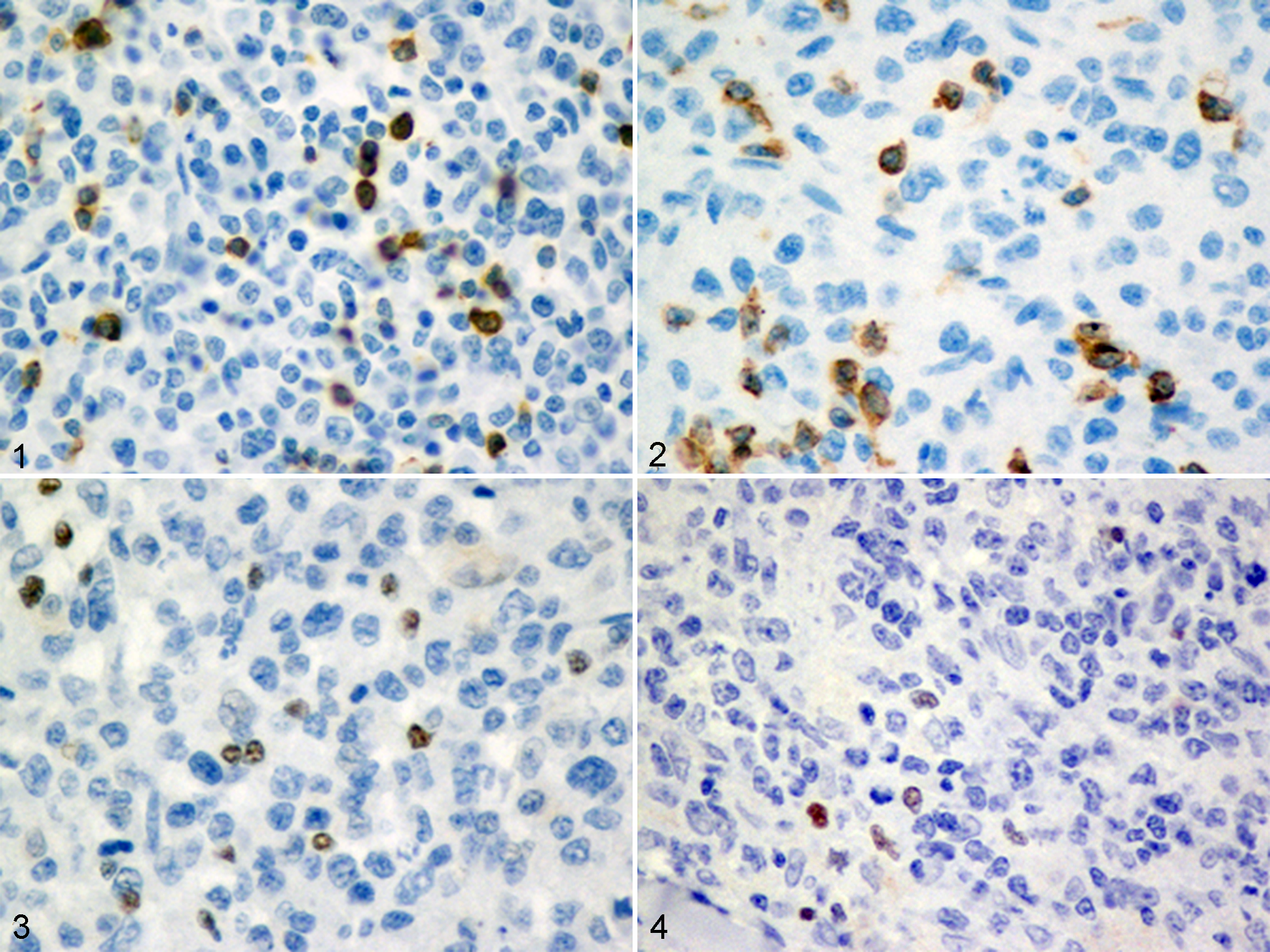
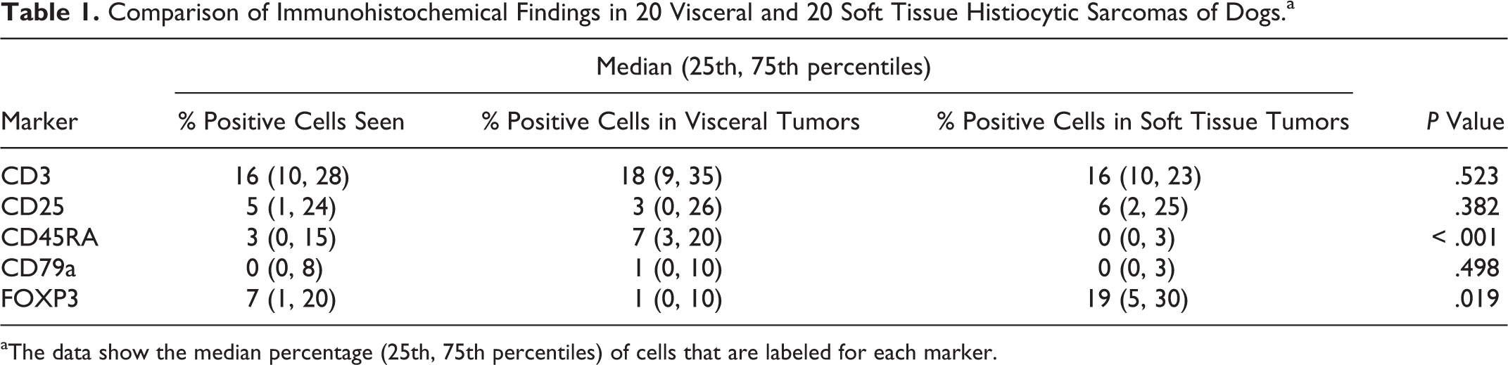
For all antibodies, immunopositive labeling was present in the positive control sections but not detected in the negative controls. Despite the variable neoplasm morphology and different locations, all 40 tumors contained lymphocytes expressing CD3 and CD45RA scattered through the neoplastic tissues, including the periphery of the neoplasm. CD45RA labeling was membranous and CD3 labeling was both membranous and cytoplasmic (Figs. 1, 2). FOXP3 labeling was nuclear. In all tumor sections examined, FOXP3-positive cells were present and scattered between the neoplastic cells, with more than 95% of CD3-positive cells being FOXP3+ (Figs. 3, 4). Immunohistochemistry data for CD3, CD25, CD45RA, CD79a, and FOXP3 are summarized in Table 1.
Comparison of Immunohistochemical Findings in 20 Visceral and 20 Soft Tissue Histiocytic Sarcomas of Dogs.a
aThe data show the median percentage (25th, 75th percentiles) of cells that are labeled for each marker.
CD25 labeling was membranous and the location of CD25-positive cells was similar to FOXP3-positive cells (data not shown).
When the proportion of cells positive for CD3, CD79a, CD25, CD45RA, and FOXP3 were compared between neoplasms at the different sites, significant differences were identified in the proportion of F
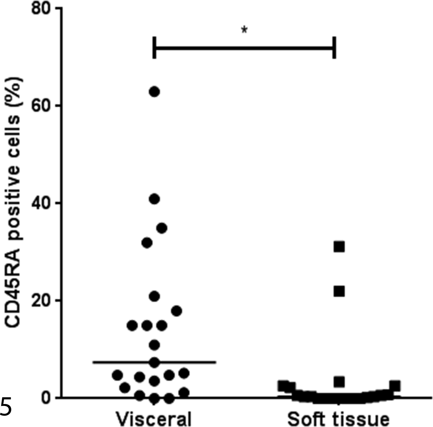
Scatterplot showing that the percentage of CD45RA-positive lymphocytes is higher in visceral compared with soft tissue HS. The line represents the median value for each group. **P < .001.
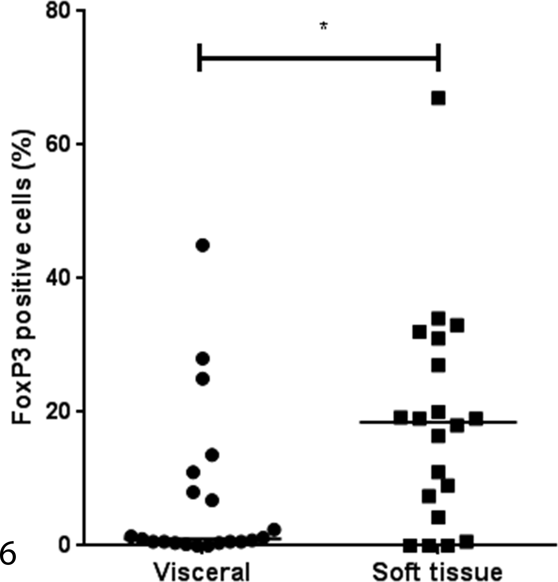
Scatterplot showing that the percentage of FOXP3-positive lymphocytes is lower in visceral compared with soft tissue HS. The line represents the median value for each group. *P = .019.
Discussion
The purpose of this study was to establish what proportion of the previously documented lymphocytic infiltrate in soft tissue and splenic HS of Flat-Coated Retriever dogs comprises regulatory T cells, to serve as a basis for future studies to assess correlation between Tregs and prognosis for Flat-Coated Retrievers with HS.
The tumor samples selected for this study represented both the soft tissue (n = 20) and splenic (n = 20) variants of HS from Flat-Coated Retrievers. The splenic neoplasms were frequently associated with severe systemic signs, particularly anemia (8/20 cases). It has been documented 13 that anemia in affected dogs may be due to a hemophagocytic variant of HS, often associated with a paraneoplastic hypoalbuminemia. 27 The severity of the clinical signs and presence of widespread visceral metastases at the time of diagnosis (often documented at the time of exploratory laparotomy) led to most of these dogs being euthanized immediately following diagnosis. Hence, the prognosis for this group of dogs was universally poor. In only 1 dog was a splenic mass noted during routine examination; this dog underwent splenectomy and survived a further 14 months. This may provide some justification for routine screening of dogs older than 7 years.
The dogs with the soft tissue form of the disease, all of which presented with a solitary mass, had a marginally better prognosis than those with splenic neoplasms. However, even in this group, survival times were poor, with 13 of 20 being euthanized at or within 6 months of diagnosis due to the extent of the primary neoplasms and their rapid and often painful progression. In those dogs where treatment was attempted, survival was still poor and did not exceed 6 months in most cases. This is consistent with previously reported survival times (median = 167 days) for dogs receiving any kind of chemotherapy, radiotherapy, surgery, or any combination. 14 However, there were 4 long-term survivors in this group living up to 1095 days, which supports the suggestion that the localized form of HS may have a better prognosis than the splenic/disseminated form. Two reports have recently suggested that dogs with the localized form of HS, with no evidence of metastasis at presentation, treated with aggressive local therapy and/or chemotherapy, can achieve longer survival times: 980 days and 568 days, respectively. 18,26 However, only 2 of 19 reported by Klahn et al 18 and none of the 16 dogs reported in Skorupski et al 26 were Flat-Coated Retrievers.
We have previously documented a high proportion (10%-20%) of infiltrating T cells in both splenic and soft tissue HS from Flat-Coated Retrievers. The present study has demonstrated that the majority of these tumor-infiltrating lymphocytes are FOXP3-positive, suggesting them to be regulatory T cells. 9
In dogs, HSs arise from interstitial DCs that occur in most tissues of the body (except the brain), and these cells are potent antigen presenting cells.
It is known that dendritic cells play a pivotal role in determining tolerance versus immunity, so determining the nature of the infiltrating T cells seemed a key step in better understanding the relationship between the neoplasm, its microenvironment, and the immune system. The finding that the majority of infiltrating T cells expressed FOXP3, suggesting them to be regulatory T cells, raises interesting questions of cause and effect, and whether there is a correlation between high numbers of FOXP3-positive cells and prognosis, which lie beyond the remit of the present study but could direct future studies.
The proportion of cells labeling positive for FOXP3 was higher in the soft tissue form compared with the splenic form of HS. This may reflect the different microenvironment provided by a soft tissue versus splenic site of origin. A recent study has suggested that hypoxia in canine neoplasms may result in an increased infiltration by Tregs. 16 We have previously demonstrated that HSs show higher Glut-1 immunoreactivity and angiogenesis than low-grade soft tissue sarcomas, suggesting that hypoxia plays an important role in the biology of these neoplasms. 1
Although it has been well documented that increased numbers of regulatory T cells in human patients with diverse solid neoplasms and hematologic malignancies can be a positive prognostic factor, studies on Tregs and F
In the present study, the soft tissue form of HS showed a higher proportion of FOXP3 cells, which is interesting as this form of HS has been demonstrated to carry a marginally improved prognosis when compared with the disseminated form, even though both variants are highly malignant. 3 Too few cases in this study received any form of treatment to allow a meaningful assessment of the prognostic significance of the proportion of cells labeling positive for FOXP3, and further studies are warranted.
The proportion of FOXP3- and CD45RA-positive cells differed between splenic and soft tissue neoplasms. CD45RA is a marker for naïve T cells but is also a common marker for hematopoietic cells, except erythrocytes and platelets. 25 It detects basophils, granulocytes, lymphocytes, macrophages/histiocytes, mast cells, monocytes, and plasma cells. 25 The higher proportion of cells positive for CD45RA in splenic compared with soft tissue HS can be explained by a more heterogeneous hematopoietic cell population in the canine spleen where erythrocytes, granulocytes, and circulating mononuclear cells are commonly seen.
A limitation to this work is that some animals were lost to follow-up and some information was not available. The sample size for both groups was relatively small, which limited the statistical power of the study. The use of formalin-fixed paraffin-embedded tissues limited the range of antibodies available for IHC, particularly with respect to CD4 and CD8.
This work demonstrated the presence of FOXP3-positive cells, which are most likely regulatory T cells in the localized and disseminated forms of HSs of Flat-Coated Retrievers, and showed a lower percentage of FOXP3 and higher percentage of CD45RA expressing cells in splenic compared with soft tissue HS of Flat-Coated Retriever dogs. These findings are a first step in the evaluation of tumor-infiltrating lymphocytes in the microenvironment of HS.
Footnotes
Declaration of Conflicting Interests
The author(s) declared no potential conflicts of interest with respect to the research, authorship, and/or publication of this article.
Funding
The author(s) received no financial support for the research, authorship, and/or publication of this article.
References
Supplementary Material
Please find the following supplemental material available below.
For Open Access articles published under a Creative Commons License, all supplemental material carries the same license as the article it is associated with.
For non-Open Access articles published, all supplemental material carries a non-exclusive license, and permission requests for re-use of supplemental material or any part of supplemental material shall be sent directly to the copyright owner as specified in the copyright notice associated with the article.