
Abstract
Sharp-force injuries are injuries caused by a mechanical force using sharp objects against the skin. Sharp-force injuries are mainly classified as stab, incised, chop, and therapeutic wounds and are less frequent than blunt-force injuries in animals. The analysis of the edges of the wound is crucial, especially if more than one type of lesion is involved. It may be difficult to differentiate between sharp trauma and blunt trauma, because lacerations can resemble incised wounds. The accurate documentation and examination of these injuries may indicate the instrument involved, the relationship between the animal and the perpetrator, and the force of the stab. Situations in which this type of trauma occurs may involve social violence, accidents, hunting, veterinary medical management, and religious rituals. The causes of death related to this type of trauma include hypovolemic shock, pneumothorax, or asphyxiation due to aspiration of blood. Necropsy findings should provide objective and unbiased information about the cause and manner of death to aid the investigation and further judgment of a possible crime.
Keywords
Forensic necropsies in veterinary medicine have increased in frequency worldwide due to the increasing demand for investigations of possible crimes against animals. 23,35,72 Veterinary pathologists play a crucial role in such cases because the identification of lesions and their accurate description and photographic documentation are key to criminal death investigations. 23,35,54,59
The identification of physical trauma in animals, such as contusions, perforations, and marks left by instruments, may not be readily visualized by the veterinary pathologist because of the hair coat. 60 In humans, who have glabrous skin, a visual examination may reveal all of these lesions at first glance. 85 Studying wound morphology is crucial for determining whether it was made antemortem or postmortem 15 and for differentiating between blunt and sharp trauma. 29,78 Whenever possible, it is helpful to obtain information about the circumstances of the crime, the weapon that was potentially used to produce the lesions, and a witness report. However, not all carcasses are accompanied by a complete history, nor is a detailed description of the crime scene always available to the veterinary pathologist. 35,57,72,77
Animals may present sharp injuries that result from intentional acts of violence, and such acts are sentinels of social violence since they are treated as family members; 4,5,26,57,77 such injuries to animals may also occur in traffic accidents, 3 while hunting, 80,81 in accidents with vessels involving marine animals, 16,17,19,75 in religious rituals, 36,55,57 and in therapeutic procedures. 61 In all cases, a detailed study of the position, shape, depth, and path of the wound and a comparison of the results with the information from the crime scene, such as bloodstain pattern analysis (BPA), may be helpful in determining or suggesting if the wound was accidental or intentional. 27,47,78
Epidemiology
Sharp injuries are less frequent than blunt lesions in animals 19,38,54,58 and are even less frequent than poisoning. 26 In a study of 271 criminal cases, 79 involved nonaccidental injuries, of which 52% were blunt trauma. 54 Another study reported 36 cases where the frequency of sharp injuries and shootings was the same (19% both), and the most affected species were dogs and cats. 89 In a study of nonaccidental injuries found in dogs and cats, the thorax and the abdomen were the most frequently affected areas both for blunt and sharp trauma in dogs and cats. 58 In cats, 3 of 191 cases of nonaccidental injury presented with stab wounds. 26 Marine animals may present with sharp injuries as well due to interactions with people. 16,17,46 Of the 660 cases recorded, including carcasses and live-stranded cetaceans, 13% were due to entanglement, deep propeller lesions, and boat strikes, and 5% were sharp wounds due to stabbing and spearing. 46
Definitions
Sharp-force injuries are injuries caused by a mechanical force using sharp objects against the skin. 11,29,78 The most common instruments used in sharp-force injuries are knives, 44,45 but other instruments can also potentially cause these injuries, such as axes, 51 machetes, 41 scissors, 65 screwdrivers, 65 needles, barbecue forks, broken glass, and arrows. 12,20,53,61,68
Sharp injuries are classified according to the lesion pattern as follows: (1) stab wounds, where the depth of the wound exceeds its length and results from the movement of the long axis of the blade in the plane approximately perpendicular to the surface of the body; 27,43,78 2) incised wounds, which are slashes and cuts where the length exceeds the depth; 16,18,39,43,46,49 3) chop wounds, which are produced by heavy instruments, such as cleavers, axes, and machetes, with a incised wound on the skin and bony fractures and/or a deep groove in the bone; 51,84 and 4) therapeutic/diagnostic wounds, which result from veterinary intervention. 27,35,48
Sharp- and blunt-force injuries should be differentiated to avoid misinterpretation. 71 In contrast to blunt-force injuries, sharp injuries do not present lacerations or bridges of soft tissue between the edges of the wound, and the wound edge is not ragged, 18,49 because there is a break of the continuity of the skin due to the sharp object. 22 The edges of sharp injuries are linear or angular, and contusion is rare. If contusion occurs, it may appear as an approximately square shape because of the dull parts of the knife, depending on the force of the thrust and the overlying tissue. 29,78 Blunt-force injuries produce 4 main types of lesions: contusions, abrasions, lacerations, and bony fractures, and the edges of such injuries are irregular; tearing of the tissue is common. 22,79
Crime Scene Findings and Trace Evidence
A proper investigation in the case of a suspicious or violent death of an animal requires a thorough crime scene investigation. 35,40,56,67,74,76 In cases of death by killing, blood is the most common body fluid found at crime scenes, and BPA may indicate the angle and distance of the blood fall. The bloodstain may be present in different shapes and patterns, and it may be possible to predict the source of the bloodstain, 7,25,48 which is the point of origin. To analyze the timeline of the events before and after death, it is important to calculate the localization of the point of origin from the radiating fluid, 7,64 which can be achieved using complex calculations and the use of computer software. 25 Blood spatter indicates the direction of the origin of the bleeding, and all length and width spatter measures should be taken, in addition to the distance of the drops from their point of origin. 1 BPA may provide information about the type of weapon used, the position of the weapon(s), the objects, the victim and the perpetrator, the movement of the individuals present at the crime scene, and an estimation of the number of stabs, blows, or shots. 1 Another important parameter of blood spatter is its velocity. Although this parameter may have some limitations, it can be a reliable indicator of the speed of the force that caused the bleeding. In the case of sharp and blunt trauma, the blood spatter is considered of medium velocity (between 1.5 and 7.6 m/s) and occurs in stabbing and arterial spurts. 1 The blood drops measure 1 to 3 mm in diameter. Low-velocity blood spatter indicates blood dripping from a body in movement, which can be from a weapon or from a person or an animal running or walking, and can be found in the body’s path or as a large amount of blood when the body stops its motion. 1 A cast-off pattern may be found in cases of sharp trauma, where the blood is projected from an object or a bloody source. 66 The information provided by these parameters may assist in the reconstruction of the events that culminated in death, and BPA should be paired with necropsy findings and molecular biology, because the DNA of the blood should be examined to determine whether the blood is from the animal or from the perpetrator, who may have received injuries during the stabbing or shooting. 56,64,66
The necropsy report provides important information, because the combination of the crime scene and necropsy findings may reveal whether the death was related to a crime or not, such as what amount of blood could be expected from a lesion of the viscera or wounded vessels or the mechanism of blood deposition. 64 Therefore, an exchange of information may assist both the pathologist and the crime scene expert, because it is unusual that a veterinary pathologist be invited to examine the crime scene. 60 The examination of the crime scene may contribute to the elucidation of the cause and manner of death, as well as the interpretation of particular postmortem findings. 60,74,76
External Examination: Protocol, Documentation and Specific Features of Sharp Injuries
In cases where there is suspicion of cruelty, the veterinary pathologist should consider radiographic examination to search for bone fractures, which are common in such cases, 58,62 and also to look for blade fragments, which may exist if the blade entered a bony structure. 24,32 The radiograph may also indicate distinct tool marks, which may match or indicate the crime weapon. 69
Unlike in people where skin wounds are typically obvious, sharp-force wounds in animals may not be readily visible due to the hair coat. 19,60,85 Therefore, the fur and skin should be carefully inspected and palpated to find dry, crusted blood. It is not uncommon for the animal to present multiple injuries, which should be individually numbered, and the topographic location of all lesions should be recorded on a diagram of the animal (Suppl. Fig. S1). 35,57,61,77
Each wound should be individually photographed with its identification number and a scale. 29,61,78 Photographing the wound before and after shaving the affected area is mandatory. 57 Whenever the veterinary pathologist has access to the crime scene photographs, it is useful to compare the wound morphology at the crime scene to that presented at necropsy. 15,60
Descriptions of the lesions should include the edges, shape, and color and marks from sharp instruments around the wounds, if present. The underlying tissue should be assessed by reflecting the skin. 27,77
To obtain an estimation of blade width, the edges of the wounds should be reconnected and measured. The length of the stab wound is equal to or greater than the width of the blade. If there is more than one wound, they should all be measured, since the average length may give a better approximation of the blade width. 12,19 However, the elasticity of the dermis may affect the width of the wound; thus, the wound may look less wide due to contraction.
The track of the stab should be traced. This includes examining solid viscera (eg, heart and liver) 53 because they may be affected in deep wounds. The shape of the resulting skin wound is affected by the nature of the overlying tissue, the movement of the victim, the force of the thrust, and the sharpness of the instrument.* Nevertheless, atypical sharp wounds may occur, such as when the victim moves or the knife twists (Fig. 1). 11,15,29,53,61,78 In such cases, lesions may present a Y- or L-shaped pattern, that can be produced by a secondary path produced by the exit of the knife, or a V-shaped pattern, which is produced by a slight movement of the victim or the knife. 29
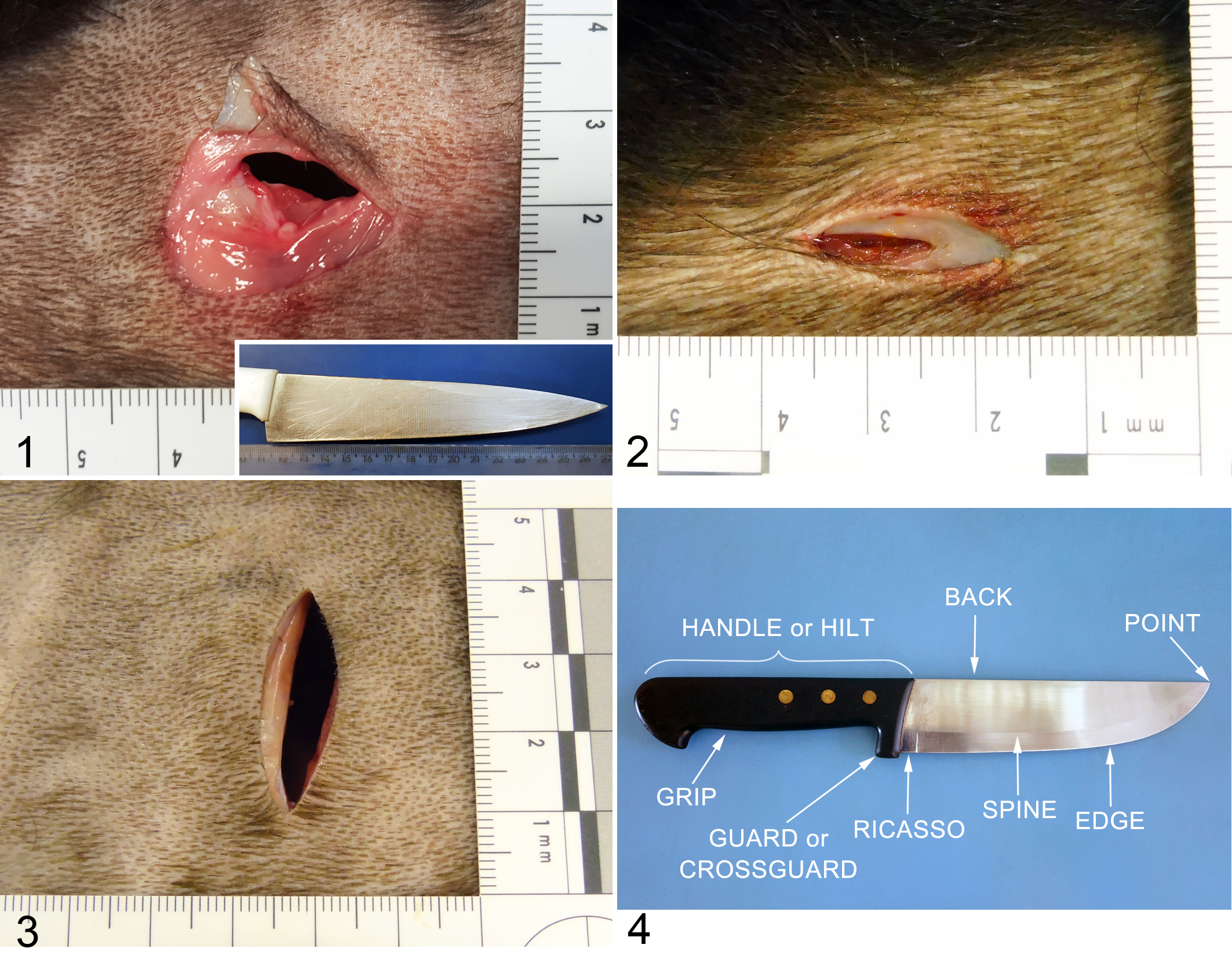
Cleavage lines or Langer’s lines are patterns of intradermal elastic and collagen fibers that produce the elasticity of the skin, and the shape of a stab wound will be correlated with the direction of the cut in relation to these lines. 15,42,52 If the stab is almost perpendicular to the fibers, these fibers pull the edges of the wound apart, creating a gaping wound (Fig. 2). If the stab is parallel to the Langer’s lines, a narrow slit-like wound is produced (Fig. 3). Between these 2 extremes are oblique wounds, which, depending on the pattern of the fibers, may be asymmetrical or semicircular. 29 However, the lesion should be documented both at the crime scene and in the necropsy room because modifications in the position of the carcass in both situations can modify the shape of the lesion and can lead to misinterpretation of the actual morphology. 15 Therefore, the knowledge and experience of the veterinary pathologist are crucial in identifying true lesions that occurred during life. 15,27
Finally, representative fragments of both sides of the stab wound should be collected in formalin to determine if there is evidence of hemorrhage and to identify wounds that may be misinterpreted as lacerations. 13,17,18 If bone or cartilage was stabbed, these specimens should also be collected for possible tool mark evaluation. 24,69
Stab and Incised Wounds
Stab and incised injuries are mostly caused by knives. 2,15,21,39,88 The parts of a knife are the ricasso, the guard, the edge, and the point or tip (Fig. 4), and each part may leave characteristic marks in the skin depending on the force and depth of the thrust. When both ends of the wound are squared, the stab penetrated up to the ricasso, the skin marks left by the ricasso may present a square-like appearance. 29,37,39,63,65 When the force of the stab is greater, a mark from the guard will be visible on the skin. In wild animals, incised wounds are commonly inflicted to the neck to provoke bleeding or to the site of a bullet wound to hide the wound or to remove the projectile from the tissue. 81 In some cases, the perpetrator may hesitate to stab the animal or miss the stab due to the movement of the animal, and such situations may also provoke superficial marks, which may be quite similar to hesitation marks in attempted suicide in humans. 29,57,77,78
Serrated knives may only be distinguished from common knives when they leave skin marks around the wound, such as linear and parallel scratches. 24,69,83 Such marks can be difficult to identify in animals during gross examination due to the presence of fur but may be distinguishable upon microscopic evaluation only in cases that affect bone or cartilage. In such cases, the pattern of striation left on the bone or cartilage may indicate the type of weapon that produced it. However, the angle of the impact was found to be the most influential aspect of the distance between striations left by blade teeth. 24
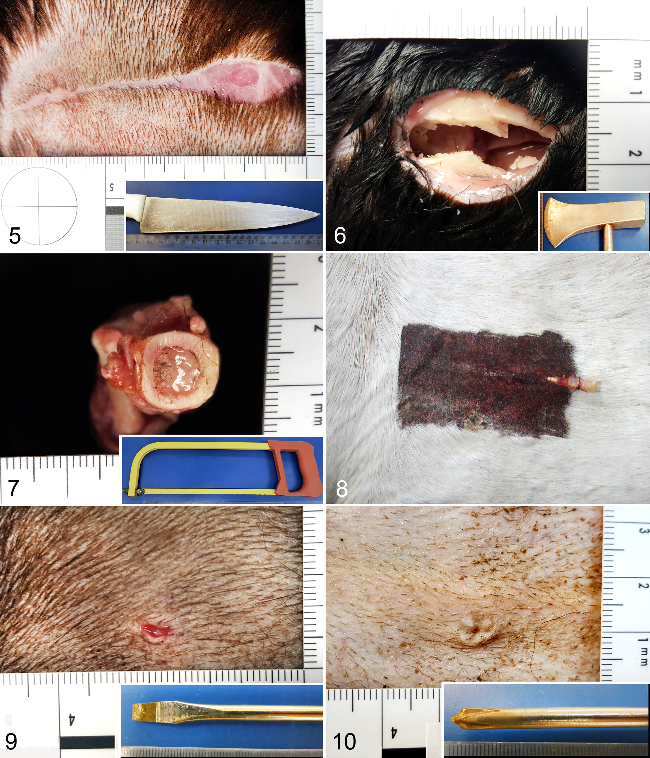
In incised wounds, if the blade contacts the skin at an oblique angle, one margin will be beveled, whereas the other will be undermined. If the angle is extreme, a skin flap may be produced. 27,29 The cut or incised wound may exhibit some characteristics that identify the direction the knife was moved in, because the cut may be more superficial at the terminal segment of the wound (Fig. 5). 34,78 However, conclusions about the shape and/or depth of the wound should be made with care due to the dynamic interaction between the assaulter, the victim, and the characteristics of the stabbed area, such as the topographical localization and the underlying tissue. 37,47,63
The force and angle of the thrust may determine the patterns of the wound edges or other changes around the wound, and the impact of the object against the body may affect internal organs and provide clues about the positioning of the aggressor in relation to the animal. 9,27,39,53,63,65 If the blade enters the body at a perpendicular angle, the mark on the skin made by the guard of the knife will likely be symmetrical, whereas with other angles of entry, the skin mark will be located below or above the wound. 29,39,65,78 However, the skin mark caused by the guard of the knife can be difficult or even impossible to detect in animals with thick hair coat or pigmented skin. 19 If the guard is not symmetrical, the wound may be misinterpreted as a blunt injury; thus, this assessment should be made in conjunction with the histopathologic and crime scene findings. The angle of the lesion may indicate the positioning between the animal and the attacker, similar to the situation that occurs in human assaults and homicides. 27,29,39,61,65,78 Similar to homicides, 11,53,65 some attackers immobilize the head of the animal and cut the ventral part of its neck. If the affected structures are on the left side of the neck, it is possible that the attacker was right-handed, and vice versa if he or she was left-handed. 61,78 In cases in which a forensic veterinary pathologist is summoned as an expert witness, he or she may be asked to quantify the force involved in a sharp-force injury. It is impossible to exactly quantify such forces, although qualitative assessments can be made by the veterinary pathologist based on the condition of the blade, the morphology of the wound, the type of tissue damage, and the depth of the wound. 2,9,29,39,47,63,65,85 Such evaluation may consider the biomechanical factors, such as properties of the knife, movement of the knife from the skin to its termination within the body, the speed and direction of delivery of the blow, the properties of the knife (such as weight, shape, and sharpness), velocity and type of thrust, and the movement of the knife within the body, which may be affected by skin and organ resistance (viscera, bone) and movement of the victim. 82
Chop Wounds
The instruments that result in chop wounds, such as axes, machetes, and meat cleavers, produce lesions with characteristic patterns that are primarily on bones. 20,24,41,51,83 The bones may present striations that are unique to each type of weapon. 20,41,51,83,84 Axes cause crushing of the bones (Fig. 6), 41,51,84 and machetes produce small bony fragments in wider and irregular wounds. 41,51 Marine animals may also be injured by boat propellers, 16 –19,38 which cause chop-like wounds. In such cases, it is not uncommon to observe a combination of blunt and sharp traumas 38,49 because the animal may be hit by a boat and/or have its skin cut and even suffer amputation, although such lesions may not be readily visualized. 19 Animals may die, depending on the site and/or viscera or vessels affected, 49 or experience further complications, such as infections, 19 due to this type of trauma. When the animal is struck by a high-speed boat propeller, it may leave closely parallel lesions, which may present characteristics of both incised and chop wounds, or even blunt and incised ones, since propellers may not present sharp edges. 17 Upon exhumation of an animal killed with a sharp instrument, examination of the remains may reveal bone fractures and marks left by the instruments in the same manner that occurs in humans. 8,50,86
Animal dismemberment is not common but may occur for ritualistic purposes or meat consumption. In some cases, in addition to the utilization of axes or machetes, the dismemberment may also be performed using a saw (Fig. 7). Upon examination, the edges of such postmortem injuries are dry and lack evidence of bleeding. 78 Some religions practice animal sacrifice in rituals in which animals are offered to the devotees’ gods based on their beliefs. In Brazil, some African-Brazilian religions have such habits, such as the Candomblé, Batuque, and Omolokô. 55 Animals that are frequently used in these rituals include pigeons, poultry, and ovine and caprine species. In these rituals, the animal is decapitated with a sharp-edged instrument, the blood is applied to some body parts of the participants, and the flesh is later consumed as food, which is seen as part of the ritual. 55 In the United States, Palo Mayombe and Santeria are Afro-Caribbean religious traditions that practice the use of animal and human remains as ritual offers. 36,77 There are other beliefs that involve animal sacrifices, such as Satanism, with several categories that are recognized by law enforcement authorities. Among Satanist cults, groups of individuals practice animal and human sacrifice, in addition to other types of crimes. Among self-styled Satanists, rituals may involve animal sacrifice, torture, or mutilation. 77
Therapeutic Sharp Wounds
Therapeutic sharp injuries 27 (those that are produced by needles) are mainly caused by medical management, which includes venipuncture, catheters, and incisions. Incisions may be surgical or part of evaluation and/or therapy of another injury, such as incision to access a bullet or incision to debride a laceration prior to suturing (Fig. 8), 57,77 and may go unnoticed because a very narrow instrument generally causes only small hemorrhages in the subcutaneous tissue. 34 Depending on the force used to produce such lesions, or if the animal has a coagulative disorder, 33,70 therapeutic wounds may range from spots of blood over the fur to large hematomas in the subcutaneous tissue. Furthermore, in cases involving multiple sharp lesions or lesions that reach organs and/or large blood vessels, the animal may die due to blood loss.
Other sharp objects, such as scissors and screwdrivers, may cause wounds with abraded margins, and the dull surfaces left by such objects exhibit scrape marks. In the case of scissors, the pattern of the wound may vary depending on whether the scissors were open or closed; if they were open and fully held in the finger holes and by the handle, 2 stab wounds may be observed. 27,29 Flathead screwdrivers produce slit-like stab wounds with abraded margins and small squared and regular ends (Fig. 9). Phillips screwdrivers may produce X-shaped lesions if the wound is more superficial, and such wounds may be circular, with 4 abraded and proportional cut edges (Fig. 10). 29,78 Bayonets may cause similar wound patterns. 78
Sharp Wounds Produced by Miscellaneous Objects
Broken glass produces incised wounds with sharp and jagged edges. When multiple wounds are present, they may exhibit distinctive depths, sizes, and shapes. 29 When wounds produced by glass are suspected, the wounds should be inspected for glass fragments. 78 Barbecue forks with 2 or 3 prongs can cause sharp injuries, and such injuries can be identified as groups of 2 or 3 wounds, with regular or irregular distances between the wounds caused by each prong depending on the angle of the stabbing. 29,77,78
Arrows and crossbow bolts can produce sharp wounds and are most commonly observed in wild animals due to hunting activities. 29,81 However, even domestic animals may suffer penetrating wounds from arrows or other nonidentified sharp objects, as revealed in a study of cats. 26 Wounds from arrowheads may exhibit several shapes and sizes that vary from circular to X-shaped lesions. 29,57,77 The wounds may exhibit hemorrhage, bony fractures, and injuries to internal organs. 29,57,77,81 The differentiation between arrowhead and bullet wounds is important for the identification of the weapon that killed the animal. Such differentiation can be performed by analyzing the track of the wound because an arrow will slice the underlying tissue, and a bullet may leave an abrasion ring. 80,81 A useful hint to an arrow wound is the adjacent hair, which will have a sharp, even line of cut from a broad-point arrowhead. Radiographic examinations may aid in the detection of lead or fragments of sharp instruments in the tissue because metals leave radiopaque traces and remain embedded in the bone. 57,72,82 Ice picks may cause sharp trauma, and similar to arrows, 80,81 such lesions may mimic bullets or shotgun pellets because they result in small, round, or slit-shaped wounds. 29,78 Analysis of microtraces by using scanning electron microscopy in combination with energy-dispersive X-ray spectrometry (SEM/EDS) may reveal some particles that may be compatible with knives, such as stainless steel or carbon steel, which can be found in or on the bone surface. The cutting edge and roughness of the blade may be evaluated by the number of particles found. 87
Cause, Manner, and Mechanism of Death
Sharp lesions may or may not be lethal, and this can be determined only by performing a detailed necropsy with a careful external and internal examination. The cause of death should be stated as a sharp injury with an indication of the topographical region(s) affected. 6,30 Lesions in the neck and thorax are most likely to be fatal. 27,78 Defining the manner of death can be challenging, and information about the crime scene (such as BPA and weapons found) and laboratory tests may be helpful in determining the manner of death. 6,30 In putrefied bodies or when the necropsy findings are inconclusive, the manner of death can be stated as undetermined. The mechanism of death depends on the site and vessels/organs involved, but the most frequent cause of death is hypovolemic shock resulting from hemorrhage, which occurs when major blood vessels are severed. When the limbs, head, or neck are affected, there may be a large amount of blood at the crime scene; however, when abdominal or thoracic stabbing occurs, bleeding may occur mainly within body cavities. 27,29
When a deep stab wound occurs in the neck area and the trachea or larynx is exposed, the mechanism of death may be asphyxiation due to the aspiration of blood. Deep stab wounds affecting the lungs may lead to hemothorax and/or pneumothorax. 27,29 Conversely, if a deep stab in the neck involves larger blood vessels, air may enter the bloodstream due to the negative pressure of the veins. A thoracic X-ray may reveal air in the heart. 3,14,28 However, in an experimental study, dogs seemed to be more resistant to air embolism due to vessel laceration. 31
When multiple wounds are observed, it may be difficult to determine the timing of wounds relative to death based on their gross appearance, and a histopathological analysis can be valuable. 16,71 The amount of hemorrhaging may not be a reliable indicator because unless the circulation has ceased for some time, some bleeding is likely to occur from injuries that are inflicted near death or shortly thereafter. In cases of sharp trauma to lung, liver, spleen, or kidney, bleeding in the internal cavities is also likely to occur for several hours after death until all of the blood from the area has drained. 18
For forensic purposes, it may be important to determine the age of lesions, if they were inflicted antemortem or postmortem, and if they were or were not lethal. 16,18 If the lesion was inflicted antemortem, intentionally or unintentionally, hemorrhage will occur and be detected by gross and/or microscopic examination. Postmortem hemorrhage is generally distinguished microscopically by confinement of the blood to a single fascial plane. In contrast, antemortem hemorrhage extends through several tissue planes. 10,53 However, if the body is putrefied, such determination is difficult. 73
Infection is a late complication of stab wounds and requires several days or weeks to develop. When a stab wound occurs in the abdomen, fecal content may spread and cause peritonitis. 78 Such situations are observed in cases in which the animal suffers nonlethal and undiagnosed accidental stab wounds, such as in car crashes, 3 or intentional wounds, such as torture or aggression. 26,35,54,58,61
Concluding Remarks
Animal wound analyses are challenging due to the difficulty of prompt observation and, in some cases, the lack of information from the crime scene about the weapon or object used to injure or kill the animal. Veterinary pathologists should be aware of the possibility of sharp wounds that may or may not be directly involved in the animal’s death and should understand proper protocol for thorough wound examination. Additional examinations, such as radiographs, can be very useful in determining the nature of some lesions. Microscopic examinations can be required in cases of stab and chop wounds because such examinations may aid in the identification of the object that caused the lesion. However, such interpretations should be performed only in conjunction with the information provided by the police report and crime scene.
Footnotes
Acknowledgements
We thank Dr Beverly McEwen for her valuable contribution to this manuscript and José Cesar Menk Pinto Lima and Raquel Gonçalves Gomes, who are residents in anatomic pathology at FMVZ/USP, for their assistance with the documentation of the lesions.
Declaration of Conflicting Interests
The author(s) declared no potential conflicts of interest with respect to the research, authorship, and/or publication of this article.
Funding
The author(s) disclosed receipt of the following financial support for the research, authorship, and/or publication of this article: The authors received support from FAPESP (Sao Paulo Research Foundation, grant number 2013/23645-4) CAPES (Coordination for the Improvement of Higher Level Personnel) and CNPq (National Council of Technological and Scientific Development).
References
Supplementary Material
Please find the following supplemental material available below.
For Open Access articles published under a Creative Commons License, all supplemental material carries the same license as the article it is associated with.
For non-Open Access articles published, all supplemental material carries a non-exclusive license, and permission requests for re-use of supplemental material or any part of supplemental material shall be sent directly to the copyright owner as specified in the copyright notice associated with the article.