
Abstract
Projectile injury represents an estimated 14% of reported animal cruelty cases in the United States. Cases involving projectiles are complicated by gross similarities to other common types of injury, including bite wounds and motor vehicle injuries, by weapons and ammunition not commonly recognized or understood by veterinary medical professionals, and by required expertise beyond that employed in routine postmortem examination. This review describes the common types of projectile injuries encountered within the United States, as well as firearms and ammunition associated with this form of injury. The 3 stages of ballistics—internal, external, and terminal—and wounding capacity are discussed. A general understanding of firearms, ammunition, and ballistics is necessary before pursuing forensic projectile cases. The forensic necropsy is described, including gunshot wound examination, projectile trajectories, different imaging procedures, collection and storage of projectile evidence, and potential advanced techniques for gunpowder analysis. This review presents aspects of projectile injury investigation that must be considered in tandem with standard postmortem practices and procedures to ensure reliable conclusions are reached for medicolegal as well as diagnostic purposes.
Keywords
Veterinary forensics is a relatively new discipline within the forensic sciences. The importance of veterinary pathology to the discipline cannot be overstated, particularly in cases involving projectile injury. Projectile injuries are often lethal or result in euthanasia, and they tend to have a medicolegal component. Veterinary pathologists have advanced training in postmortem examination and access to purpose-built facilities and specialized equipment, often including digital radiography and computed tomography. Access to these resources can be essential in recovery, interpretation, and preservation of projectile evidence obtained from an animal. While veterinary pathologists are in the best position to conduct forensic postmortem examinations, they do not typically have all of the expertise these cases require. Thus, it is important that veterinary pathologists who engage in forensic casework integrate themselves into a team that can supply the expertise needed. In cases of projectile injury, such teams might include forensically trained veterinarians, law enforcement personnel, scene investigators, and ballistic and tool mark experts.
This article provides veterinary pathologists with a useful projectile injury resource. It includes an overview of the types of weapons and ammunition used to produce gunshot projectile injury in animals. Ballistics theory, patterns of tissue injury, methods to determine and interpret projectile trajectory, recovery and preservation of ballistic evidence, and additional techniques used in projectile case investigations are also provided. While the discussion is restricted to gunshot injury, most of the investigation techniques and considerations provided are applicable to other forms of projectile injury such as arrow and captive bolt. Throughout this article, the various facets of a projectile case investigation are related to the role of the veterinary pathologist and relevance to the postmortem examination.
General Considerations
While a projectile injury may be a relatively straightforward problem to solve in terms of a medical diagnosis, these cases can be extremely complex and should not be entered into without a full understanding of the case circumstances and the expectations of the submitter. There is currently no minimum standard of documentation to be completed if the legal status of a case is unknown. In this article, the authors recommend steps that mirror human forensic protocols and suggest that these be performed for every case involving a projectile injury.
First, not all projectile injuries in animals are illegal. A shooting must be determined to have been done without the owner’s permission, out of season, or without statutory protection, or it must be the cause of unnecessary suffering before being investigated as a crime. 17 Projectile injuries to animals may be accidental or intentional, they may involve complex crime scenes and investigations beyond the injured animal, and the perpetrators can range from juveniles to adults. Cases may include public endangerment or property damage, and they may involve legal or illegal firearms.
To understand and interpret projectile injuries, veterinarians and veterinary pathologists need to understand wound ballistics, and they need to be aware that these types of cases may require analysis and documentation of large quantities of evidence in preparation for prosecution. In a postmortem examination, the pathologist must be able to distinguish entrance from exit wounds and understand factors that can affect or alter their appearance. The pathologist may need to assist in the determination of the approximate distance a projectile was fired from based on wound examination and should determine the wound path “trajectory” into and through the body. Proper collection and preservation of ballistic evidence and assisting with other aspects of an investigation may also be necessary. Finally, proper documentation and preparation of a forensic report involve an awareness of medicolegal needs and requirements. 16
Projectile Injury Cases in the United States
There is limited data on the number of veterinary forensic projectile cases investigated or prosecuted annually. A national database for entry of these types of crimes does not exist. Profile data on suspects that commit animal cruelty using firearms are also scant.
A study to establish the Factors in the Assessment of Dangerousness of Perpetrators of Animal Cruelty (FADPAC) was developed by Randall Lockwood of the American Society for the Prevention of Cruelty to Animals (ASPCA). It is intended to evaluate the significance of an individual’s involvement in a particular act of animal cruelty as an indicator of dangerousness or possible risk for involvement in future acts of violence against others. 14 In this assessment, 33 factors were established based on threat assessment criteria used by the National Center for the Analysis of Violent Crime and on studies of animal cruelty offenders and habitual violent offenders. The perpetrator risk assessments consider victim vulnerability (small, harmless, or nonthreatening animals), number of victims, severity of injury inflicted, close proximity to the victim at the time of injury, duration of abuse, degree of premeditation, risk of exposure when performing the act of abuse, history of positive interactions with an animal victim, and documentation of the act through photographs or video-recording. The presence of 5 or more of these factors is considered cause for concern about future acts. 13
Projectile injury profile features that have been reported indicate that shooting incidents almost invariably involve male perpetrators, that animal shooting from a vehicle typically involves 2 suspects (one to operate the vehicle and the other to shoot), and that the type of weapon used may reflect the age of the suspect. 13,14,22 For example, preteen and teen shooters are more likely to use air- or gas-powered weapons that use BB or lead pellet projectiles, and adults are more likely to use higher power weapons and shotguns. Data on the types of weapons used were presented in a study of 121 projectile injuries treated at Boston’s Angell Memorial Animal Hospital over an 11-year period. In this study, 86% of wounds were caused by handguns, 3% by rifles, 5% from shotguns, and 15% from BB or pellet guns. 18 Tendencies in weapon type have also been reported, with handgun injury predominating in urban areas, rifles and shotguns in rural areas, and air- or gas-powered guns (including pellet and BB guns) in suburban areas. 18 Historical and signalment data reported on dogs with projectile injuries in a University of Pennsylvania study indicated that the majority are young and sexually intact. These included German Shepherds (18%), Pit Bull mixes (13%), Rottweilers (13%), Doberman Pinchers (10%), and Mastiffs (2%). 13 Companion animals shot with firearms also tend to be “outside and unsupervised,” with most shootings occurring in animals roaming at large. 16,18
Pet-Abuse.com (http://www.pet-abuse.com) is an online resource that gathers data on animal cruelty investigations in the United States. According to this source, in 2013, projectile injuries accounted for over 14% of recorded animal cruelty cases, and from 1998 to 2010, 10.3% involved shootings of companion animals (dogs, cats, and horses).
Firearms
To effectively evaluate projectile injuries, a basic knowledge of firearms and the ammunition they carry is necessary. There are 5 categories of small arms: handguns, rifles, shotguns, submachine guns, and machine guns. Projectile injuries in veterinary species are commonly caused by handguns, rifles, and shotguns, and thus the following discussion will be limited to these types of firearms.
Handguns, rifles, and air- or gas
In the United States, the caliber of a weapon is determined based on the diameter of the bore of the weapon measured from land to land, so it is the diameter of a barrel before the rifling is cut. Caliber specifications can be given as US customary measure or as metric units. Examples of the former using inches include the .38 special, .38 Smith & Wesson, or a .357 Magnum and of the later metric units include 9-mm × 19-mm Parabellum or 9-mm × 18-mm police cartridges.
Handguns have rifled barrels and are generally considered low-velocity weapons. The speed at which a projectile leaves the barrel of a weapon is called the muzzle velocity. The muzzle velocity for pistol projectiles ranges from 183 to 580 m/s (600–1900 ft/s). 20 This velocity is dependent on the type and caliber of ammunition, the length of the barrel, the propellant (gunpowder) burn rate, and the gas produced after initial ignition of the primer.
The most common types of handguns currently in use are auto-loading pistols (automatics) (Fig. 1) and revolvers (Fig. 2). To fire an auto-loading pistol, the trigger must be pulled each time to fire a cartridge (bullet). Firing of the cartridge generates a gas pressure that initiates a process of extracting and ejecting the empty cartridge case and loading a fresh one, returning the gun to a position to fire the next round. The cartridges are stored in a removable magazine in the handgrip of the gun (Fig. 1). 4 The most common firing mechanisms in automatic pistols are “blow-back” and “recoil.” Blow-back action involves the pressure of the gas that is produced by the combustion of the gunpowder forcing the slide of the weapon to the rear, starting the cycle of extraction, ejection, and reloading of each projectile. In a recoil-operated automatic, the barrel and the slide are locked together when the weapon is fired. When the bullet leaves the barrel, the reverse thrust of the propellant gas on the cartridge case causes the barrel and the slide to move to the rear of the gun. The barrel is halted after a short distance and the locking mechanism is released from the slide. The slide continues to the rear of the gun, ejecting the fired cartridge case and starting the reloading cycle. 4
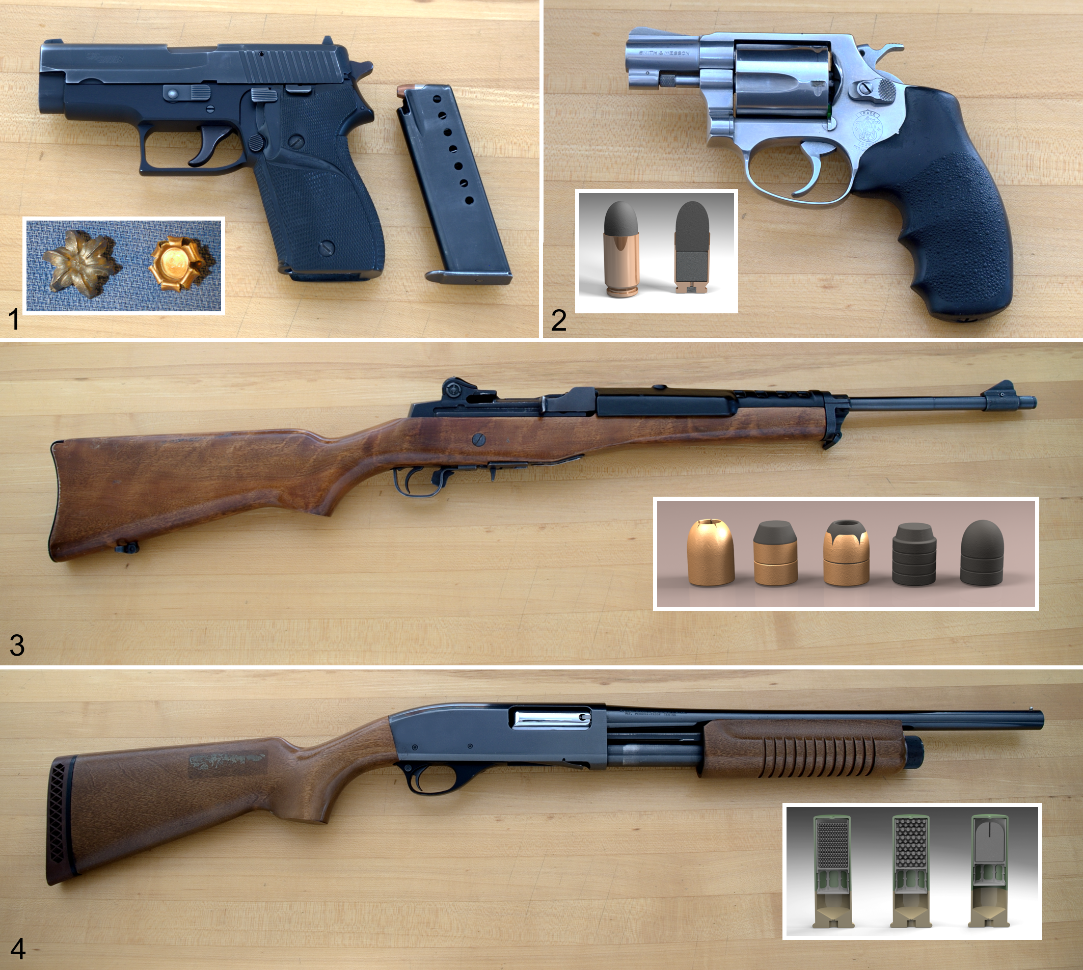
A revolver has a revolving cylinder containing multiple chambers, each containing 1 cartridge. The cylinder rotates mechanically to align each chamber with the barrel and the firing pin. There are 3 types of revolvers: the swing out, break top, and solid frame. The most common is the swing out. In this type of revolver, there is a cylinder latch on the left side of the gun frame. When this is pressed and the cylinder pushed to the left, it will swing out, exposing the chambers. Each chamber is loaded with a cartridge (Fig. 2). The cylinder is replaced back in the frame of the weapon and the revolver is ready to be fired. 4 Revolvers are either single action or double action. In a single-action revolver, the hammer is cocked back manually each time the weapon is fired. Pressure is applied to the trigger releasing the hammer, discharging the gun. A double-action revolver applies continuous pressure to the trigger. Each time the trigger is pulled, the weapon is cocked, meaning the hammer is drawn back. When the hammer is released, the weapon is fired. 4
A rifle is a type of weapon designed to be fired from the shoulder (Fig. 3). Rifles are used for longer distances and have greater precision and accuracy than handguns. The barrel of this weapon is rifled. In the United States, rifles are required to have a minimum barrel length of 16 in. (40.6 cm). Rifles are generally considered a high-velocity weapon, with projectiles reaching speeds of 762 to greater than 1160 m/s (2500–3800 ft/s). 20
There are multiple types of rifles: single shot, lever action, bolt-action, pump-action, and auto-loading. A single-shot rifle has 1 firing chamber (barrel) that is manually loaded each time the rifle is fired. A lever action rifle has a lever under the grip used to open the rifle action and extract the cartridge case. When the action is closed, a fresh cartridge is placed and ready to fire. In a bolt-action gun, the handle extends from a bolt. Moving the handle back and forward causes the bolt to extract and eject a cartridge, allowing a new cartridge to be placed and ready to fire. A pump-action uses the manual movement of a slide under and along the barrel. This will open the action, extract and eject the used cartridge, and load a new cartridge. An auto-loading or semiautomatic rifle extracts, ejects, reloads, and readies a weapon with each trigger pull, using the force of gas pressure or recoil to operate the action. The trigger must be released and pulled for each shot, repeating the cycle. 4
A shotgun is fired from the shoulder and the barrel is smooth bore, so no barrel rifling is present (Fig. 4). Shotguns are used for dispersion shots on moving targets at relatively short distances. It can fire multiple types of shells (ammunition), including pellets, buck shot, and slugs. In general, velocities range from 1200 to 1300 ft/s (366–396 m/s), but barrel length, presence of a choke, powder charge, weight of shot, and the type of shell will affect the velocity. 7 For example, the velocity of pellets will be relatively low compared with that achieved with solid slugs. In the United States, federal law mandates a minimum barrel length of 18 in. (45.7 cm). The barrel of a shotgun may have a device known as a choke that can influence the dispersion of shotgun pellets. This can alter interpretation of the range of discharge in a shot gun injury. The shotgun may be identified as a single shot, over-and-under, double-barrel, lever action, pump-action, or auto-loading. An over-and-under shotgun has 2 barrels one above the other, and the double-barrel shotgun has 2 barrels alongside one another. 4
Air- or gas-powered weapons range from toys to highly sophisticated air rifles. An air rifle uses the expanding force of compressed air or gas to move a projectile down a rifled barrel. An air gun is differentiated from an air rifle by its smooth-bored barrel. The same projectiles may be used in either type of weapon. Speeds reached by projectiles from these weapons are less than 304 m (1000 ft) per second, with standard velocities ranging from 61 to 122 m/s (200–400 ft/s). 4,20
Ammunition
Ammunition is a propellant and projectile developed to achieve specific parameters of distance, target penetration, and wounding capacity. To accomplish this, ammunition design must take into consideration the caliber of the weapon it will be used in, composition of the projectile, and the amount and burn rate of the propellant.
A cartridge consists of a cartridge case, a primer, propellant (gunpowder), and a bullet (projectile). The cartridge case is usually composed of some type of metal. The purpose of the cartridge case is to expand and isolate the chamber of the weapon against a backward escape of gases when the cartridge is fired. The primer is the cartridge ignition component, igniting the propellant (gunpowder) in the cartridge when it is struck. In the United States, primer compounds may consist of lead styphnate, barium nitrate, and antimony sulfide, and there is an increasing use of lead- and barium-free primers such as Sintox (Dynamit Nobel, Troisdorf, Germany). Primers are classified as a center fire or a rim fire, with the primer in the center or in the rim of the cartridge base, respectively. The propellant is the gunpowder within the cartridge that is ignited to propel the projectile forward. The bullet (projectile) is the segment of the cartridge that exits the barrel of the weapon when it discharges. 4
Modern bullets generally consist of 2 categories: lead and metal-jacketed. These categories can be further subdivided into pure lead, full-jacketed, and hollow-point semi-jacketed bullets, with modifications of the bullet tips like round nose, semi-wad cutter, and wad cutter. There are 4 configurations of lead bullets: round nose, wad cutter, semi-wad cutter, and hollow-point. Round-nose bullets consist of a partially blunt, conical shape, and a flat or beveled base. The wad cutter bullet resembles a cylinder with a base that is either beveled or hollow. The semi-wad cutter bullet is a truncated cone with a flat surface at the tip. At the base of the cone, there is a shoulder lip the diameter of the bore. The hollow-point bullet is similar to the semi-wad cutter with a cavity in the nose portion of the bullet used to enhance expansion (mushrooming) of the bullet when it impacts the target. 4 Jacketed bullets are used in high-velocity weapons. At high velocities, lead bullets will melt or fragment. Jacketing provides a protective metal casing around the bullet. 4 Projectiles may be semi-jacketed (partial) or full metal jacket. All of the above bullets can be semi-jacketed rounds. An example of this would be a semi-jacketed lead round nose, called a soft tip. After entry in the body of an animal, the metal jacketing can separate from the lead bullet. The jacketed segment is essential for ballistics. This portion of the projectile may retain rifling marks that can be used to identify the weapon used.
Shotgun shells are composed of a primer in the base, powder, wadding, and shot enclosed in a cardboard or plastic casing. Terminology to describe shells will vary in different parts of the world. In the United States, there are 3 standard sizes of shells based on length: 2¾ in., 3 in., and 3½ in., and the most popular caliber sizes are 12 (approximately equal to an 18.5-mm bore diameter), 16 (16.8-mm bore), and 20 (15.7-mm bore). The length of the shell will determine the amount of powder, and the volume of shot varies depending on the size and number of shot pellets. A shotgun shell may have birdshot pellets ranging in size from .08 to 0.2 in. (2–5 mm). BBs are spherical projectiles generally ranging from 0.177 to .22 caliber (4.5–5.6 mm). A shotgun shell may also contain buckshot pellets ranging from .24 to 0.36 in. (6.1–9.1 mm) or slugs, which are solid pieces of metal. 4,7 Air guns or air/gas-powered guns fire pellets or BBs. Pellets come in various shapes, including round nose, pointed, wad cutter, or flat. The most common pellet has a “wasp waist,” also called diablo style. 4
The Process of Firing a Gun
The discharge of a firearm is initiated by pulling the trigger, releasing the firing pin within the gun. When the pin strikes the primer of the cartridge case, it ignites, causing a flame. The flame enters the chamber of the cartridge case, igniting the powder and producing large quantities of gas and heat. One gram of propellant produces 1 liter of gas under high temperature and pressure. The intense heat of the gas exerts pressure on the projectile and the sides of the cartridge. The gas pressure on the base of the projectile propels it down the barrel of the weapon. 4 After the bullet exits the barrel, a burst of flame, gas and unburnt powder, soot, primer residue, and vaporized metal from the projectile and cartridge case all follow the projectile. In revolvers, similar material will emerge from both the cylinder-barrel gap and the barrel. 4
Ballistics and Wounding Capacity
Ballistics is the science of projectile travel. The flight path of a projectile can be broken down into 3 stages: the path of a projectile within the weapon, called internal ballistics; the projectile’s flight through the air, external ballistics; and the projectile pathway through an object, terminal ballistics. When a projectile enters an animal, terminal ballistics may also be referred to as “wound ballistics.” 5 This phenomenon is partially expressed in the following equation:

The equation describes the influences that mass and velocity have on the amount of kinetic energy that can be delivered to tissues by a projectile. 2 When the mass of a projectile is doubled, the kinetic energy is doubled, but if the velocity is doubled, the kinetic energy is quadrupled. 16
In addition to the variables described in the kinetic energy equation, the effects of distance traveled and changes in media along the path will influence the capacity of a projectile to cause tissue damage. Projectile velocity changes with the distance traveled before contact and thus the amount of kinetic energy transferred to tissue. The resistance of tissues to the projectile will also influence the amount of energy delivered; for example, skin provides enough resistance that a minimum bullet velocity of 50 to 60 m/s is required for penetration. Thus, ballistics influences the final kinetic energy transferred to tissues, and it is the kinetic energy transfer, in combination with weapon type, 4,21 that best predicts the wound capacity of a projectile.
Tissue damage through projectile injury can occur in 3 ways: through laceration or crushing, by cavitation, and through shock waves. In the first instance, tissue damage will occur along the path that the projectile or the path its fragments take through the body. A temporary wound cavity is formed by outward expansion and resultant stretching and tearing of tissue along the trajectory. Pulsating collapse of the temporary cavity results in the formation of a permanent cavity. The type of ammunition, previously detailed, has the greatest influence on the cavity formed, but the varying ability or tendency of the projectile to fragment or to partially or completely flatten (mushroom) on impact, in combination with the caliber of the projectile and the presence and design of an outer jacket, also significantly affects wound capacity. 2,6 While the shape of the frontal surface of a projectile largely determines the size of the cavity, current measurement techniques are too variable to allow postmortem caliber derivation. 4 Advances in the use and evaluation of computed tomography to characterize cavity dimensions have the potential to improve measurement accuracy such that cavity dimension measurement may contribute data useful for weapon and projectile identification in the future. 12
In formation of a wound cavity, both crush and stretch forces act on tissues, and a multiphase medium is created around the projectile. The phases include fluid, composed of blood, lymph, and cellular fluids, surrounded by a radially dispersed phase of solid tissue. The radially accelerated tissue will cause an abrasion collar (see entry wound description below) and may cause backspatter if tissue is pushed out of the wound cavity. There is also potentially a gas phase, which may be formed by air within the tissue and/or gas produced by the projectile. Formation of the cavity also creates a vacuum that can pull hair and debris into a wound. 16
Tissue characteristics influence the degree of damage a projectile generates. Thicker soft tissue, such as liver and brain, and dense rigid tissue such as bone offer greater resistance, increasing the energy transfer. Elastic tissues that can give on impact, such as lung or muscle, are more resistant to damage, particularly that caused by cavitation. 2,21 Bone fracture is further dependent on the type of bone and the angle of impact. Cancellous bone is softer, enhancing its ability to absorb energy, resulting in less fragmentation. Cortical bone, due to its greater density, tends to fracture and fragment. If a projectile hits the skull, intracranial increase of pressure may contribute to concentric fractures that form a target-shaped arrangement of fracture lines. When a bullet makes contact with bone, there are deceleration, deformation, and potential fragmentation or tumbling effects on the projectile and the bone itself. Bone fragments can act as additional projectiles and can influence the direction and range of the original projectile. 2,4 The nature of the tissue also influences drag on the projectile’s terminal ballistics, altering the velocity it travels through the tissue.
Examination and Necropsy: General Considerations
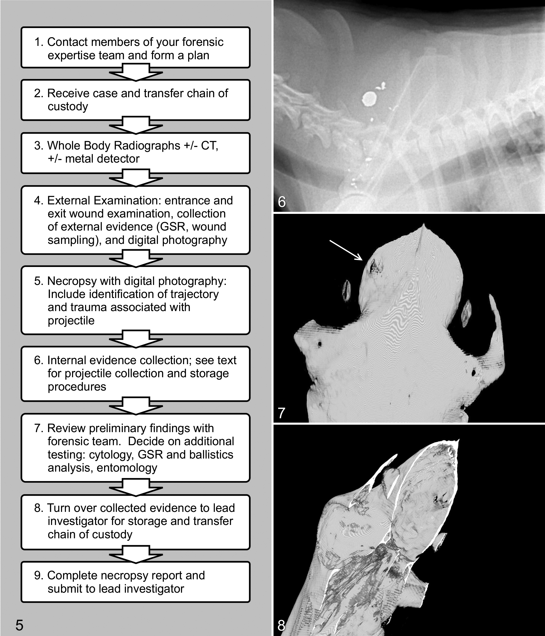
Determination of the cause and manner of death in projectile injury cases is typically insufficient to address all of the questions these cases raise. In addition to standard necropsy procedures and reporting, the pathologist must provide information on entry and exit wounds, projectile trajectory, and related forms of tissue injury. The pathologist must also be aware of external evidence that needs to be collected prior to the postmortem examination and must, whenever possible, retrieve ballistics evidence. Before manipulating the carcass, make a plan that takes into account the best sequence for gathering evidence. Figure 5 provides a suggested stepwise procedure that will address most case scenarios. Prior to receiving the case, communicate with the law enforcement agency crime scene investigative (CSI) unit or state crime laboratory, and be familiar with the types of evidence they can process and how they want evidence submitted. These units can offer a great deal of expertise in evidence handling and collection and may be able to provide trained personnel, for example, to obtain and analyze gunshot residue (GSR). Image analysis (see specific examples below) should also be completed before in-depth physical manipulation of the body is conducted. Manipulation of the body during removal from a body bag or during the course of the external exam should be minimized until the presence and positioning of a projectile are established and external evidence has been collected. Investigation teams may check a body with a metal detector prior to turning it over to a pathologist, but pathologists may also consider having this piece of equipment in their necropsy facility.
Following image analysis, additional planning should occur for the recovery of each projectile and its components or for identifying a projectile’s path in the event of complete penetration through the body. The pathway (trajectory) within the body needs to be photographed and documented. Documentation will describe any organs or structures involved in the path of the projectile and provide measurements of any injuries sustained. Pathway descriptions should be kept simple and succinct, listing organs or structures involved using anatomically correct terms and using the directional terms up, down, left, right, forward, and backward or, alternatively, the anatomical terms dorsal, ventral, rostral, cranial, caudal, medial, and lateral.
Photography
Before manipulation of a carcass, a complete set of digital images should be taken. A minimum database of external digital photography should include full-body right and left lateral images of sufficient quality that the animal can be identified. These images need to document the state of the carcass as it was received and should capture body position before rigor mortis is broken down. Images should also capture all external evidence, including blood and other fluids on the body, collars and tags, ropes, chains, leaves, or other environmental or foreign material that might be trapped in the fur or received with the body. Closer images of external lesions and environmental evidence should also be taken at this time. All images should include a minimum amount of information to allow case continuity, including identification number, date, and measurement.
Radiology
When a projectile injury is suspected, whole-body radiographs prior to postmortem examination will be necessary. Therefore, if undertaking forensic projectile casework, one should either have radiographic equipment available on-site or have a working relationship with a facility that does. Radiographic images need to be taken with a minimum of 2 views with standard markers and identification. Radiographs can show the shape and density characteristics of the projectile, as well as location of the projectile within the body, and often will better define bone fractures than gross postmortem examination (Fig. 6). Radiographs can also help to define the trajectory of the projectile within the body. Examination of radiographs should include identification and localization of the projectile and any related fragments in the body. Wound tracks may also be identified based on the presence of gas, hemorrhage, bone, or metal opacities. The direction of fragments or beveling of bone in images may help determine the direction of the projectile and contribute to entry and exit wound identification. 11
If a projectile hits a bone, the majority of the bone fragments normally will be found behind the altered bone, indicating the direction of the projectile, and between the altered bone and any exit wound that may exist.
Computed Axial Tomography
Computed axial tomography (CT) operates on the same principle as radiography but offers improved full-body scanning and visualization for projectile retrieval and fracture description. 8 CT imaging is a rapid and increasingly affordable technology that allows postmortem examination without disturbing anatomical structures. While CT imaging has similar soft tissue limitations to radiography, it greatly improves 3-dimensional localization of projectiles and projectile paths prior to necropsy (Figs. 7, 8). 9 In general, digital imaging is conducive to storage and use of images for legal proceedings, and the comprehensive nature of CT scans offers in-depth, full-body medicolegal documentation of trauma that is superior to standard radiography.
Collecting External Evidence
Because protocols may differ between laboratories, collection of external evidence should be discussed with the responsible forensic laboratory before proceeding. Some procedures include excision of the entry wound and surrounding tissue (to a specified diameter) untouched and as a whole. Skin and the adjacent underlying soft tissues can be placed on a piece of cork or Styrofoam to keep the shape, size, and anatomical orientation for further analysis. These types of samples can be analyzed using a scanning electron microscope and x-ray spectrometry to determine the quality and quantity of soot particles, information that can be used to help determine the range of discharge. In contrast, histologic examination of entry and exit wounds is often consistent with mechanical and thermal insult but is nonspecific. There are several simple techniques used to collect evidence during the external examination. When powder particles associated with gunshot wounds are found, they can be scraped into a Post-it note and placed in a sealed paper envelope. Entry and exit wounds should be shaved and the fur placed in a cellophane or waxed paper envelope. Clear tape can be used to retrieve stippling from the skin and then placed on a glass slide. Sections of skin containing contact wounds with soot or stippling can also be taken at necropsy and either forwarded to a ballistics expert or photographed, sampled for residue, and fixed in formalin. 16,19
Gunshot Wound Examination
On examination, projectile injury needs to be differentiated from other forms of trauma, including wounds from captive bolt stunners, motor vehicles, animal bites, and lacerations. In the absence of an accurate history that accounts for wounds observed, one should consider whole-body radiographs prior to proceeding with the examination. Examination findings consistent with trauma, including that caused by projectiles, include hemorrhage, fractured organs such as the liver and spleen, bone fractures, and emboli. Entrance, exit, and intermediary wounds are the specific lesions that indicate projectile injury, and thus it is essential that these lesions are accurately identified and documented during the external examination.
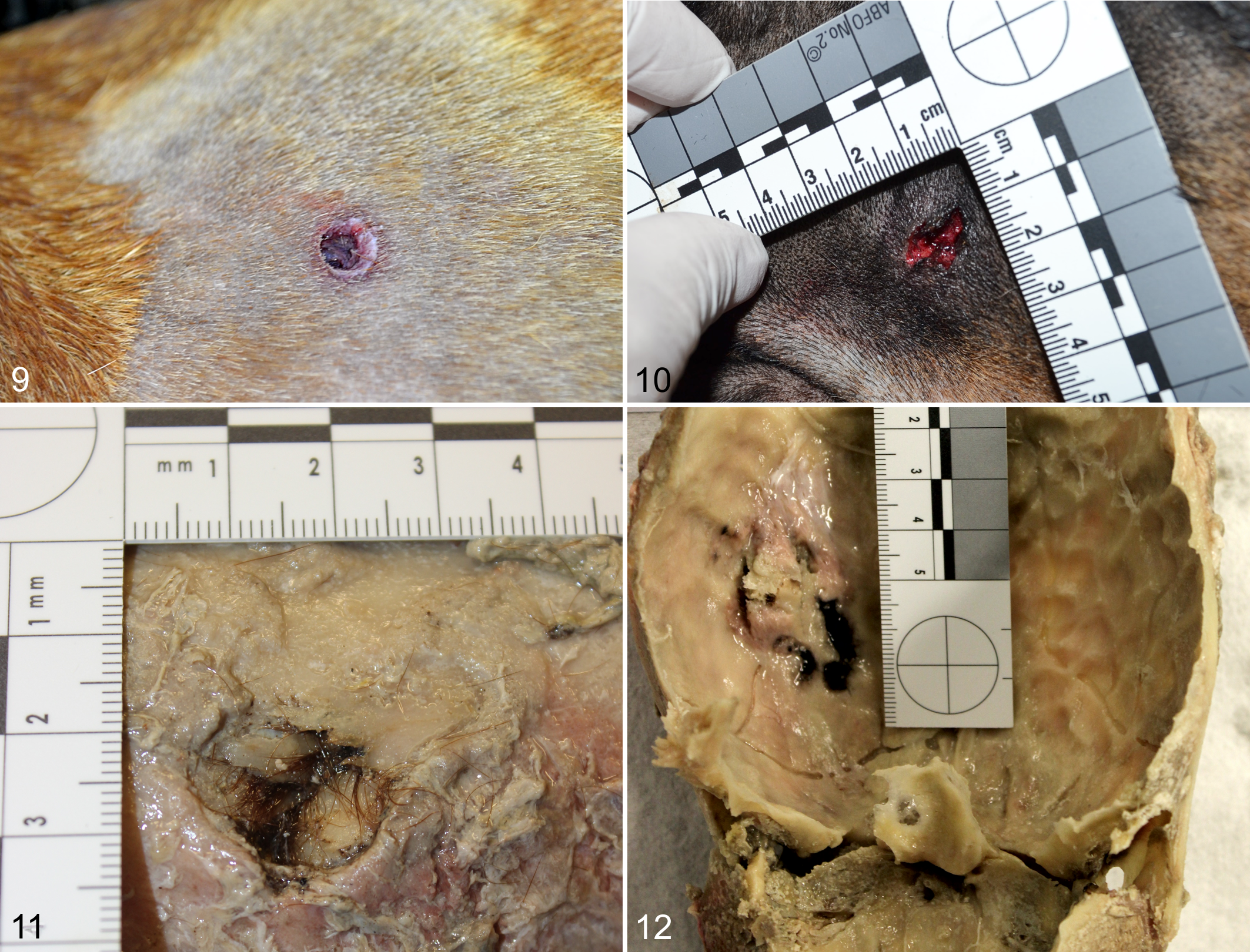
Determination of entrance vs exit wounds and establishing if there are any intermediary wounds is a necessary early step when conducting a complete projectile injury postmortem examination. This determination is critical for crime scene re-creation and trajectory analysis. An entrance wound typically has sharp margins, and the diameter of the wound is similar to that of the projectile (Fig. 9). Entry wounds may have soot marks (also called stippling or tattooing) or an abrasion ring from the expulsion of the projectile if it was shot at close to intermediate range. During the discharge of a weapon, in addition to the powder, soot from the ignition of the powder emerges from the muzzle. The soot is carbon and contains vaporized metals from the primer, projectile, and cartridge case. If the muzzle of the barrel is held close to the body of the animal, soot can be deposited on the animal. These marks are typically found on exposed skin, and thus long or dense hair coats may obscure these findings. The “flame,” also called the muzzle flash, produced when firing a gun, is composed of superheated incandescent gases. These gases can cause searing of skin or fur around contact and near-contact gunshot entrance wounds. 4 Soot or burned hair may still be present in these cases, and thus it is important to sample hair over projectile wounds prior to further examination of the entrance wound. The size, intensity, and appearance of the soot pattern and the range at which it occurs will depend on multiple variables, including range of fire, type of gunpowder, angle of the muzzle to the target, barrel length of the weapon, weapon caliber, type of weapon, and the type of target. Additional factors influencing the soot pattern include weather conditions such as wind, air pressure, or rainfall. Because most areas of the animals that will be examined are haired, it will be necessary to sample hair for powder and soot. It will then be necessary to shave the area around wounds to identify any evidence on the skin.
Intermediary wounds occur when a projectile passes through one part of the body, then reenters another part, causing a reentry wound. A reentry wound normally does not have a grease (abrasion ring), which can help differentiate it from an entry wound. In addition, an intermediary wound may develop when a projectile passes through an intermediary object, such as a door, wall, or window, before hitting the animal’s body. Either circumstance can change the appearance and number of entrance wounds and will complicate wound interpretation. The penetration of an intermediate target can affect the morphology of the entrance wound in the animal’s skin. This results in an atypical entry wound due to deformation of the projectile and/or tumbling. An intermediary object may fragment and can cause a wider dispersal of shotgun pellets, thus altering the gunshot range determination. The object may become embedded in the projectile, causing decreased stability and deformation. This may result in a larger or more irregular entrance wound, causing a wider or irregular abrasion ring. 4,11,16
Exit wounds typically have jagged margins and a larger diameter than that of the entrance wound (Fig. 10). Low-velocity projectiles can cause a slit-like exit wound resembling a stab wound. Exit and intermediary wounds, while very important in understanding the trajectory of a projectile through the body, are unlike entrance wounds in that their appearance will be less dependent on the size of the projectile, and their surface will hold none of the evidence related to the firing of the weapon. Therefore, the following discussion focuses on the characteristics and interpretation of entrance wounds. A summary of the features of entry and exit wounds is provided in Table 1.
Characteristics of Entry and Exit Wounds.
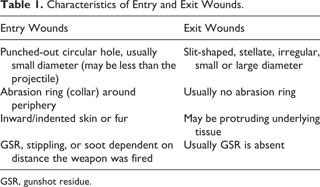
GSR, gunshot residue.
Contact wounds are caused by placing the end of the muzzle in direct contact with the skin at the time a gun is fired. There are hard-, loose-, angled-, or incomplete-contact wounds. 4 In hard-contact wounds, the barrel is pushed hard against the skin, so that the skin rises up around the end of the barrel. The edges of the wound are seared by the hot gases exiting the barrel. In this type of wound, soot is forced into the skin . 4 In skin areas supported by bone, a separation of the skin from the subcutis may occur, forming a “soot cavity.” In loose-contact wounds, the barrel is in contact with the skin but held lightly against the skin. Any gas preceding the projectile pushes the skin inward, creating a gap between the skin and the end of the barrel, allowing the gas to escape. Soot is deposited in a zone around the entrance. 4 Angled-contact wounds occur when the barrel is placed at an acute angle to the skin; the circumference of the end of the barrel (muzzle) is not in complete contact with the skin. Gas and soot radiate outward from the muzzle where contact is not complete. This causes an eccentric pattern of soot with 2 zones. The inner zone, most distinguishable, is a blackened seared area of skin or fur having a pear, circular, or oval shape. The larger outside zone is shaped like a fan of light gray soot. All or the majority of the inner zone will be on the opposite side of the wound from the muzzle, pointing in the direction the gun was fired. 4 Incomplete-contact wounds are similar to angled-contact wounds. The muzzle of the gun is in contact with the skin; however, the surface of the body may not be completely flat, causing a space between the end of the barrel and the skin. Soot and gas escape from this space, causing an area of seared skin or fur. 4
As a weapon moves from contact to varying distances away from a target, the deposition of soot or powder residue will change, but accurate interpretation of these differences may require additional analysis using scanning electron microscopy or spectrometry. The morphology involving skin defects, margins, or abrasions usually will remain the same. A ballistics expert will consider the appearance of an entrance wound in light of known variations in types of weapons and ammunition and may also need to test fire a specific weapon to determine the distance from which a weapon was fired.
Near-contact wounds are created when the firearm is not in contact with the skin but is held a very short distance away. The distance is so small that most of the particles escaping cannot disperse and they mark the skin. In near-contact wounds, the entrance wound is surrounded by a zone of powder soot covering seared and blackened skin or fur. The zone of searing is larger than that observed in a loose-contact wound. The soot is embedded in the skin of the seared zone. 4 In near-contact angled wounds, soot radiates outward from the end of the barrel, creating 2 zones similar to angled-contact wounds. In a near-contact angled wound, the majority of the blackened seared zone is on the same side of the barrel pointing toward the weapon. The direction of fire is in the opposite direction.
The transition from near-contact to intermediate-distance wounds is subtle. Intermediate-range wounds occur when the muzzle of the weapon is held at a distance away from the body when fired yet is close enough that the powder grains discharged with the projectile cause “powder tattooing” of the skin. Powder tattooing involves both embedding of gun powder particles in the skin and hemorrhage. The hemorrhage associated with tattooing appears as numerous reddish-brown to orange-red punctate lesions surrounding an entry wound. The distribution around the wound is either symmetrical or eccentric depending on the angle that the gun was fired. Powder tattooing is an antemortem event, and thus its presence indicates that an animal was alive when it was shot. 4 The distance for an intermediate range wound can be over 1.0 M depending on the weapon fired, but an intermediate range from a handgun is generally 10.0 mm. 4 These wounds can assist in determining direction of fire based on tissue beveling and angle of an abrasion ring or soot marks.
At ranges greater than intermediate range, soot or powder tattooing is not present near the entrance wound, and the only marks are those produced by the projectile perforating the skin. Wounds inflicted from these longer ranges may be referred to as distance range wounds or, more recently, as “indeterminate range.”
With shotgun wounds, as the muzzle-to-animal separation distance increases, the diameter of the pellet pattern will increase and the margins of the entry wound will become more indented. At extreme distances, 41 to 100 m, pellets separate, creating individual injuries or striking surrounding inanimate objects. If the angle of a shotgun discharge is perpendicular to the body, the pellet pattern is usually circular, while an angled discharge will cause an eccentric pattern. In both instances, the diameter of the pattern is range dependent. 4,7
The postmortem report involving a projectile injury should include the number, appearance, and locations of all entrance and exit wounds, as well as some description of the path of the projectile (trajectory), the injuries produced, and the site of lodgment if a projectile failed to exit the body. It should also include the description of any retrieved projectile. Retrieval of all projectile segments should be done with gloved hands, plastic instruments, or forceps with rubber tubing to prevent damage of forensic evidence.
Projectile wounds may be penetrating or perforating. A penetrating wound occurs when a projectile enters a body and does not exit, in which case the projectile may be referred to as a “retained missile.” A perforating wound occurs when the projectile passes completely through the body. In addition, projectile wounds can be considered graze or tangential. A graze wound involves the projectile striking the skin at a shallow angle, causing an abrasion. In a tangential wound, the skin is torn to the level of the subcutaneous tissue. 4,16
It is relatively common to receive projectile wound cases for which there is no intact entrance wound in the skin. This may be due to predation, postmortem decomposition, or removal of tissue under some other circumstances. In the absence of this critical evidence, interpretation of fractures in bones, with the aid of previously described imaging tools, can be very important in identifying projectile injury. For example, identifying beveling of fractured bone edges in the direction of the projectile’s path and retention of hair or skin driven into fracture sites by the projectile can be very helpful in directing an investigation of a poorly preserved carcass (Figs. 11, 12). Funnel-like bone loss caused by a projectile passing through a flat bone can assist with the determination of the trajectory direction of a projectile. Similar findings are regularly encountered in captive bolt gun lesions of slaughtered cattle.
Angles and Trajectories
Defining a projectile path or trajectory is a critical component in the reconstruction of crime scenes. Trajectories can give information on the location of the shooter, including distance from the victim; identify intermediate objects in the path; aid in the interpretation of the order of shots fired; and help to determine the weapon and ammunition used. Trajectory can also be used to corroborate or refute testimony provided by an assailant or witnesses. To establish a trajectory, the pathologist must first find the entrance wound or wounds. Measurements of the length and width of the bullet entrance wound are taken to establish the degree of the angle of the wound.
Trajectory rods can help in the visualization and characterization of projectile trajectories in some cases. Rods must be straight and can be made of wood, fiberglass, or plastic. Metal rods are also available, but they may cause scatter if being used with CT imaging. The animal can be placed in sternal recumbency or in the position in which it was found at the crime scene. The rods are meant to simulate the path of the projectile into and potentially through a body (Fig. 13). Once placed, photographs should be taken from the front, back, left, right, top or bottom, and above the body. These directional photos contribute to the development of theories based on investigative findings and should be discussed with the rest of the forensic team. Trajectory rods should be used with caution. If used incorrectly, a rod can manipulate valuable and relevant forensic evidence located in the wound canal and may cause an artificial canal. Placement of a trajectory rod and/or probing of a wound canal should only be done after exploration of the wound canal has taken place with thorough documentation. In addition, not all entry wounds lend themselves to placement of trajectory rods. For example, entry wounds in bone can be rigid enough to prevent placement of rods without physically altering the wound, and thus rods should not be used in these cases.
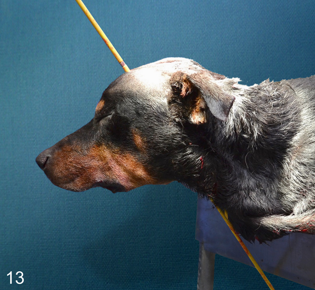
Trajectory rod, projectile injury through the top of the head; canine. Lateral view illustrating the placement of a trajectory rod. The rod extends from an entry wound in the left dorsal skull to the exit wound in the right ventral neck caudal to the ramus of the right mandible.
Additional techniques used with trajectory rods include the addition of a centering piece that can be placed first to ensure that the weight of the trajectory rod does not alter the angle of the wound. Once placed into the centering piece or directly into the wound, angles can be measured from the rods by centering a protractor against the middle of the entry wound. An angle finder is used to ensure the protractor is level, and a plumb line is hung from the rod and bullet hole to determine horizontal and vertical angles. There are several types of trajectory kits that provide these tools (Kaleidoscope System Fusion Trajectory Kit, Tritech Forensics, South Port, NC; Evi-Paq trajectory kit, Safariland Group, Ontario, CA).
Lasers can be used in conjunction with rods to improve the examiner’s understanding of trajectory. Once a trajectory rod has been placed and measurements taken if so desired, a laser can be attached to the rod and the light followed to an area of probability for the origin of the shot. Photographic fog can be sprayed over the laser beam to assist in visualizing the light beam and to improve photographic evidence. Lasers can also be mounted on a tripod in the area of probability and shown into transparent trajectory rods to establish the likely location of the shooter.
The trajectory techniques described above can be very helpful, but they are not uniformly applicable. Physical proportions, normal range of motion, and differences in locomotion for the species under investigation can influence likely paths of trajectory and should also be considered. Current concepts in trajectory analysis are largely drawn from 2 sources: individual case analysis in humans 2,3 and research studies, many of which were conducted by the military in pigs and dogs. The studies conducted on dogs 1 are most applicable to veterinary forensic trajectory analysis, but often the weapons and ammunition used in these studies are not consistent with that encountered in a nonmilitary context. Ballistics data available from human civilian case studies tend to better represent the types of weapons and ammunition seen in veterinary forensic pathology. While data on projectile trajectory analysis in veterinary species are limited, some physical commonalities support extrapolation of data across species. For example, during steady motion, there is universality to animal patterns and the extent to which anatomical parts bend. 15 For this reason, one should be able to factor in relative proportionality and predict rough bending patterns and maximum angles using data across species.
Repeatability and reliability of a technique should be considered when producing discoverable evidence in a legal case, and the production of trajectory angles may be the most controversial technique introduced in this review. Angles obtained using rods are dependent on positioning of the animal. The exact position of the victim prior to death is often unknown and can be particularly difficult to determine if a projectile penetrates a victim while the victim is in motion or if there is evidence of intermediary wounds. In addition, angles may be inaccurate or differ depending on who acquired them. The practicality and relevance of the information that could be gained from trajectory rods and angles should be considered on a case-by-case basis and in consultation with the forensic team.
Projectiles as Evidence: Collection and Storage
The following are best practices for handling projectile evidence. Once a projectile is recovered, it should be examined for the presence of tissue or markings. The projectile can be cleaned for examination and photography and prepared for cytological analysis by placing it in a test tube with sterile saline. Once the debris has been rinsed off, place it on a surgical towel to dry. The saline from the container can then be forwarded to law enforcement agency CSI personnel for cytological analysis. Projectile cytology may be necessary if the projectile went through something or someone prior to entry into the animal under examination. 4 Once cleaned and dried, the projectile should be photographed. Identification, date, measurement, police report number, and location of recovery should be included in these images. Finally, the projectile should be measured and weighed before storing in wax paper in a dry match box-type container with identifying information. Projectiles are considered evidence, and thus they require an intact chain of custody. If a projectile or any other evidence obtained during a postmortem investigation is given directly to an officer or investigator, a date and signature in an evidence log will maintain this chain. The movement of evidence should also be documented in your report, including the name and identity number of the officer or investigator who received the evidence. While evidence is in the possession of the pathologist, it must be stored in a secure location.
Advanced Techniques in Gunpowder Analysis
The main component of modern gunpowder is nitrated cellulose. During the firing of a weapon, particles of unburned, partially burnt, and burnt gunpowder containing nitrite and nitrate compounds are expelled from the barrel. Use of infrared photography can be beneficial in demonstrating the nitric acid present in ammunition on the fur of an animal. This may help distinguish a residue pattern, which can assist in determining the distance from which a projectile has been fired. Digital cameras can be enhanced with an infrared lens attachment to facilitate taking these types of entrance wound pictures. Ballistics experts can also use microscopy of fur from entrance wounds to distinguish particles of burned and unburned gunpowder residue or use chemical analysis, called a Griess test, to detect organic nitrite residue left on hair or other materials. 7,10
Conclusion
Deciding whether to accept any form of forensic pathology case, projectile injury or otherwise, can be challenging for veterinary anatomic pathologists. Understanding what additional tasks this form of postmortem examination may require and what expectations exist beyond the typical diagnostic postmortem report is an important part of the decision-making process. This article was intended to provide background information on the nature of projectile injury and describe the examination technique to clarify the additional steps and expertise required by medicolegal cases involving projectile injury. We hope that it has also conveyed the necessity for establishing relationships with an extended team of forensic experts and that it will spark interest among veterinary anatomic pathologists to look deeper into the subject and to begin taking on these challenging and rewarding cases.
Footnotes
Acknowledgements
We thank Mike Haag, Leo Egar, VMD, Mary Dudley, MD, Dustan Englin, and Randy Anglin for their consultation and contribution to the development of this article. We also thank Les Siemens and Sasha Willis for their technical expertise and assistance with figure images.
Declaration of Conflicting Interests
The author(s) declared no potential conflicts of interest with respect to the research, authorship, and/or publication of this article.
Funding
The author(s) received no financial support for the research, authorship, and/or publication of this article.