
Abstract
Pheochromocytoma, a rarely reported adrenal gland neoplasm in Old World primates, was diagnosed in 5 rhesus macaques (Macaca mulatta) and 2 African green monkeys (Chlorocebus aethiops) from 3 research institutions. Age and sex were available for 6 primates. Two males and 4 females were affected, ranging in age from 9 to 31 years. All neoplasms were unilateral and, in the cases reporting the affected gland, 4 involved the right adrenal gland and 2 involved the left. Diagnosis was established by characteristic histologic features. Immunohistochemically, neoplastic cells in all cases expressed chromogranin A and met-enkephalin and were negative for melan-A and inhibin. Six of 7 tumors were positive for β-endorphin. Pulmonary metastases were present in 2 rhesus macaques and portal vein invasion in 1 African green monkey. To the authors’ knowledge, this is the first report of malignant pheochromocytoma in Old World primates.
Keywords
Pheochromocytomas are neuroendocrine neoplasms originating from catecholamine-secreting chromaffin cells of the adrenal medulla. They have rarely been reported in macaques (Macaca sp) 4,5,8 –10 and have not been reported in African green monkeys (Chlorocebus aethiops).
In humans, pheochromocytoma is an uncommon tumor that affects all ages. Most are sporadic, unilateral, solitary, and benign. There is no sex predilection. Many pheochromocytomas are functional and secrete catecholamines, causing vasoconstriction and thus sustained or paroxysmal hypertension, tachycardia, cardiac hypertrophy, pulmonary edema, sweating, and headaches. Detection of urinary catecholamines or catabolites allows diagnosis of functional pheochromocytomas. 7 Grossly, pheochromocytomas vary in size and are dark red-brown often with foci of hemorrhage, necrosis, and cystic degeneration. Microscopically, tumors generally exhibit a characteristic neuroendocrine pattern. Prediction of the clinical behavior can be challenging unless metastases are present. 7 Nuclear pleomorphism is not reliable as a determinant of malignancy. Hematogenous or lymphatic metastases to the liver, lymph node, lung, and bone have been reported in humans. Reported cardiovascular changes secondary to the effects of elevated catecholamine levels include cardiomyocyte necrosis, interstitial myocardial fibrosis, myocarditis, and arteriosclerosis. 7 Histochemical staining and ultrastructural analysis to detect neurosecretory granules aid in the diagnosis of pheochromocytoma. Immunoreactivity is expected for catecholamines, chromogranin, catecholamine-synthesizing enzymes, neuron-specific enolase, synaptophysin, and opioid peptides (met-enkephalin, leu-enkephalin, β-endorphin, dynorphin B). 7 In humans, pheochromocytomas may carry germline or, less frequently, somatic mutations in multiple genes. 2,7 Several familial tumor syndromes such as multiple endocrine neoplasia type 2 (MEN-2), type 1 neurofibromatosis, and von Hippel-Lindau syndrome may include pheochromocytoma. Genetic forms tend to occur at a younger age and may be multifocal. 7 Germline mutation of the succinate dehydrogenase complex, subunit B (SDHB) gene is considered a strong risk factor for malignancy. 2 GATA binding protein 3 (GATA3), a transcription factor, is used immunohistochemically to identify pheochromocytomas and is used to differentiate them from tumors exhibiting similar morphologic features. 6
This report describes 7 pheochromocytomas diagnosed in Old World primates at necropsy, affecting 5 rhesus macaques (Macaca mulatta) and 2 African green monkeys. The animals were maintained in research colonies at the Oregon National Primate Research Center (ONPRC), the Harlow Center for Biological Psychology (HCBP), and the Behavioural Science Foundation (BSF). Animal care and procedures were approved by the Institutional Animal Care and Use Committee at the ONPRC and HCBP and were conducted to ensure compliance with the US Animal Welfare Act and other prevailing statutes, regulations, and guidelines. Animal care and use at the BSF were approved by the Animal Care Committee of the BSF acting under the auspices of the Canadian Council on Animal Care. Complete necropsies were performed and representative tissues were collected for microscopic examination. Tissues were fixed in 10% buffered formalin, embedded in paraffin, sectioned at 5 microns, and stained with hematoxylin and eosin; 2 were stained with the Churukian-Schenk method for argyrophil granules. Tissue microarrays were constructed with tissue from each case, and immunohistochemistry for chromogranin A, met-enkephalin, β-endorphin, melan-A, and inhibin was performed (Suppl. Methods 1). Separately, immunohistochemistry was performed for synaptophysin, chromogranin A, GATA3, and SDHB on case No. 7 (Suppl. Table S1 and Suppl. Methods 2).
Case summaries are presented in Table 1. All neoplasms were unilateral, with 4 affecting the right adrenal gland, 2 affecting the left adrenal gland, and 1 not specified (case No. 4). Adrenal gland enlargement was observed grossly in 5 cases (Nos. 1, 2, 5–7) and ranged from 3.0 to 4.5 cm in the greatest dimension. Affected glands were distorted and exhibited a variegated appearance with foci of hemorrhage and necrosis (Fig. 1). On microscopic examination, neoplastic cells often replaced the medulla and compressed the cortex; tumors were composed of polygonal cells arranged in nests, packets, and cords supported by a fine, fibrovascular stroma (Fig. 2). Cell borders were distinct with a moderate to abundant, granular, eosinophilic to basophilic cytoplasm. The nucleus was irregularly round with lightly stippled chromatin and a solitary, variably distinct nucleolus. Anisocytosis and anisokaryosis ranged from mild to marked. Occasional cells had giant, irregular, hyperchromatic nuclei (case Nos. 1, 5–7) or multiple (up to 4) nuclei (case Nos. 5–7). Cytoplasmic invagination was frequent in case No. 7 (Fig. 2, inset). Mitoses ranged from ≤1 to 3 per 10 400× fields. Variably sized, blood-filled spaces were present in all but case No. 3. Metastatic pheochromocytoma was observed in the lungs of 2 rhesus macaques (case Nos. 1, 5; Fig. 3), and portal vein invasion occurred in 1 African green monkey (case No. 6).
Signalment and Clinical and Pathologic Findings in 7 Old World Primates With Pheochromocytoma.
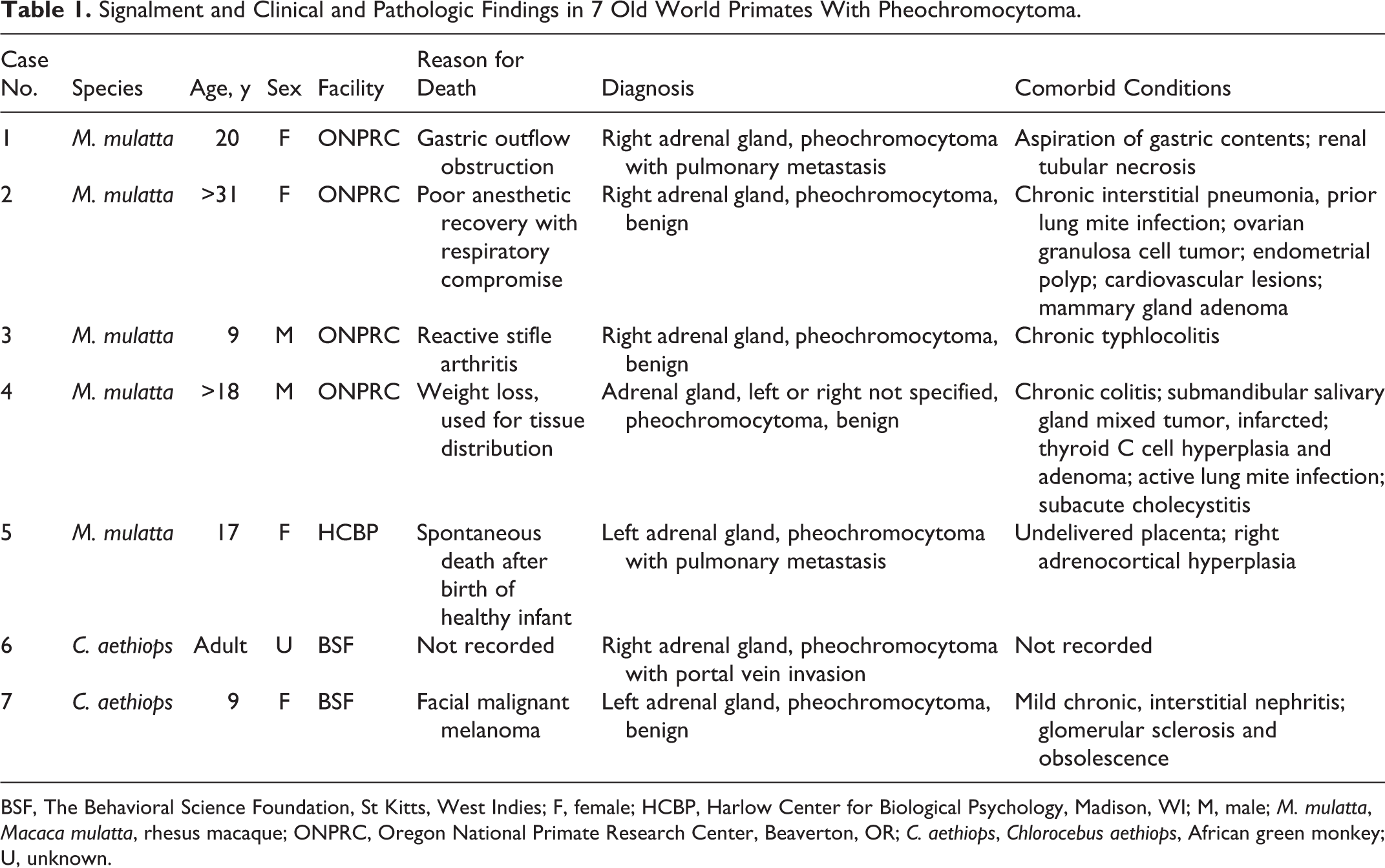
BSF, The Behavioral Science Foundation, St Kitts, West Indies; F, female; HCBP, Harlow Center for Biological Psychology, Madison, WI; M, male; M. mulatta, Macaca mulatta, rhesus macaque; ONPRC, Oregon National Primate Research Center, Beaverton, OR; C. aethiops, Chlorocebus aethiops, African green monkey; U, unknown.
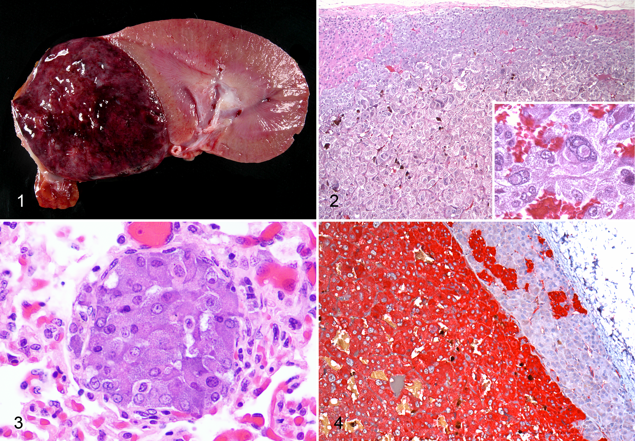
The Churukian-Schenk method demonstrated myriad brown to black cytoplasmic granules in the 2 cases evaluated (case Nos. 1, 2). Immunohistochemically, neoplastic cells of all cases exhibited cytoplasmic labeling for chromogranin A and met-enkephalin, and 6 cases were positive for β-endorphin (case Nos. 1–5, 7; Suppl. Table S2). Adjacent adrenocortical tissue did not label with these markers. Neoplastic cells failed to label with inhibin and melan-A, whereas adrenocortical cells labeled well. In case No. 7, neoplastic cells had diffuse intense immunopositivity for chromogranin A (Fig. 4) and variably intense labeling for synaptophysin, while reactivity for melan-A was limited to adrenocortical cells. GATA3 expression was not detected in case No. 7; SDHB expression was intact.
Available tissues were examined microscopically for cardiovascular abnormalities to help identify catecholamine-secreting pheochromocytomas. Mild to moderate myocardial fibrosis (case Nos. 2, 7), aortic atherosclerosis (case Nos. 1, 2) with mineralization (case 2), mild intimal sclerosis and fibromyxomatous medial change and fragmentation of the elastic lamina of intrarenal arteries (case Nos. 1, 2), afferent glomerular arteriolar changes including hyperplasia of the tunica media (case Nos. 1, 2, 5, 6), intimal sclerosis (case No. 2), and minimal splitting of the internal elastic lamina (case No. 5) were observed. Systemic arteriosclerosis affecting the kidneys, heart, lung, gastrointestinal tract, mesentery, uterus, right adrenal gland, and pancreas was characterized by hyalinosis of vessel walls and variable medial hyperplasia, hypertrophy, and fibrosis in case No. 7. No vascular lesions were identified in case No. 3 or 4.
Few pheochromocytomas have been reported in Old World primates. 1,3 –5,8 –10 Most were identified as incidental lesions at necropsy, their endocrine activity unknown. Malignant pheochromocytomas have not been reported. Three retrospective studies of nonhuman primate neoplasms include 8 pheochromocytomas in macaques. 4,5,8,9 One report described an angiomatous pheochromocytoma in an aged rhesus macaque with a history of chronic heart disease. 10 Pheochromocytomas have been reported in 6 baboons (Papio cynocephalus) and a patas monkey (Erythrocebus patas). 1,3,4 Cardiovascular lesions noted in the rhesus macaque diagnosed with an angiomatous pheochromocytoma included cardiomyofiber atrophy, hypertrophy, and lysis with associated inflammation and replacement fibrosis. 10 On the basis of these findings, the authors presumed a functional pheochromocytoma with catecholamine excess and a consequent cardiotoxicity. Clinical signs of hypertension were observed in most of the baboons diagnosed with pheochromocytomas at necropsy; pulmonary congestion with increased heart rate and dyspnea were exhibited by 4 animals. 3 Cardiovascular histologic findings were not discussed.
Diagnosis for all cases in this series was established by identification of characteristic microscopic features on routine hematoxylin and eosin staining and was supported by special histochemical and immunohistochemical staining. The Churukian-Schenk method demonstrated argyrophilic granules consistent with neuroendocrine granules in the 2 cases evaluated. Neoplastic cells were positive in all cases for chromogranin A, confirming the neuroendocrine nature of the tumors. In addition, neoplastic tissue was immunoreactive for opioid peptide markers produced by the adrenal medulla: met-enkephalin in all cases and β-endorphin in 6 of 7 cases. GATA3 has been shown to be a useful marker for pheochromocytomas in humans, with 1 study reporting immunohistochemical positivity in 20 of 21 tumors. 6 In contrast, it was not expressed in the 1 pheochromocytoma tested (case No. 7). Intact SDHB expression in this same tumor suggests that there was no germline mutation of SDHB.
As the pheochromocytomas in this series were identified at necropsy, no antemortem hormonal assessments were performed to confirm excessive catecholamine production. However, cardiovascular lesions were variably present in 5 cases and included regional and systemic arteriosclerosis, myocardial fibrosis, and aortic atherosclerosis. These morphologic changes suggest hypertension and possible catecholamine hypersecretion, particularly in the relatively young, 9-year-old African green monkey (case No. 7) with systemic arteriosclerosis and myocardial fibrosis. Cardiovascular changes observed in 3 rhesus macaques are likely multifactorial. The average age of these affected animals was 22.9 years; thus, age-associated vascular changes and stress were considered potential contributing factors to the cardiovascular lesions as well as the possibility of elevated catecholamines. The spontaneous death in case No. 5 was attributed to the combination of presumptively high circulating catecholamines from the pheochromocytoma and high circulating glucocorticoids at parturition.
In this series, an ovarian granulosa cell tumor (case No. 2) and thyroid gland parafollicular cell (C cell) adenoma and multifocal C cell hyperplasia (case No. 4) were concurrently diagnosed, suggesting the possibility of multiple endocrine neoplasia-like syndrome. Multiple endocrine neoplasms, including adrenal carcinoma, pituitary adenoma, parathyroid adenoma, and ovarian granulosa cell tumor, have been associated with pheochromocytomas in baboons. 1,3 MEN-2, a heritable condition caused by mutation of the receptor tyrosine kinase proto-oncogene (RET), is associated with 3 primary types of tumors in humans: pheochromocytoma, medullary thyroid carcinoma, and parathyroid tumor. 7 None of our cases match the current definition of MEN-2, and mutations in RET have not been described in nonhuman primates.
Histologic diagnosis of pheochromocytoma in this case series was straightforward on routine evaluation of hematoxylin and eosin–stained sections. Immunohistochemical panels were useful in confirming adrenal tumors of medullary origin. Cardiovascular lesions suggesting possible hypertension secondary to a functional pheochromocytoma were noted in 5 of 7 cases. This case series should heighten awareness of pheochromocytomas in Old World primates and reviews the associated signs that may be detected during clinical assessment. It adds 7 cases of spontaneous pheochromocytoma to the existing literature and documents the occurrence of this neoplasm in 2 African green monkeys, a species in which pheochromocytoma has not been previously reported. To the authors’ knowledge, this is also the first report of malignant pheochromocytoma in Old World primates.
Footnotes
Acknowledgements
We thank Dr Amy Beierschmitt for providing case Nos. 6 and 7 in this series, Natalie Keirstead for her microscopic evaluation of case No. 6, Dr Josepha DeLay for immunohistochemical analysis of tumor 7, and Dr Michelle Hirsch for performing and interpreting SDHB and GATA3 immunohistochemistry on tumor 7. Case No. 5 was presented at the 40th Annual Workshop of the Association of Primate Veterinarians, November 2012, St Paul, Minnesota. Case No. 7 was presented as a poster at the combined 65th Annual Meeting of the American College of Veterinary Pathologists (ACVP) and 49th Annual Meeting of the American Society for Veterinary Clinical Pathology, November 2014, Atlanta, Georgia. An oral presentation of case No. 1 was given during the Primate Pathology Post-Meeting Workshop of the same meeting.
Declaration of Conflicting Interests
The author(s) declared no potential conflicts of interest with respect to the research, authorship, and/or publication of this article.
Funding
The author(s) disclosed receipt of the following financial support for the research, authorship, and/or publication of this article: This work was supported in part by grant P51OD011092 from the National Institutes of Health.
References
Supplementary Material
Please find the following supplemental material available below.
For Open Access articles published under a Creative Commons License, all supplemental material carries the same license as the article it is associated with.
For non-Open Access articles published, all supplemental material carries a non-exclusive license, and permission requests for re-use of supplemental material or any part of supplemental material shall be sent directly to the copyright owner as specified in the copyright notice associated with the article.