
Abstract
Choroid plexus tumors (CPTs) are reported with an increasing incidence in dogs, and they call for a reexamination of histologic features and criteria of classification corresponding to their biological behavior. In this study, the human World Health Organization classification was applied to 16 canine CPTs, and the expression of molecules involved in neoplastic cell adhesion (E-cadherin, N-cadherin), invasion (doublecortin), and proliferation (Ki-67) was investigated. Mitotic index was found to be the main criterion for grading CPTs. Cell density and multilayering of papillae were also statistically associated with histologic grade. Intraventricular spread and parenchymal invasion was observed for tumors showing histologic benign features. E-cadherin was expressed in all CPT grades, independent of tumor invasion. N-cadherin immunolabeling was more expressed in grade I than high-grade CPTs, whereas doublecortin expression was not detected in CPTs. An increasing proliferative activity was observed in relation with histologic grade.
Choroid plexus tumors (CPTs) are uncommon neoplasms in humans as in dogs, arising from choroid plexus epithelium of the cerebral ventricular cavities. The present renovation in veterinary diagnostics call for an update of the current histopathologic classification of canine CPTs, including a grading system predicting the biological behavior of these tumors. Currently 2 major grades of CPTs are recognized in domestic animals: choroid plexus papilloma (CPP) and choroid plexus carcinoma (CPC). 8 On the contrary, in humans, World Health Organization (WHO) histologic classification recognizes grade I (CPP), grade II (atypical CPP), and grade III (CPC) tumors associated with different outcomes. 11 Recently, several attempts have been made to reclassify canine CPTs using the human classification system. However, these efforts have produced contradictory results. 10,12,14
Normal adult choroid plexus strongly expresses E-cadherin 5 and N-cadherin 9 as adhesion molecules. E-cadherin is found mainly in the epithelia, where it promotes tight cell-cell associations known as adherens junctions. 13 In contrast, N-cadherin is found primarily in neural tissue and fibroblasts, where it is thought to mediate a less stable and more dynamic pattern of cell-cell adhesion. 1 If loss of E-cadherin expression is considered one of the key steps in tumor progression and invasion, the overexpression of N-cadherin is an acquired change occurring in the epithelial-to-mesenchymal transition affecting epithelial neoplasms. 15 Doublecortin (DCX) plays a crucial role in neuroblast migration during the development of the cerebral cortex, 4 and it is highly expressed in invasive brain tumors. 4 In this study, we classified a canine CPT case series according to the human WHO histologic grading system, and we compared the results with those previously reported in the veterinary literature. Moreover, we investigated E-cadherin, N-cadherin, and DCX expression, and Ki-67 by immunohistochemistry (IHC), in relation to the assessed histologic grade.
Sixteen tumors previously recorded as CPTs were included in the study. Pathologic materials used in this study came from biopsies or necropsies performed upon owners’ request. Formalin-fixed, paraffin-embedded tumor material was stained with hematoxylin and eosin for routine diagnosis and graded according to the criteria of the human international WHO histologic classification of central nervous system (CNS) tumors as papilloma, atypical papilloma, or carcinoma. 11 Local aggressive brain invasion was not considered among histologic diagnostic criteria. 11 Intraventricular or subarachnoid dissemination of neoplastic cells (drop metastases) and extraneural metastases were also assessed in 14 cases.
In 14 cases, additional 4-µm sections were used for IHC, performed with the ABC method and aminoetile-carbazole substrate (Suppl. Table 1). Normal canine choroid plexuses (NCCPs) were also submitted to IHC. For each tumor, area of labeling was assessed in 5 grades (–, absent; +, <25% of tumor; ++, 25%–50%; +++, 50%–75%; ++++, >75%). Moreover, membranous, cytoplasmic, and nuclear staining pattern was determined. As for Ki-67 immunolabeling, a direct count of positive nuclei was performed on 10 high-power fields (HPF) for each tumor. Histologic and immunohistochemical findings were compared with histologic grade of tumors via Fisher exact test and Kruskal-Wallis test, respectively (SPSS 20.0; SPSS, Inc, Chicago, IL). P < 0.05 was considered statistically significant.
The affected dogs included 12 male and 4 female (Suppl. Table 2). The age of animals ranged from 4 to 11 years (mean age, 7.3 years), and the most representative breeds were of large size (Suppl. Table 2). Based on the human WHO classification system of CNS tumors, we identified 6 of 16 CPPs (37.5%), 8 of 16 atypical CPPs (50%), and 2 of 16 CPCs (12.5%). The most common localizations of CPTs were the third (33.3%) and lateral ventricles (33.3%). In 2 cases, CPTs were identified outside the ventricular system as of ectopic origin. Tumors classified as CPPs were histologically characterized by a papillary pattern occupying 100% of samples, with a single layer of neoplastic cells covering the papillae (Fig. 1). CPPs generally showed a low cellular density, and nuclear atypia was mild to severe. In all grade I tumors, the mitotic index was 1 mitotic figure per 10 HPF, while signs of local infiltration were observed in 1 case. One case showed intraventricular spread in the form of drop metastases. Extraneural metastases were not observed for grade I tumors. Tumors classified as atypical CPPs showed a papillary pattern occupying 100% of samples, with multilayered papillae (2–4 cell layers). Areas of necrosis were observed in 4 cases (Fig. 2). Cell density tended to increase, and nuclear atypia was mild to moderate. Except for 1 case showing >4 mitotic figures per 10 HPF, in all grade II tumors the mitotic index was 2 to 4 mitotic figures per 10 HPF. Periventricular neoplastic infiltration was observed in 2 atypical CPPs. Intraventricular diffusion was present in 2 cases. Extraneural metastases were not found in grade II tumors. As for CPCs, the papillary pattern was present in 100% of the tumor in 1 case and in 25% to 50% of the remaining case. Necrosis was present in both tumors. Both CPCs showed high cellularity with multilayered papillae (>4 cell layers), moderate nuclear atypia (Fig. 3), and mitotic index ≥5 mitotic figures per 10 HPF. For these cases, periventricular neoplastic infiltration could not be estimated. Intraventricular diffusion or extraventricular metastases were not reported for CPCs.
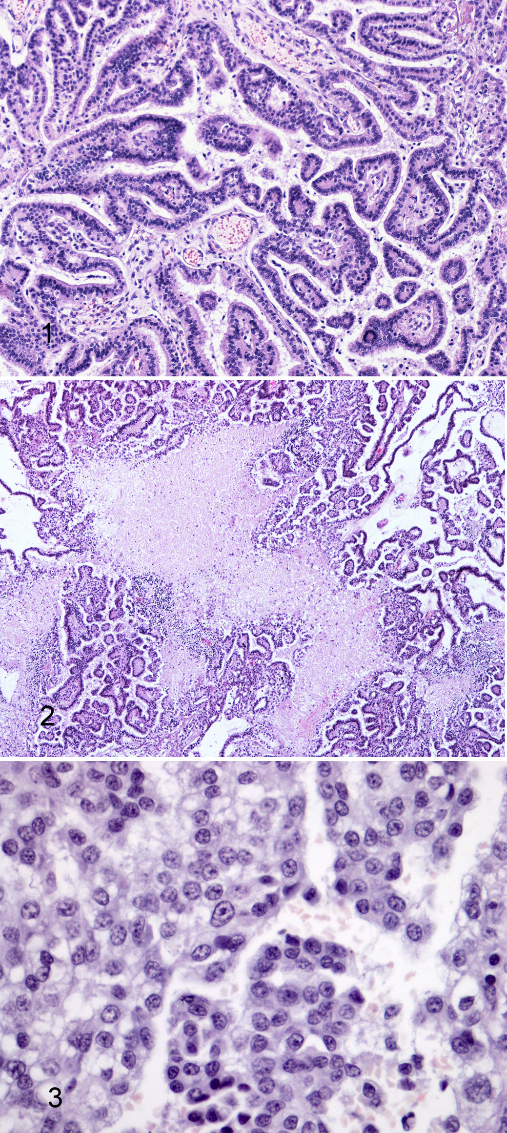
Choroid plexus tumors, dog (hematoxylin and eosin).
E-cadherin expression was observed in NCCPs with a membranous pattern (Suppl. Fig. 1) and in 12 of 14 tumors (85.7%). The negative tumors consisted of a CPP and in a CPC. Seven CPTs—4 CPPs, 2 atypical CPPs, and a CPC—maintained the membranous pattern of staining observed in NCCPs (Suppl. Figs. 2–4). Four cases—a CPP and 3 atypical CPPs—showed a cytoplasmic E-cadherin expression (Suppl. Fig. 3), whereas the remaining case (an atypical CPP) showed a mixed membranous and cytoplasmic pattern of staining. N-cadherin expression was observed in NCCPs as well as in all CPTs, regardless of histologic grade. NCCPs showed a mixed cytoplasmic and membranous pattern of staining (Suppl. Fig. 5), whereas in most of the CPTs (71.4%), the immunoreactivity was confined to the cytoplasm of the neoplastic cells (Suppl. Figs. 6–8). Contrary to the positive control (cerebral cortex from a canine fetal brain), expression of DCX protein was observed neither in NCCPs (Suppl. Fig. 9) nor in CPTs (Suppl. Figs. 10–12). Eleven CPTs showed nuclear immunoreactivity for Ki-67. The mean value of positive nuclei per 10 HPF was 2.6 (median = 1.0) for CPPs, 7.6 (median = 3.5) for atypical CPPs, and 17.7 (median = 17.75) for CPCs (Suppl. Figs. 14–16). NCCPs did not show any immunoreaction (Suppl. Fig. 13).
Statistical analysis confirmed mitotic index (P = .000), cell density (P = .008), and layers of papillae (P = .008) as the main significant markers for grading tumors. N-cadherin expression was the only significant IHC result, with grade I tumors showing an immunolabeling stronger than high-grade tumors (P = .038). Although not statistically significant, an increase in the proliferative activity was observed with higher grade tumors.
In 1989, based on the exclusion of metastasis to extraneural sites, Ribas et al. determined that most CPCs should be renamed atypical CPPs, concluding that CPC is a rare event. 12 However, based on histologic criteria more recently, Westworth et al. did not meet criteria for atypical CPPs and identified CPCs in up to 64% of cases in dogs. 14 In the same way, atypical CPPs were not identified by Nentwig et al. 10 In the current study a high number of neoplasms were classified as atypical CPPs. Certainly contributing to this result is the fact that in the human classification system, (1) the role of the peripheral infiltration is downsized, and (2) the presence of necrotic areas is no longer assumed as an unquestionable index of malignancy. 11 Therefore, in the human classification, the mitotic rate remains the most important index to determine the histologic grade of CPTs. 11 Supported by the statistical analysis, our study indicates mitotic index, cell density, and multilayering of papillae as the main significant histologic markers of malignancy for grading CPTs in dogs. The intraventricular spread that we observed in CPP and atypical CPP supports the idea that spread within the ventricular system and the subarachnoid space are not indicators of CPCs. 11 However, the biological significance of a such a revision of the histological criteria of malignanacy remains to be assessed in canine CPTs. 8
Although a statistically significant correlation with histologic grade was not found, E-cadherin expression tended to be expressed more in grade I than grade II CPTs. More recent opposite results have supported that mechanisms other than the loss of E-cadherin expression confer invasive and metastatic properties to canine CPTs. 6,10 Furthermore, we were not able to correlate aberrant cytoplasmic E-cadherin expression to adjacent brain tissue invasion, as has conversely been done for abnormal nuclear E-cadherin expression. 10 Several studies indicated that a non-tissue-specific expression of N-cadherin in tumors plays a crucial role in cell migration, invasion, and metastasis, as part of the epithelial-to-mesenchymal transition. 15 In this study, NCCPs appear to constitutively express N-cadherin, with a mixed membranous and cytoplasmic pattern. However, the decrease of N-cadherin-expression observed in high-grade tumors seems to suggest a partial dedifferentiation of malignant forms from the original tissue. Again, a change from a mixed membranous and cytoplasmic labeling to an exclusive cytoplasmic pattern of staining observed in most of the tumors remains to be interpreted. Contrary to our results, the lack of N-cadherin expression has been associated with dissemination and invasion in canine CPCs. 6 However, this contradiction could be more apparent than real, since the authors classified the tumors using the WHO classification of domestic animals. 6 DCX has been suggested to serve as a regulator of microtubule polymerization and as a bridge between the cytoskeleton and the membrane-organizing protein neurofascin, 4 and it has been suspected to play a role in invasion of neuroepithelial tumors. 4 In this study, we confirmed the absence of DCX expression in canine CPTs, 7 maybe supporting their direction to epithelial differentiation as compared with other neuroepithelial neoplasms. 7 In humans, Ki-67 is a well-documented prognostic indicator for patients with CPTs, as demonstrated by the indirect correlation between survival time and the proliferative potential of tumors. 3 On the contrary, few data about the application of proliferative markers to canine CPTs are currently available. 2 Albeit not supported by the statistical analysis, the proliferative potential of canine CPTs investigated by IHC suggests an increasing immunoreaction for Ki-67 in relation to histologic grade.
Although the data given in this study are not conclusive due to the low number of cases, the application of the human grading system to canine CPTs revealed an intermediate histologic grade between papilloma and carcinoma for the first time. Prospective studies are necessary to validate this grading system in association to the expression of cell adhesion and invasion molecules, as well as proliferating index.
Footnotes
Declaration of Conflicting Interests
The author(s) declared no potential conflicts of interest with respect to the research, authorship, and/or publication of this article.
Funding
The author(s) received no financial support for the research, authorship, and/or publication of this article.
References
Supplementary Material
Please find the following supplemental material available below.
For Open Access articles published under a Creative Commons License, all supplemental material carries the same license as the article it is associated with.
For non-Open Access articles published, all supplemental material carries a non-exclusive license, and permission requests for re-use of supplemental material or any part of supplemental material shall be sent directly to the copyright owner as specified in the copyright notice associated with the article.