
Abstract
AstraZeneca ran a bespoke study to generate age-matched clinical pathology and histopathology data from a cohort of Beagle dogs aged between 25 and 37 months to support the use of these older animals in routine preclinical toxicology studies. As the upper age range of Beagle dogs routinely used in toxicology studies does not normally exceed 24 months, there is an absence of appropriate age-matched historical control data. The generation of such data was crucial to understand whether age-related differences in spontaneous findings might confound the interpretation of toxicology study data. While the majority of the histopathology findings in all the older dogs occurred at a similar prevalence as those expected in young adult dogs (<24 months), a number of differences were observed in the thymus (involution), bone marrow (increased adiposity), testes (degenerative changes), and lung (fibrosis, pigment and alveolar hyperplasia) that could be misinterpreted as a test article effect. Minor differences in some clinical pathology values (hemoglobin, alkaline phosphatase, absolute reticulocytes) were of a small magnitude and considered unlikely to affect the interpretation of study data.
Keywords
While Beagle dogs were originally developed for hunting, they have been extensively used as a species for the toxicity testing of agrochemicals and pharmaceuticals. 2,4,22 Olson 21 provided data that supported the use of nonrodents in safety testing by comparing human toxicities with those identified in animals. The results showed the true positive concordance rate of 71% for rodent and nonrodent species combined, with nonrodents alone being predictive for 63% of human toxicities.
Chronic toxicity testing in dogs is normally confined to studies with a maximum duration of 1 year, with animals typically being 6 to 12 months old at study start. As a result, publications detailing the incidence of spontaneous pathology findings in Beagles are confined to animals no older than 2 years. 12,16 –18,28 While chronic lifetime studies in Beagle dogs have been undertaken at the Lovelace Respiratory Research Institute (LLRI), 19 published data from these studies does not include detailed observations of the spontaneous pathology noted in dogs aged between 2 and 5 years.
AstraZeneca had a cohort of older beagle dogs available for use on routine toxicity studies, with the age of all animals exceeding the upper age limit of Beagles routinely used in toxicity testing (>24 months), raising concerns about a lack of suitable age- and strain-matched historical control data and potential impact on the interpretation of histopathology findings. AstraZeneca has a commitment to fully implement the principles of replacement, reduction, and refinement (3 Rs) for the use of animals in research; therefore, the aim of the study described here was to generate a complete “guideline compliant” set of hematology, clinical chemistry, and anatomic pathology end points to support the use of these older dogs in preclinical toxicity testing. For the purposes of this article, we define young adult dogs as being aged between 6 and 24 months and older dogs, between 25 and 37 months.
Materials and Methods
The study was reviewed by the Institutional Animal Welfare and Ethical Review Body and was conducted in accordance with the animal care and ethics described in “Guidance on the Operations of the Animals (Scientific Procedures) Act 1986” issued by the UK Home Office. A good statistical practice was applied to the design of this study (details are included in the supplementary data).
Animals
Thirty-two purpose-bred beagle dogs (16 males and 16 females) were used that originated from Kennel Rååhöjden, Örkelljunga, Sweden. Animals were acclimatized for approximately 2 weeks prior to the start of the study. They were housed individually or in groups (to minimize the potential for fighting) in environmentally controlled rooms with 12-hour light cycles, relative humidity of 55% ± 15%, and a temperature of between 15°C and 24°C. They were fed a set weight of commercially available diet (SDS Dog-D3 [E] SQC; supplied by Special Diet Services Ltd, Witham, UK) and allowed ad lib access to water.
Study Design
At the study start, dogs were assigned to 2 groups according to age (8 males and 8 females per group). Group sizes were based on the AstraZeneca good statistical practice review applied to the study. 24 At the start of the study, animals in the first group (group 1) were between 25 and 30 months of age, while those in the second group (group 2) were at least 31 months of age, with the oldest being 37 months. Dogs in both groups were sham dosed by insertion of an oral gavage tube once daily for 3 days. This was undertaken to mimic the most common route of dosing for dog toxicology studies and to allow for any spontaneous pathology that might be attributed to the dosing procedure. A blood sample was taken for hematology and plasma chemistry analysis from the jugular vein prior to the first sham dose and again prior to necropsy. Urine was taken by catheter prior to necropsy for urine analysis. Hematology analysis was conducted on an Siemens Advia 2120i analyzer. Plasma chemistry and urine analysis was conducted on a Roche Modular System analyzer. For details of the measured parameters see Supplemental Table 1.
Dogs were euthanized after 3 days of sham dosing by intravenous administration of sodium pentobarbitone followed by exsanguination. All animals were necropsied, gross abnormalities recorded, and selected organs weighed. Representative samples of a full range of tissue were taken and fixed in Davidson’s (eyes and optic nerves), Bouin’s (testes and sciatic nerves), and 10% neutral buffered formalin (all other tissues). For details, see Supplemental Table 2. Tissues were trimmed, routinely processed, and embedded in paraffin wax prior to sectioning (4–6 µm) and staining with hematoxylin and eosin for routine histologic examination. The tissues were embedded according to the standard blocking pattern employed by AstraZeneca for routine dog toxicology studies. A single section of each paired organ was examined, along with 2 sections of heart and spinal cord, 3 sections of liver, 4 sections of brain, and 7 sections of lung. The tissues were examined by an experienced toxicologic pathologist with subsequent peer review of approximately 10% of the tissues by a second experienced toxicologic pathologist. Findings were recorded with descriptive terminology similar to that previously used at AstraZeneca in the evaluation of dog toxicity studies. The findings were graded with a 4-point semiquantitative scale ranging from minimal to marked. Pathology data were entered directly into a computerized database (Provantis 8.6.1.3), while clinical pathology data were captured from the automated analyzers via a validated LIMS system (GLIMS).
Historical data were analyzed retrospectively from control groups of young adult Rååhöjden beagles aged between 9.5 and 19 months at necropsy, from toxicology studies undertaken within the last 3 years. These animals were housed under similar conditions as the present study. Study data including histologic prevalence and individual animal data were extracted from the clinical pathology and pathology data entry systems and study reports to provide a comparison with the older animals from the bespoke study described here. Data were available from 15 studies providing a total of 108 controls animals. Due to minor variations in tissue list examined, the absolute number of controls varied slightly on an individual organ basis.
Results
Clinical Pathology
The majority of the hematology and plasma chemistry values of the dogs in group 1 (25–30 months of age) and group 2 (≥31 months of age) fell within the reference range for young adult beagles of the same strain aged up to 19 months. There were also no appreciable differences between groups 1 and 2 (data not shown). Minor differences were however noted for hemoglobin, total alkaline phosphatase (ALP), and absolute reticulocyte counts (Table 1).
Clinical Pathology Parameters Showing Minor Differences Between Young and Older Animals.
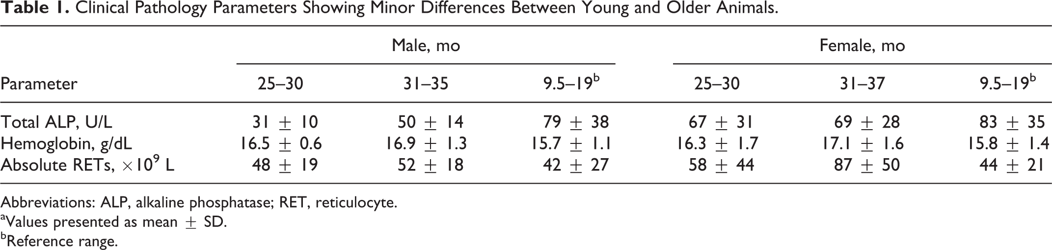
Abbreviations: ALP, alkaline phosphatase; RET, reticulocyte.
aValues presented as mean ± SD.
bReference range.
Values for hemoglobin tended to be higher in both sexes from dogs in groups 1 and 2 when compared with young adults, especially in animals aged >31 months old, and females in particular. Values for total ALP were marginally lower for both sexes from groups 1 and 2 when compared with young adults. The range for absolute reticulocyte counts in females in groups 1 and 2 showed greater variability when compared with young adults, particularly those animals aged >31 months. There were no differences noted in urinalysis (data not shown).
Anatomic Pathology
A number of macroscopic findings were observed at necropsy and consisted of red discoloration (congestion) in the small intestine, lungs, urinary bladder, and lymph nodes; pale area in the spleen (capsular fibrosis); and small thymus (involution). These were noted at a prevalence commonly observed in general toxicology studies conducted in young adult Beagle dogs (9.5–19 months at necropsy) from the test facility, with no apparent difference between groups 1 and 2.
Histopathologic findings were observed in the majority of organs within the study and were predominantly consistent with the common background spontaneous lesions routinely observed in general toxicology studies when conducted in young Beagle dogs (9.5–19 months) at the test facility. Details of the microscopic findings are provided as Supplemental Table 3. Findings noted at an increased prevalence compared with the expected norm were observed within the thymus, bone marrow, lungs, testes, epididymides, ovaries, lacrimal gland, parotid salivary gland, adrenal gland, and eye (Table 2).
Histopathology Findings in Older Dogs Compared With Historical Data From Young Adults.
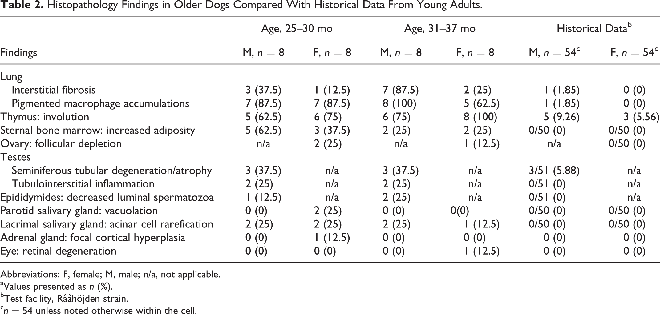
Abbreviations: F, female; M, male; n/a, not applicable.
aValues presented as n (%).
bTest facility, Rååhöjden strain.
c n = 54 unless noted otherwise within the cell.
Thymic involution was observed with an increased prevalence in both sexes from both groups 1 and 2 (Fig. 1). The incidence of thymic involution in group 2 females (8 of 8) was marginally increased as compared with group 1 (6 of 8). As part of the involution process, there was a generalized reduction in lymphoid cellularity, variable amounts of mature adipose tissue separating atrophied lobules, and the presence of multifocal epithelial-lined cysts.
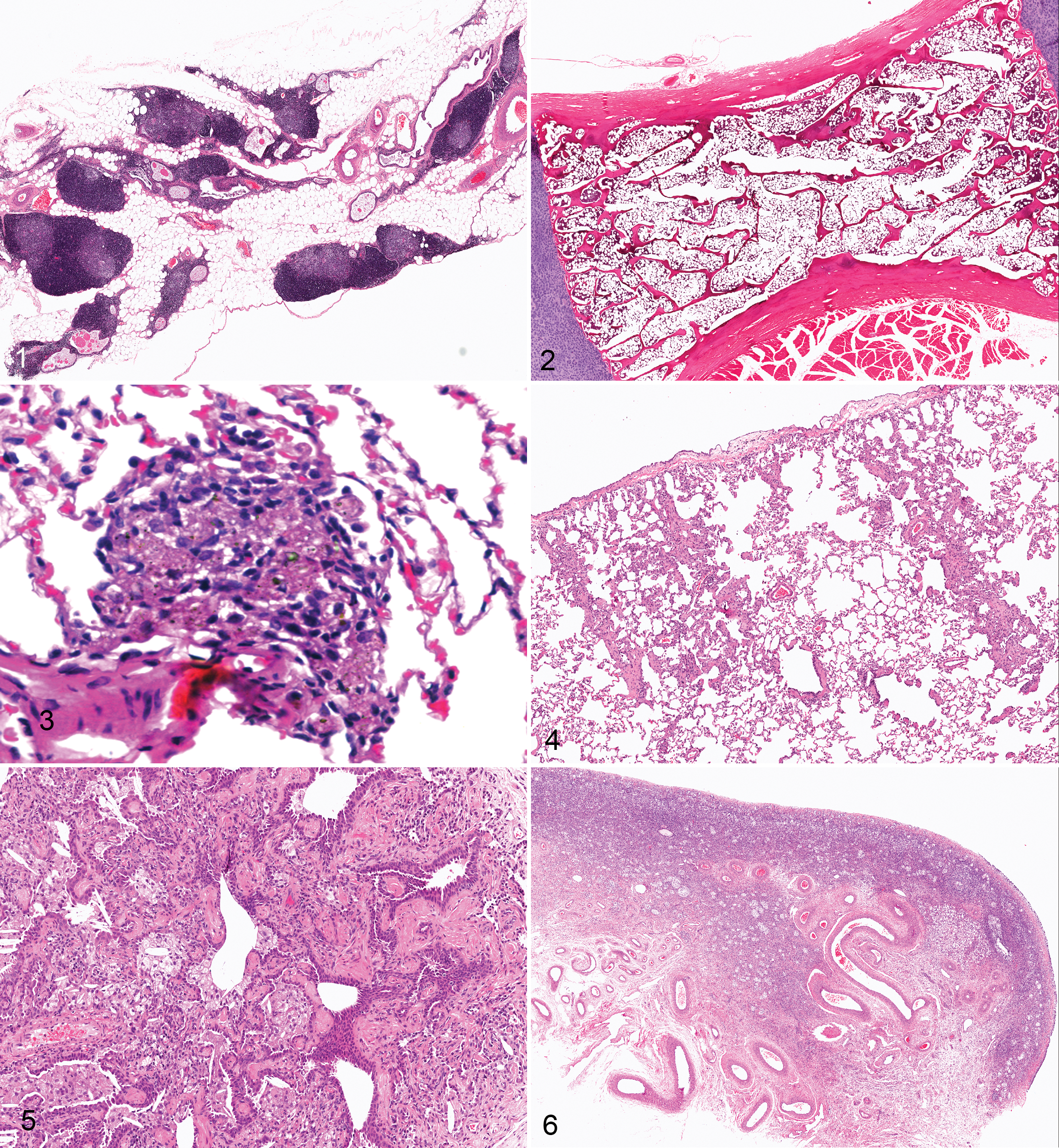
Increased adiposity (as judged by the surface area of adipose tissue compared with hematopoietic tissue) was observed in the sternal bone marrow of both sexes aged ≥25 months (Fig. 2). In our experience, the amount of hematopoietic tissue within the sternum of younger animals (typically 6–12 months of age) does show inherent variation, but the overall extent of the changes noted in this study was greater than that normally observed in young adult dogs. No attempt was made to quantify the hematopoietic tissue beyond examining the hematoxylin and eosin–stained sections, but at higher magnification, the myeloid and erythroid components (ratio) appeared similar to that normally expected in young adults.
Small focal accumulations of macrophages containing refractile golden brown pigment were observed within the lungs of the majority of animals in this study, with no apparent difference in incidence between group 1 and group 2 of either sex. These aggregates were often peribronchiolar but were also noted as clusters within alveoli (Fig. 3). No further attempt was made to characterize the pigment, but it most likely represented lipofuscin, hemosiderin, or inhaled particulates. In addition, variably sized and relatively discrete areas of subpleural interstitial fibrosis (Fig. 4), with or without accompanying bronchioloalveolar hyperplasia and/or cholesterol clefts, were observed in animals from groups 1 and 2 of both sexes (Fig. 5). The prevalence was increased in group 2 males (7 of 8) compared with group 1 males (3 of 8). Bronchioloalveolar hyperplasia was characterized by presumptive type II pneumocytes that lined alveolar walls with small bronchioles lined by a single layer or multiple layers of enlarged epithelial cells. Both these lung findings can be observed in younger dogs of this strain, but the prevalence in older dogs was increased.
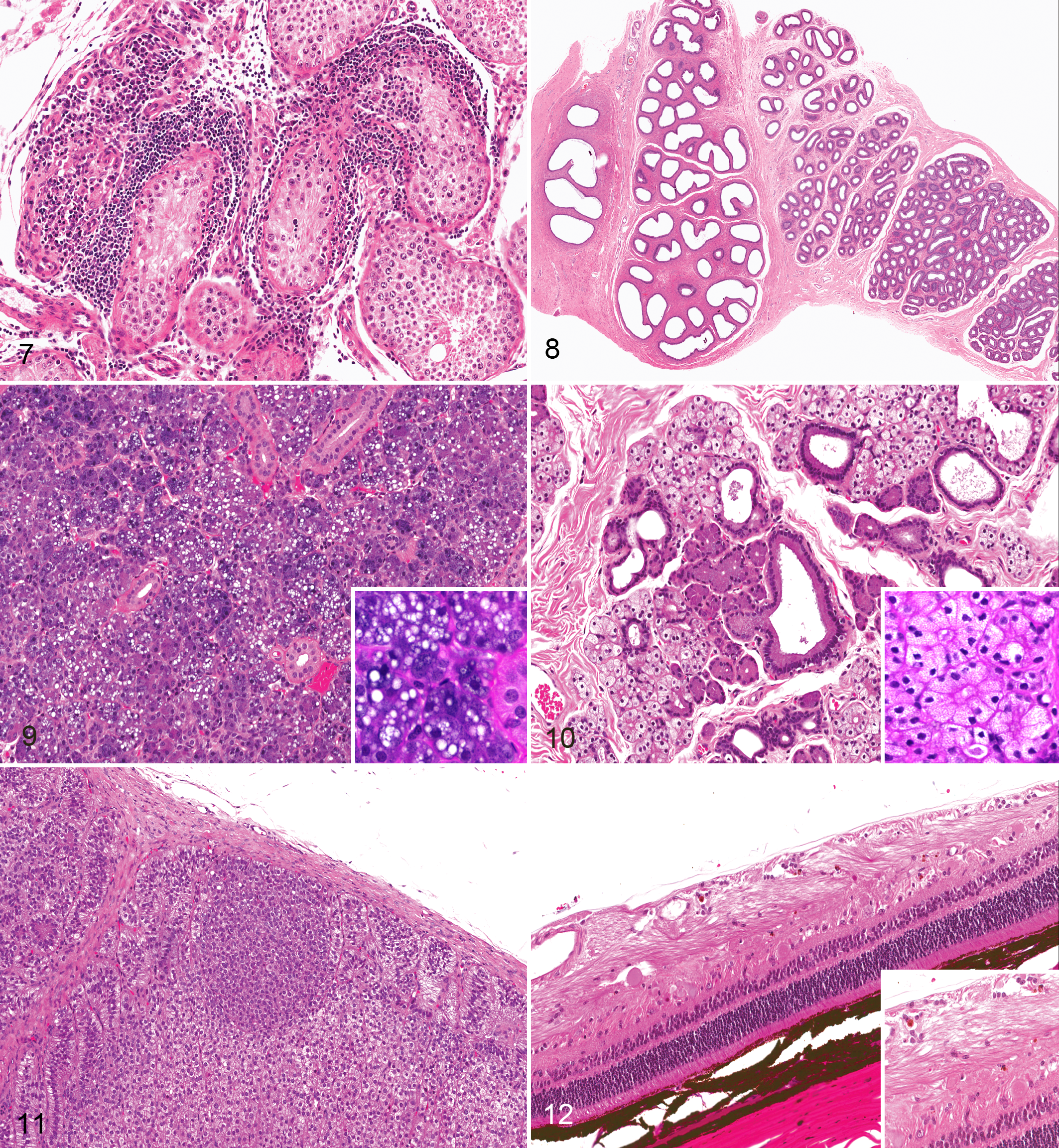
Within the ovary, there was an apparent decrease in the number of primary, secondary, and tertiary follicles within the ovarian cortex of occasional dogs from groups 1 and 2 (Fig. 6). In our experience, such a prominent lack of follicles would be unusual, even given the normal cyclical changes expected in the canine ovary. There were no notable differences in the remainder of the female reproductive tract. An increased prevalence of seminiferous tubular degeneration/atrophy was observed within the testes of groups 1 and 2. This finding was characterized by a reduction in cellularity of the seminiferous tubular epithelium, with exfoliation of effete cellular debris, multinucleate giant cells, and the variable presence of tubules containing only Sertoli cells. Seminiferous tubular changes were frequently accompanied by sperm stasis within the tubular lumen and by multifocal tubulointerstitial lymphoplasmacytic inflammation (Fig. 7). In some animals, these tubular changes were accompanied by a marked reduction of mature spermatozoa within the epididymides (Fig. 8).
Diffuse acinar cell vacuolation was observed in the parotid salivary gland of 2 group 1 female animals and was accompanied by an increased cytoplasmic basophilia (Fig. 9). Affected cells generally contained a single or occasionally multiple clear vacuoles that occupied the majority of the cytoplasm. No further attempts were made to characterize the nature of the vacuoles, but the appearance was reminiscent of lipid vacuolation. This finding has not been previously recorded in young adult animals at the test facility.
Within the lacrimal glands of a number of animals of both sexes from groups 1 and 2, a rarefication of the cytoplasm affecting contiguous clusters of epithelial cells was noted (Fig. 10). The characteristic acinar cell eosinophilic cytoplasm was replaced by a clear granular or vacuolated appearance, often expanding the cell, with displacement of the nucleus to a central location. This finding in the lacrimal gland was not associated with any clinical observations or other histopathologic findings in the eye.
Two other isolated findings were observed that are not normally noted in young adult Beagles of this strain. In the adrenal gland, a single unilateral focus of cortical hyperplasia was observed in the zona fasciculata in a single group 1 female (Fig. 11). This was characterized by a well-circumscribed and discrete area of hyperplastic cells contained within the zona fasciculata of the existing adrenal cortex.
In the eye, unilateral degeneration was observed within the ganglion cell layer of the retina in a single group 2 female (Fig. 12). Scarce pigmented cells containing a refractile golden brown pigment were noted within the nerve fiber layer of the ganglion cell layer, with occasional swollen axons.
Discussion
The aim of the present study was to generate a bespoke set of clinical pathology and anatomic pathology data from a cohort of Beagle dogs aged between 25 and 37 months to support the use of a surplus of older dogs in preclinical toxicology studies. The data were intended to be used as a direct comparator for any subsequent data generated when these older dogs were used in toxicology studies. In the present article, the findings of this bespoke study are presented and compared with spontaneous pathology findings and clinical pathology data in a cohort of young adult dogs of the same strain, aged 9.5 and 19 months at necropsy. For Beagle dogs aged >24 months, the majority of published historical control data for clinical and anatomic pathology dates from the 1970s and 1980s, with little published thereafter, highlighting the need for publication of more contemporaneous data from older animals (>24 months).
The small group sizes used in dog toxicology studies can be problematic in the interpretation of changes in the clinical pathology and anatomic pathology data from treated animals, particularly if there are no concurrent controls. In addition, more emphasis is being placed on the pathology data generated early in the drug development process 26 (ie, at the maximum tolerated dose/dose range–finding phase), in an attempt to reduce subsequent attrition as a result of unexpected findings noted in pivotal good laboratory practice studies. This has placed even more emphasis on the appropriate interpretation of early toxicology data. Routine clinical pathology data are almost exclusively generated from automated analyzers that—if maintained and subject to the appropriate quality control procedures—produce consistent and absolute numerical data. This makes it easier to generate appropriate age- and strain-matched reference ranges. In addition, blood samples are routinely taken before the commencement of dosing, providing a set of predose values for comparison. The generation of anatomic pathology data is more subjective, with the potential for the study pathologist to utilize differing thresholds and descriptive terms to record findings. Therefore, the interpretation of differences noted in treated animals is largely based on the pathologist’s prior knowledge of the anticipated spontaneous findings alongside the expected pharmacology or toxicology of the test item. It is not uncommon for sponsors to have to defend their interpretation of pathology data submitted to regulatory authorities, which may often require the provision of robust historical control data as part of a weight-of-evidence approach. Given the lack of published historical control data for Beagle dogs aged 2 to 5 years, the study described here was designed to generate a robust set of age-matched historical control data, to support the use of these older dogs in preclinical toxicology studies. The generation of the data in this study was crucial in the scientific assessment of the preclinical toxicology studies using these older dogs while implementing the principles of the 3 Rs by providing an evidence base to support using the available older dogs in research. In addition, by publishing these findings, the intention is that making the findings available may support other companies who need to use older dogs in preclinical studies.
For the clinical pathology end points, the majority of the parameters fell within the reference range for young adult dogs (typically 10 months at study start). Very minor differences were however noted in total plasma ALP, hemoglobin, and ranges for absolute reticulocyte counts. Changes in total ALP have been described with aging in dogs, 7,15 with a trend for decreases following skeletal maturity, stable values from around 1 to 8 years, and increases in old age (≥8 years). Most investigators measured total ALP, while others 32 measured ALP isoenzymes (bone, liver, and corticosteroid induced). The decreases and subsequent increases in total ALP observed during aging were reflected by decreases and subsequent increases in bone ALP. Liver-derived ALP gradually increased from puberty to old age, while corticosteroid-induced ALP showed a small fall following puberty (1–7 years) and a rise to pubertal levels from 7 years on. 7 , 31,32 The differences in the absolute values for total ALP in the older dogs from the present study were very minor and consistent with other published data. 7,15,31,32 A number of studies have evaluated age-related changes in hematology parameters in both Beagle and companion dogs. 3,7,11,15 For Beagles in which the hematocrit values were measured at yearly intervals up to 14 years, 7,11 there were no significant trends in dogs aged 2 to 5 years, but hematocrit values did decline in both studies when dogs reached ages of around 8 years. Conversely, some reports suggest that older dogs have either no change or an increase in red blood cell mass. In a study in which 57 female Beagles were evaluated from 1 to 6 years of age, 3 hematocrit remained the same, while hemoglobin increased, particularly in females. Increased hemoglobin in these Beagles is consistent with the small increases noted in the older dogs from the present study, with the oldest females (>31 months) showing the greatest increase. The females from both age groups in this study also showed a greater range of absolute reticulocyte counts compared with young adult animals. In our experience, values for absolute reticulocytes are often highly variable in dogs, with this variability exaggerated in the older females. Given that most routine toxicology study designs utilize ≥1 predose bleeds in dogs, there was less overall concern about the potential for misinterpreting age-related variations in clinical pathology and hematology data. However, the complete data set collected in this study confirmed that the majority of the values remained unchanged in animals up to 37 months old when compared with young adults.
The majority of the pathology findings noted in the present study were consistent with the common background spontaneous pathology routinely observed in young adult Beagle dogs of this strain (9.5–19 months), as well as with published descriptions of the spontaneous pathology of Beagles. 12,16,18,20,28 Changes in some organs, such as the thymus and bone marrow, could be anticipated given the well-recognized association between the degree of thymic involution and decrease in bone marrow cellularity with increasing age. In the thymus, the histologic pattern of involution was similar to that reported in young adults, 23 with animals >30 months having an increased prevalence of this change. Little evidence exists for the potential of aging to affect thymic susceptibility to toxic insults. 30 In animals >30 months old, while smaller amounts of cortical and medullary lymphoid tissue remained, it is unclear whether the residual lymphoid population would show a similar response to a toxic insult as typically seen in young adults.
The increase in adiposity noted in the bone marrow of the dogs from both age groups could have the potential to confound the interpretation of bona fide treatment-related effects, particularly in studies utilizing chronic dosing. In the present study, no attempt was made to quantify the hematopoietic tissue with flow cytometry or cytological counts, although these techniques could be employed for compounds with known or suspected bone marrow toxicity. The increased adiposity noted in older dogs was not accompanied by any discernible changes in circulating red or white blood cells. However, for compounds known to cause increased adiposity as a pharmacologic effect, the use of older Beagles such as these would not be recommended.
The findings noted in the lungs of older dogs were similar to those observed in young adults but occurred with an increased prevalence. The increased prevalence of pulmonary macrophage aggregates containing cytoplasmic pigment is consistent with the results of a study in which the lungs of Beagles aged 1 to 10 years were examined. 27 The researchers found the primary lesion in the lungs of older dogs to be the accumulation of pulmonary macrophages, containing dust and golden brown pigment, in the walls of respiratory bronchioles and alveolar ducts. These dust- and pigment-laden macrophages became more prominent with age and were sometimes accompanied by pneumonitis. The exact nature of the pigment was not described. No attempts were made to identify the nature of the pigment noted in the macrophages of the present study, although differentials could include lipofuscin, hemosiderin, or an accumulation of inhaled particulates (dust/carbon). Areas of pulmonary fibrosis, often accompanied by bronchioloalveolar hyperplasia, were also more prevalent in dogs aged >30 months. They often occurred below the pleural surface, forming wedge-shaped areas. In the majority of the cases, this segmental fibrosis was not accompanied by other changes to the pulmonary parenchyma. Segmental fibrosis with a similar histologic appearance is occasionally observed in young adult Beagles of this strain and is described in published accounts of spontaneous findings. 28 Similar-appearing areas of pulmonary fibrosis were also a common finding described in Beagles from the LLRI colony, in which it occurred at an incidence of 28% in 178 Beagles maintained for their life span. 9 In many animals from the LLRI colony, it was also not associated with other pulmonary changes and was regarded as an incidental lesion. The underlying cause of these fibrotic areas is not understood, although they likely represent a response to a previous insult, which then persists for the life span of the individual. The increased prevalence in the older dogs from the AstraZeneca colony also suggests a cumulative effect with age, but the nature of the initiating insult remains unclear. The increased prevalence of pulmonary fibrosis noted in animals >30 months of age in the present study could confound the interpretation of drug-induced pulmonary changes, particularly during recovery from a previous insult, where the presence of fibrosis could be misinterpreted as a sequel to previous damage. Given the increased prevalence of spontaneous pulmonary pathology, the use of older Beagles for inhalation toxicology studies or compounds with potential pulmonary liabilities would not be recommended.
Although dogs have estrous cycles throughout life, prolonged anestrus intervals have been observed in dogs aged >5 years. 1 The majority of published literature on aged canine ovaries deals with the occurrence of cysts and hyperplasia of the subsurface epithelial structures. 1,10 In the present study, neither of these changes was observed, but an apparent decrease in the number of follicles was noted in a small number of females. The significance of this change is difficult to assess without a proper quantification of follicle numbers. The apparent decrease in number of follicles was not accompanied by other changes in the female reproductive tract or mammary gland.
The potential for spontaneous changes in the testes of dogs to confound the interpretation of toxicology studies is well documented, 5,8,14,17,25 especially when sexually immature animals are used. Even in young adults, a number of seminiferous tubular changes can occur, such as hypospermatogenesis and segmental atrophy, which can resemble low-grade effects of some testicular toxicants. 5,8,25 In the present study, an increased prevalence of seminiferous tubular changes was observed in both the older age groups. While the majority of the tubular findings were similar to those observed in young adult Beagles, in some animals, these were accompanied by a varying degree of tubulointerstitial lymphoplasmacytic inflammation. In our experience, the presence of inflammation alongside tubular degeneration is not commonly encountered in young adult Beagles. In some individuals, tubular changes were accompanied by a marked reduction of mature spermatozoa within the epididymides. The trend of an increased prevalence of testicular findings is in agreement with a study in which the testes from aged Beagles from the LLRI colony were examined. 17 Here there was a trend for an increase in degenerative changes within the seminiferous tubules with increasing age. The increased prevalence of spontaneous testicular findings in Beagles aged >24 months could have a significant impact on the assessment of testicular toxicity, and it is recommended that such older dogs should be avoided where testicular toxicity would be an issue for the test article in question.
Relatively minor changes were noted in the epithelial components of the parotid salivary and lacrimal glands in a small number of individuals. In the parotid salivary gland, this involved the presence of discrete cytoplasmic vacuoles. These vacuoles were contained within the cytoplasm of the acinar epithelium and should not be confused with the accumulation of interstitial adipose tissue. No further attempt was made to characterize the contents of the vacuoles, so the underlying mechanism for this change is unclear. To our knowledge, this finding has not been reported in aged Beagles and had not been encountered previously in young adults of this strain. A rarefication/vacuolation of the epithelial cytoplasm of the lacrimal glands was also noted. Interestingly, this change occurred in some individuals where vacuolation was also noted in the parotid gland. Given the similar histologic appearance of parotid salivary and lacrimal gland, it is tempting to speculate on a possible association, although we have no evidence to substantiate this. So-called serous changes have been reported in the lacrimal glands of aged rats 6 but do not feature in the published accounts of spontaneous findings in Beagles.
Two isolated findings occurred that have not been previously noted in young Beagles used at the test facility. These included focal adrenal cortical hyperplasia and pigment deposition in the retina. They both affected only 1 of a paired organ. In the adrenal, the focal hyperplasia should not be confused with the accessory adrenal tissue that is occasionally encountered in young Beagles. 28 In a large survey of the spontaneous findings in Beagles aged 8 to 20 months, 12 focal adrenocortical hyperplasia was also described as a sporadic finding. Adrenocortical neoplasia is common in aged dogs, although it is not known whether the focus of cortical hyperplasia noted in the present study may represent a hyperplastic precursor to this.
Pigmentary changes in the retina of older Beagle dogs have been described. These can involve the retinal pigment epithelium as well as more superficial layers of the retina. 13,29 In the retinal pigment epithelium, they are generally considered to represent the accumulation of lipofuscin. In 1 study, 13 a clinically apparent increase in pigment was noted as an age-related change, with pigment present throughout the retinal layers. The nature of the pigment was not determined. The same authors also described degeneration in the peripheral retina in 85% of 8-year-old Beagles. In the present study, a small number of pigmented cells were confined to the ganglion cell layer of the retina and were accompanied by occasional swollen axons. No attempt was made to identify the pigment with special stains, and so the exact nature of the pigment remains unclear. The finding occurred as a focal lesion and was confined to 1 eye. It was not associated with any degeneration in the underlying tapetum. Similar retinal lesions had not been described in young adults of this strain. It was unclear whether the pigment and axonal changes in the ganglion layer noted in this dog were related to aging.
Conclusion
The clinical pathology and anatomic pathology data generated by this study provided a suitable database to allow the successful utilization of all remaining aged Beagles in preclinical toxicology studies. The results for clinical chemistry, hematology, and urinalysis confirmed that there were no significant age-related changes that were likely to confound study interpretation. For the anatomic pathology data, spontaneous findings at an increased prevalence were noted in the bone marrow, lung, thymus, and testes, which could interfere with the interpretation of histopathology data, based on the current knowledge of young adult Beagle dogs.
Footnotes
Acknowledgements
We thank Brian Rochford for technical assistance with the experimental phase and Marie South for statistical review.
Declaration of Conflicting Interests
The author(s) declared no potential conflicts of interest with respect to the research, authorship, and/or publication of this article.
Funding
The author(s) received no financial support for the research, authorship, and/or publication of this article.
References
Supplementary Material
Please find the following supplemental material available below.
For Open Access articles published under a Creative Commons License, all supplemental material carries the same license as the article it is associated with.
For non-Open Access articles published, all supplemental material carries a non-exclusive license, and permission requests for re-use of supplemental material or any part of supplemental material shall be sent directly to the copyright owner as specified in the copyright notice associated with the article.