
Abstract
Feline enteropathy-associated T-cell lymphoma (EATL) type II is characterized by infiltration of the small intestinal mucosa with small T-cells with variable epitheliotropism and is often difficult to differentiate from inflammation. Polymerase chain reaction (PCR) to assess antigen receptor rearrangements (PARR) amplifies the T- (T-cell receptor gamma, TCRG) or B-cell (immunoglobulin heavy chain, IGH) antigen receptor genes and is used to differentiate EATL from inflammation. However, PARR does not determine lymphocyte phenotype, and clonal rearrangement of either or both the TCRG or IGH genes may be detected in neoplastic T-cells. The purpose of this study was to determine the incidence of cross lineage rearrangement in feline EATL type II. Using a diagnostic algorithm combining histology, immunohistochemistry, and PARR testing, 8 of 92 cases diagnosed as EATL type II at Michigan State University between January 2013 and June 2014 showed cross lineage rearrangement (8.7%). PARR for the IGH gene facilitates the diagnosis of cases histologically highly suggestive of EATL type II in which polyclonal rearrangement of the TCRG gene is detected.
Feline small intestinal lymphoma most frequently presents as a mucosal T-cell lymphoma consistent with the World Health Organization classification of enteropathy-associated T-cell lymphoma (EATL), type II. 4 EATL type II is characterized by expansion of the lamina propria by a monomorphic population of small T-cells with variable epitheliotropism. 2,4 This epitheliotropism is commonly observed as infiltration of the villous epithelium by nests and/or plaques of small T-cells, which have been shown to be statistically associated with or specific for a diagnosis of EATL type II, respectively. 2 However, diagnosis of EATL type II remains challenging, as it is histologically similar to common inflammatory diseases of the feline small intestine, including inflammatory bowel disease. As greater than 50% of cats diagnosed with intestinal lymphoma are reportedly dead within 1 year of diagnosis compared to less than 30% of cats diagnosed with inflammatory bowel disease, differentiation between these conditions is prognostically and therapeutically relevant. 2
Based on our current understanding, the diagnosis of EATL type II is most appropriately based on a combination of histology, immunohistochemical phenotyping, and polymerase chain reaction (PCR) to assess antigen receptor gene rearrangements (PARR). 2,4 PARR has improved the accuracy of EATL type II diagnosis and amplifies the highly variable T- or B-cell antigen receptor genes, specifically the genes for T-cell receptor gamma (TCRG) and the immunoglobulin heavy chain (IGH). 5,10 PARR is used to detect clonally expanded lymphocyte populations; however, PARR does not determine lymphocyte phenotype. The phenotype of a lymphoma is based on expression of CD molecules as determined by immunohistochemistry, immunocytochemistry, or flow cytometry, as neoplastic lymphocytes may show clonal rearrangement of either or both the T- or B-cell antigen receptor genes, regardless of phenotype. 1,2,5 For example, cases of T-cell lymphoma, including EATL type II, may show clonal rearrangement of the IGH gene. This is termed cross lineage rearrangement and has been demonstrated in both human and canine lymphoid neoplasms. 1,5,7 –9 If not investigated in the appropriate cases, this cross lineage rearrangement can produce conflicting or at worst, false negative results in some cases. The purpose of this study was to determine the incidence and therefore diagnostic relevance of cross lineage rearrangement in feline EATL type II.
A total of 175 cases of suspected EATL type II submitted to the Michigan State University Diagnostic Center for Population and Animal Health between January 2013 and July of 2014 were included in this study. Cases were evaluated based on a diagnostic algorithm including histopathology, immunohistochemical phenotyping, and PARR for either the TCRG gene only (139 cases) or both the TCRG and IGH genes (36 cases) using previously published methods and primer sequences. 2,5,10 Immunohistochemistry for both CD3 and CD79a, which represent T- and B-cell markers, respectively, was performed for phenotyping. Cases were considered histologically suggestive of EATL type II if there was either or both expansion of the small intestinal lamina propria by a monomorphic population of small T-cells or infiltration of the villous epithelium by nests and/or plaques of small T-cells. 2,4 An intraepithelial nest was defined as 5 or more small T-cells clustered within the villous epithelium, and 5 or more adjacent epithelial cells obscured by infiltrates of small T-cells was considered an intraepithelial plaque. 2 For cases histologically and immunophenotypically suggestive of EATL type II, PARR for the TCRG gene was performed. If PARR for the TCRG gene showed polyclonal rearrangement and the case was histologically strongly suggestive of EATL type II, PARR for the IGH gene was performed.
Of the examined cases, 92 were diagnosed as EATL type II by the diagnostic algorithm previously described. Eight of these 92 cases showed cross lineage rearrangement of the IGH gene. The histologic features of all 8 of these cases were strongly suggestive of EATL type II, including expansion of the small intestinal lamina propria by a monomorphic population of small lymphocytes, which were identified as T-cells by immunohistochemistry for CD3 (Fig. 1). Rare, scattered lymphocytes within the small intestinal mucosa of these sections labeled for the B-cell marker CD79a. Marked epitheliotropism, defined by prominent intraepithelial nests and/or plaques of small T-cells, was observed in serial sections routinely stained with hematoxylin and eosin or immunohistochemically labeled for CD3. For all 8 of these cases, PARR for the TCRG gene demonstrated multiple, short, broad peaks on an electropherogram produced by capillary gel electrophoresis following the PARR assay, consistent with polyclonal rearrangement of the TCRG gene. However, the capillary gel electropherograms of the PARR assays for the IGH gene showed a single, tall, thin peak consistent with clonal rearrangement of the IGH gene (Fig. 2).
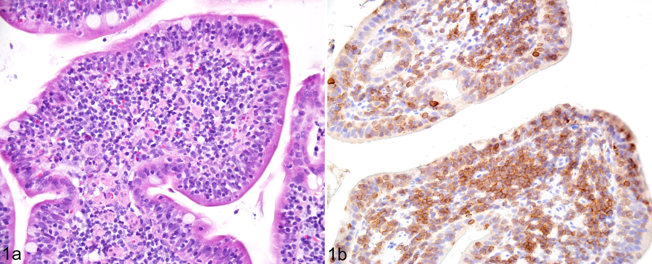
Enteropathy-associated T-cell lymphoma (EATL), type II, small intestine, cat. Representative case of feline EATL type II with cross lineage rearrangement showing expansion of the lamina propria and prominent epitheliotropism by a monomorphic population of small lymphocytes with (a) hematoxylin and eosin staining and (b) immunohistochemistry for CD3e.
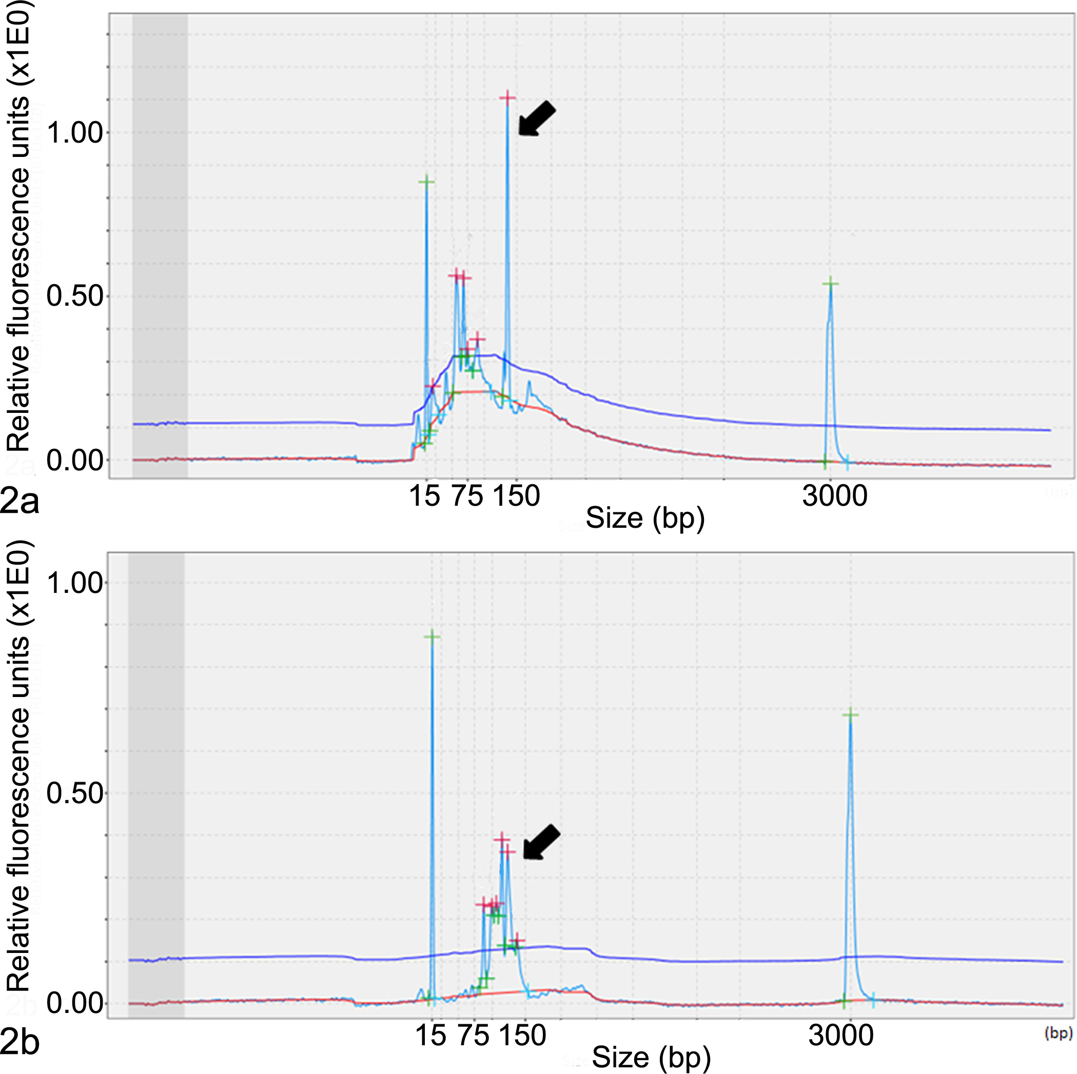
Results of polymerase chain reaction (PCR) to assess antigen receptor rearrangements (PARR) for the immunoglobulin heavy chain (IGH) and T-cell receptor gamma (TCRG) genes depicted in capillary gel electropherograms. The tall narrow peaks (15 and 3000 bp) on either side of the peaks designated by arrows represent alignment markers. (a) Electropherogram showing clonal rearrangement of the IGH gene as indicated by a single tall peak (arrow). (b) PARR failed to detect clonal rearrangement of the TCRG gene, demonstrated by multiple short peaks on the capillary gel electropherogram (arrow).
Cross lineage rearrangement of the IGH gene was detected in 8.7% of cases diagnosed as EATL type II in the present study; however, this percentage may actually be underestimated. The sensitivity of the PARR assay for the IGH gene is 68.2%, so a proportion of positive cases may have been falsely negative due to the relatively low sensitivity of this assay. 10 Additionally, the cases reported herein represent diagnostic cases subject to the financial limitations of the submitting owners and the need to only obtain prognostically relevant information. As a result, in cases for which PARR for the TCRG gene initially demonstrated a clonal population, PARR for the IGH gene was not performed. Consequently, cases in this study that may have shown clonal rearrangement of both the TCRG and IGH genes (bigenotypic) would have remained undetected. The percentage of bigenotypic cases potentially left undetected in the present study may not be trivial: in a study of human T- and B-cell malignancies, 6.7% of T-cell neoplasms evaluated were bigenotypic, and this rate rose to 11% when both T- and B-cell neoplasms were evaluated. 6 As a result, future studies evaluating clonality of both the TCRG and IGH genes in cases of EATL type II may provide a more accurate assessment of the rate of cross lineage rearrangement in this entity.
The incidence of cross lineage rearrangement in this study is similar to that reported in both human and canine T-cell malignancies. The reported rate of cross lineage rearrangement in various human mature T-cell malignancies is 0% to 7%, 9 while a study of canine T-zone lymphoma reported a rate of 13%. 8 Interestingly, similar to the present study, clonal rearrangement of the TCRG gene was only detected within a portion of cases diagnosed as T-cell lymphoma in both of these studies. 8,9 This may reflect either the sensitivity of the TCRG PARR assay (89% in the present study) or interference by concurrent inflammatory T-cell infiltrates within the small intestine. 5
In all cases of cross lineage rearrangement in the present study, PARR detected polyclonal rearrangement of the TCRG gene. This inability to detect a clonal rearrangement of the TCRG gene may be due to concurrent inflammatory T-cell infiltrates within the evaluated tissues masking the clonal T-cell population. Additionally, there may have been exclusive amplification of the TCRG gene within either resident or inflammatory T-cell populations within the small intestine. This exclusive amplification could be due to failure of the PARR assay primers to recognize the specific rearrangement within the neoplastic T-cells, or there may have been chromosomal alterations within the neoplastic T-cells that distorted the sequence of the primer binding site and resulted in binding failure. 5
Detection of a clonal lymphocyte population is not always indicative of neoplasia and can be seen as a response to pathogens or a concurrent neoplasm. 2 In the current study, diagnoses of EATL type II were only made by the collective results of histologic examination, immunohistochemistry, and PARR testing, greatly reducing the possibility of a misdiagnosis of clonality due to a nonmalignant etiology.
The precise mechanism for cross lineage rearrangement in lymphocytes is unclear. Rearrangement of the antigen receptor genes is regulated by chromatin accessibility; however, the conditions permitting recombination enzyme access to both antigen receptor loci and the point(s) in cellular differentiation this may occur have not been identified. 3 It has been speculated that cross lineage rearrangement could be an effect of malignant transformation, 6 but the demonstration of cross lineage rearrangements in normal human and murine lymphocytes suggests that if malignant transformation can produce these rearrangements, it is not the only mechanism. 3,6 The effects cross lineage rearrangements may have on the biologic behavior of lymphoid neoplasms and their response to treatment is yet to be determined.
The results of this study demonstrate that cross lineage rearrangement of the IGH gene is present in at least 8.7% of feline EATL type II cases. PARR for both the TCRG and IGH genes is not recommended on a routine basis; however, PARR for the IGH gene can support a diagnosis of EATL type II in cases with strong histologic and immunohistochemical evidence for EATL type II in which polyclonal rearrangement of the TCRG gene is detected.
Footnotes
Authors’ Note
Dr Caroline Andrews is a molecular pathology fellow in the NIH Comparative Biomedical Scientist Training Program supported by The National Cancer Institute in partnership with Michigan State University.
Declaration of Conflicting Interests
The author(s) declared no potential conflicts of interest with respect to the research, authorship, and/or publication of this article.
Funding
The author(s) received no financial support for the research, authorship, and/or publication of this article.