
Abstract
The present study describes a generalized congenital skin condition in 14 Great Dane puppies. Macroscopically, all dogs showed generalized gray to yellow scaling and skin wrinkles on the head and all 4 extremities. Skin sections were histologically examined using hematoxylin and eosin, Heidenhain’s Azan, and Sudan red III staining methods and by conducting the alcian blue/periodic acid Schiff (AB/PAS) reaction technique on sections. Furthermore, incubation with hyaluronidase was performed. Skin samples were ultrastructurally analyzed using transmission electron microscopy. All affected Great Dane puppies had epidermal and follicular orthokeratotic hyperkeratosis, enlarged keratohyaline granules, vacuolated keratinocytes, and accumulations of an eosinophilic and alcianophilic, lipid-rich material within dilated hair follicular lumina and the cytoplasm of sebocytes. The macroscopic, histopathologic, and ultrastructural skin changes in all 14 Great Dane puppies indicate a new variant of a primary disorder of cornification with congenital, non-epidermolytic, lamellar ichthyosiform appearance.
Introduction
Ichthyoses are a heterogenous group of rare congenital disorders of cornification that are characterized by hyperkeratosis and scales. This condition has been reported in cattle, dogs, pigs, chickens, laboratory mice, and a llama. 3,11,20 Based on histologic findings, ichthyoses can be subdivided into epidermolytic and non-epidermolytic forms. In epidermolytic ichthyosis, vacuolization and lysis of keratinocytes together with hypergranulosis and hyperkeratosis occur within the spinous and granular cell layers. 7,22 Non-epidermolytic ichthyosis is mainly characterized by marked, lamellar, orthokeratotic hyperkeratosis and mild acanthosis. 19 Ichthyosiform disorders in dogs have strong breed predilections although they can occur in mixed-breed dogs as well. 1,10,14,19,21 Syndromic ichthyoses, characterized by extracutaneous lesions, have been described in dogs, such as in the Cavalier King Charles spaniel with keratoconjunctivitis. 4,15,16 Congenital, non-epidermolytic forms of ichthyosis with clinical and histomorphological features resembling autosomal recessive congenital ichthyosis in humans (ARCI) have been described in several purebred dog breeds, such as Golden Retriever, American Bulldog, and Jack Russell Terrier. 11,12,14,20,21
The aim of this study was to describe the macroscopic, histopathologic, and ultrastructural features of a new variant of a non-epidermolytic, congenital ichthyosis observed in Great Dane puppies associated with accumulations of an alcianophilic and lipid-rich material within sebaceous glands and dilated hair follicle lumina.
Materials and Methods
Animals and Clinical Findings
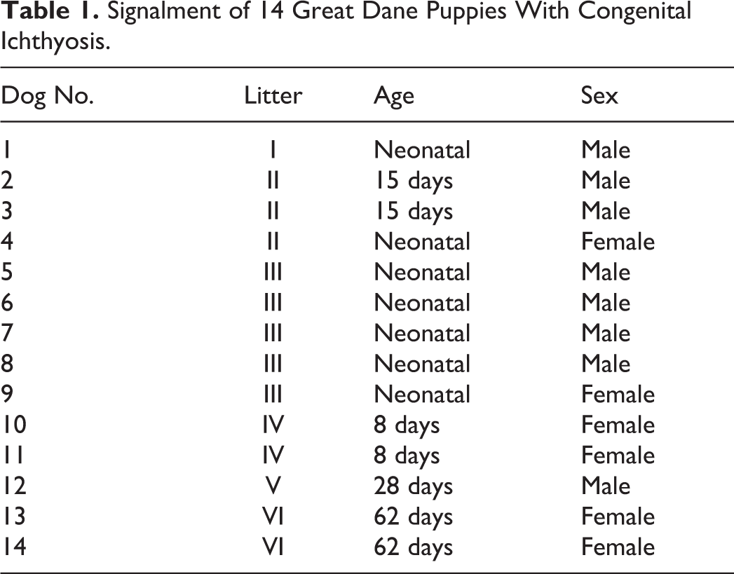
Fourteen Great Dane puppies, 6 female and 8 male, aged from a few days to 5 weeks and originating from 6 different litters (Table 1) had been submitted for necropsy between 2006 and 2014 to the Department of Pathology, University of Veterinary Medicine Hannover (dog Nos. 1–11, 13, and 14) or to the CVUA-Westfalia in Arnsberg (dog No. 12). Dog Nos. 1 through 12, after clinical examination, were euthanized at veterinary practices with the consent of the owners. Two puppies (dog Nos. 13 and 14), which were referred to the Small Animal Clinic, University of Veterinary Medicine Hannover in 2014, were examined clinically by 1 of the investigators (R.M.) and were euthanized with the consent of the owners. All animals had marked skin wrinkles on the head and legs since birth. The skin of the head and legs and other regions showed moderate to severe scaling with fine white to yellow scales. Furthermore, the skin was covered with a yellow, greasy material, especially on the skin of the head and legs. Glabrous skin regions, such as axillar and inguinal areas, were dry and had a leathery appearance.
Signalment of 14 Great Dane Puppies With Congenital Ichthyosis.
Skin Samples From Affected and Control Dogs
At necropsy, skin samples were collected from different wrinkled locations (head and fore and hind limbs), non-wrinkled locations (back, abdomen, sides, shoulders, navel), and various other organs and tissues. The samples were fixed in 10% formalin within approximately 24 to 36 hours after death and embedded in paraffin. Paraffin sections, 3 to 5 µm in thickness, were routinely stained with hematoxylin and eosin (HE). From 2 dogs (dog Nos. 13 and 14), 8-mm punch samples of skin were taken approximately 2 hours postmortem, and either processed as above, embedded in Tissue-Tek O.C.T. compound (Sakura Finetek, Alphen aan den Rijn, the Netherlands) and snap frozen in 2-methylbutane, or fixed in 2.5% glutaraldehyde in 0.1M cacodylate buffer (pH 7.2) for 24 hours before processing for transmission electron microscopy.
As normal control tissue, skin samples from the head, fore and hind limbs, back, and navel were obtained from 7 Great Dane puppies that were unrelated to the diseased dogs and had died after Caesarian section. Samples were obtained within 24 hours after death, histologic sections were prepared as above, and skin samples from 1 of the puppies were prepared for transmission electron microscopy as described above.
Histochemistry and Transmission Electron Microscopy
On skin sections from all diseased and control puppies, the combined alcian blue (pH 2.5)/periodic acid Schiff (AB/PAS) reaction was performed. Furthermore, to evaluate hyaluronan, sections were digested with 80 μl/mL hyaluronate lyase from Streptomyces hyalurolyticus (Sigma-Aldrich, St Louis, MO, USA) in 50 mM sodium acetate, 0.15 M NaCl, pH 6.7, at 37°C, for 3 hours followed by staining with AB/PAS at pH 2.5 (modified, according to Zanna et al 29 ). Control sections were incubated in parallel with the same solution without the enzyme. On representative skin sections from several diseased dogs and control dogs, a Heidenhain’s Azan stain was performed. Frozen and previously formalin-fixed skin sections (5 µm in thickness) from dog Nos. 13 and 14 were routinely stained with Sudan red III.
Transmission Electron Microscopy
Electron microscopic examination of skin samples from dog Nos. 1, 2, 8, 9, 10, and 12 was performed using the pop-off technique. 17 From skin samples obtained from dog Nos. 13 and 14 and from the control dog, first semi thin sections were cut (1-2 µm) and stained with 0.1% toluidine blue. Subsequently, ultrathin sections were cut at 60 to 90 nm and stained with uranyl acetate and lead citrate. These were then examined with a Zeiss EM 10 A transmission electron microscope (Zeiss, Oberkochen, Germany).
Results
Pathologic Findings
All 14 Great Dane puppies had markedly wrinkled skin on the head and on the distal extremities (Figs. 1–3). The skin surface had moderate to severe fine, dry, white to yellow scales. Furthermore, the skin had a greasy appearance, especially affecting the head and legs. In 1 puppy (dog No. 12), glabrous skin was noted on the muzzle (Fig. 1). In 2 puppies (dog Nos. 10 and 11), bilateral, mild to moderate hydronephrosis was found. Except for agonal changes, no other gross abnormalities were seen in any of the dogs.
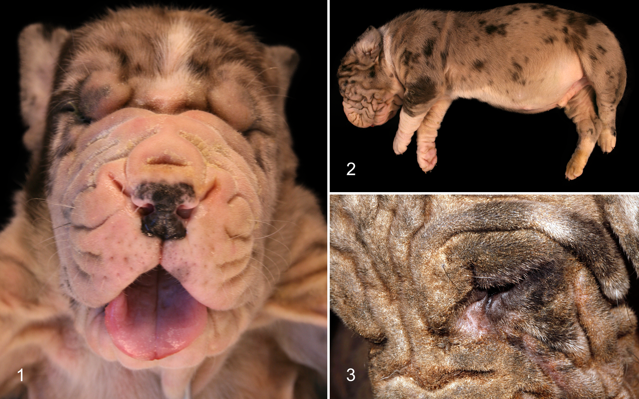
Ichthyosis, dog.
HE-stained paraffin sections from wrinkled and non-wrinkled skin of all 14 puppies had similar histopathologic changes of moderate to marked, diffuse, orthokeratotic, lamellar, epidermal, and follicular hyperkeratosis and mild, regular acanthosis (Figs. 4, 5). In all 14 cases, vacuolated keratinocytes with occasionally slightly shrunken nuclei were present within the stratum granulosum, the stratum spinosum, and the hair follicle epithelium. Some keratinocytes had empty perinuclear vacuoles, while others had vacuoles with slightly eosinophilic homogenous material (Fig. 5). The keratohyaline granules of the stratum granulosum of the epidermis and of the hair follicle epithelium were normal in number and distribution. However, often enlarged, spherical keratohyaline granules were noted (Fig. 5). There was marked, orthokeratotic hyperkeratosis of dilated hair follicle infundibula with frequent keratotic plugging (Fig. 4). In all 14 dogs, some sebaceous gland appeared normal with mature sebocytes surrounded by a peripheral layer of basal cells, while other sebaceous glands were enlarged and had numerous degenerating sebocytes with shrunken nuclei and homogenous unstained cytoplasmic material. Large amounts of such material were also found within dilated hair follicle lumina (Fig. 6).
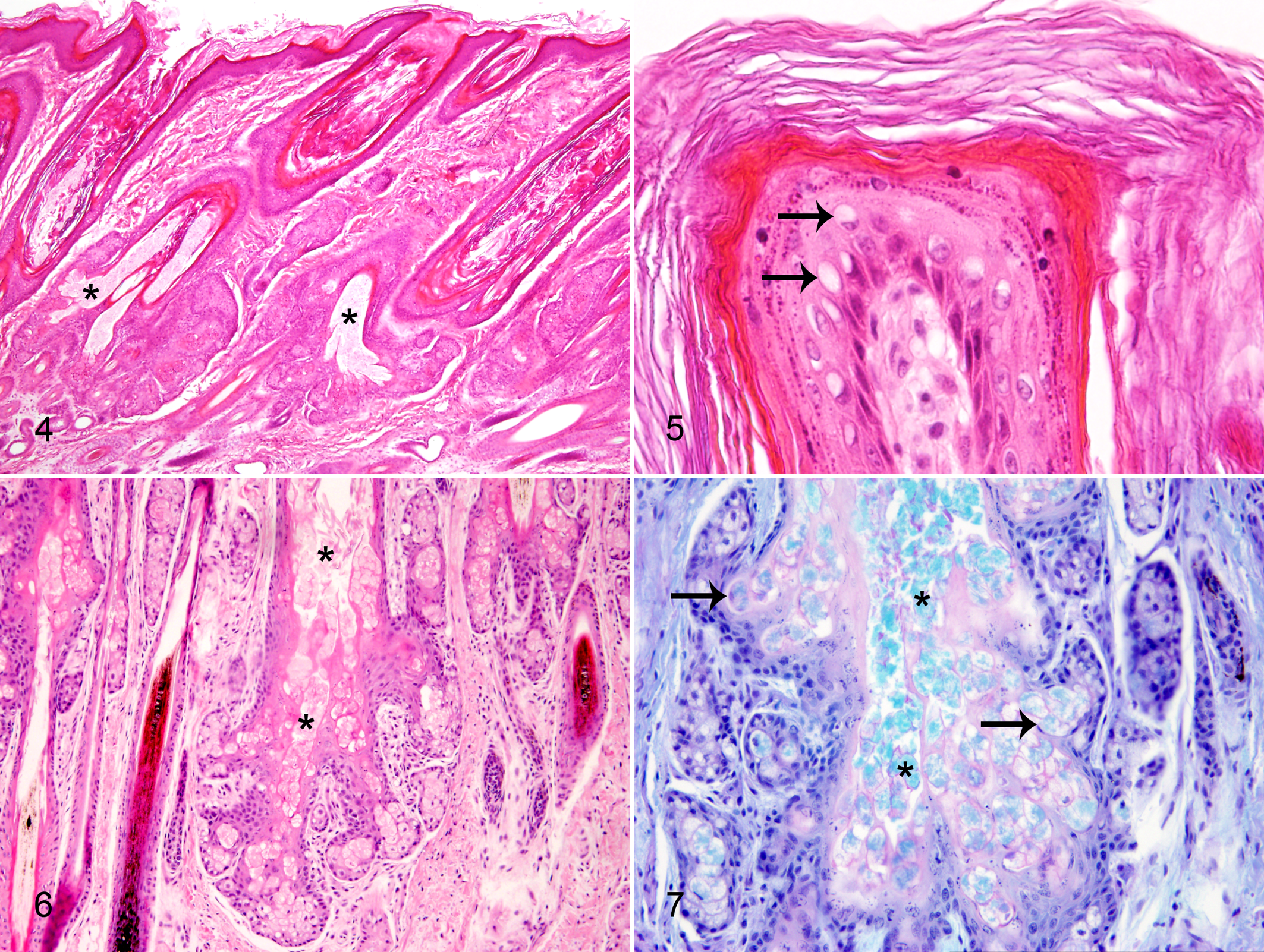
Ichthyosis, skin, dog.
Except for 3 cases (dog Nos. 1, 12, and 13), inflammatory skin lesions were absent. In the skin of dog No. 1, few scattered lymphocytes and plasma cells were found within the superficial dermis. Dog No. 12 focally had bacterial colonies within dilated hair follicle lumina and in the stratum corneum, in samples of the muzzle and of the deep pyogranulomatous dermatitis on the lip. This dog also had a mild focal intracorneal infiltration of neutrophils together with bacterial colonies. A moderate focal pyogranulomatous furunculosis including the sebaceous gland tissue as well as a mild focal intracorneal infiltration with neutrophilic granulocytes and few intralesional bacteria were seen in a section from the muzzle of dog No. 13. Dermal collagen and subcutaneous tissues were within normal limits for all 14 dogs. Additional findings were restricted to a diffuse mild lymphohistiocytic interstitial pneumonia in dog Nos. 10 and 13, with a marked alveolar histiocytosis and edema in the latter. The 2 dogs with bilateral hydronephrosis (dog Nos. 10 and 11) had focal, mild fibrosis of the renal medulla and mild dilation of tubules. In the remaining organs and tissues of the 14 dogs, no histologic abnormalities were seen.
In the skin of the control dogs, only few individual vacuolated keratinocytes were present both in the epidermis and the hair follicle epithelium. Otherwise, no histologic abnormalities were present.
Histochemical Findings and Results of Special Stains
In skin sections of all 14 dogs, the AB/PAS reaction revealed that the homogenous material within the deep portion of hair follicles and the sebaceous gland epithelium was strongly alcianophilic (Fig. 7). Furthermore, in sections from all puppies, accumulations of such alcianophilic material were present within and on the surface of the stratum corneum directly located at the hair follicle infundibula (Fig. 8). In all 14 cases, small amounts of slightly alcianophilic material were seen within peri- and interfollicular areas of the deeper dermis and in the subcutis. In sections treated with hyaluronidase, the alcianophilic material was absent in peri- and interfollicular dermal areas while the material within sebaceous glands and hair canals was still present. Small amounts of alcianophilic material digestable with hyaluronidase were also found in skin samples from the 6 control puppies, but in none of them was such material seen within the sebaceous glands or within the hair follicle lumina or infundibula.
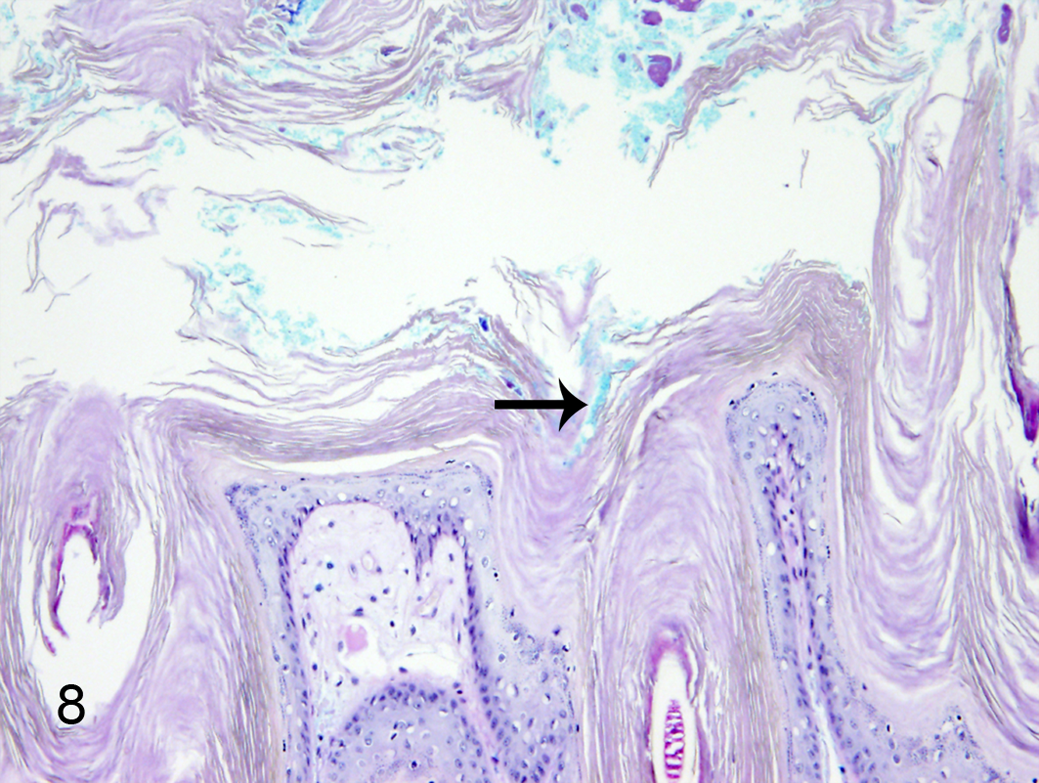
Ichthyosis, skin, distal forelimb, dog No. 12. Within the epidermal stratum corneum close to hair follicle infundibula, accumulations of alcianophilic material are present (arrow). AB/PAS.
In sections from all 14 dogs, the homogenous material within hair follicle infundibula was intensely stained by Sudan red III. In histologically normal appearing sebaceous glands, the Sudan red III stain revealed accumulations of lipids showing a droplet-like staining pattern in the cytoplasm of mature sebocytes while the sebocytes in enlarged sebaceous gland lobules exhibited a homogenous staining pattern. On sections stained by the Heidenhain’s Azan method, differences in quality and distribution of collagen fibers were not seen between affected and control dogs.
Transmission Electron Microscopic Findings
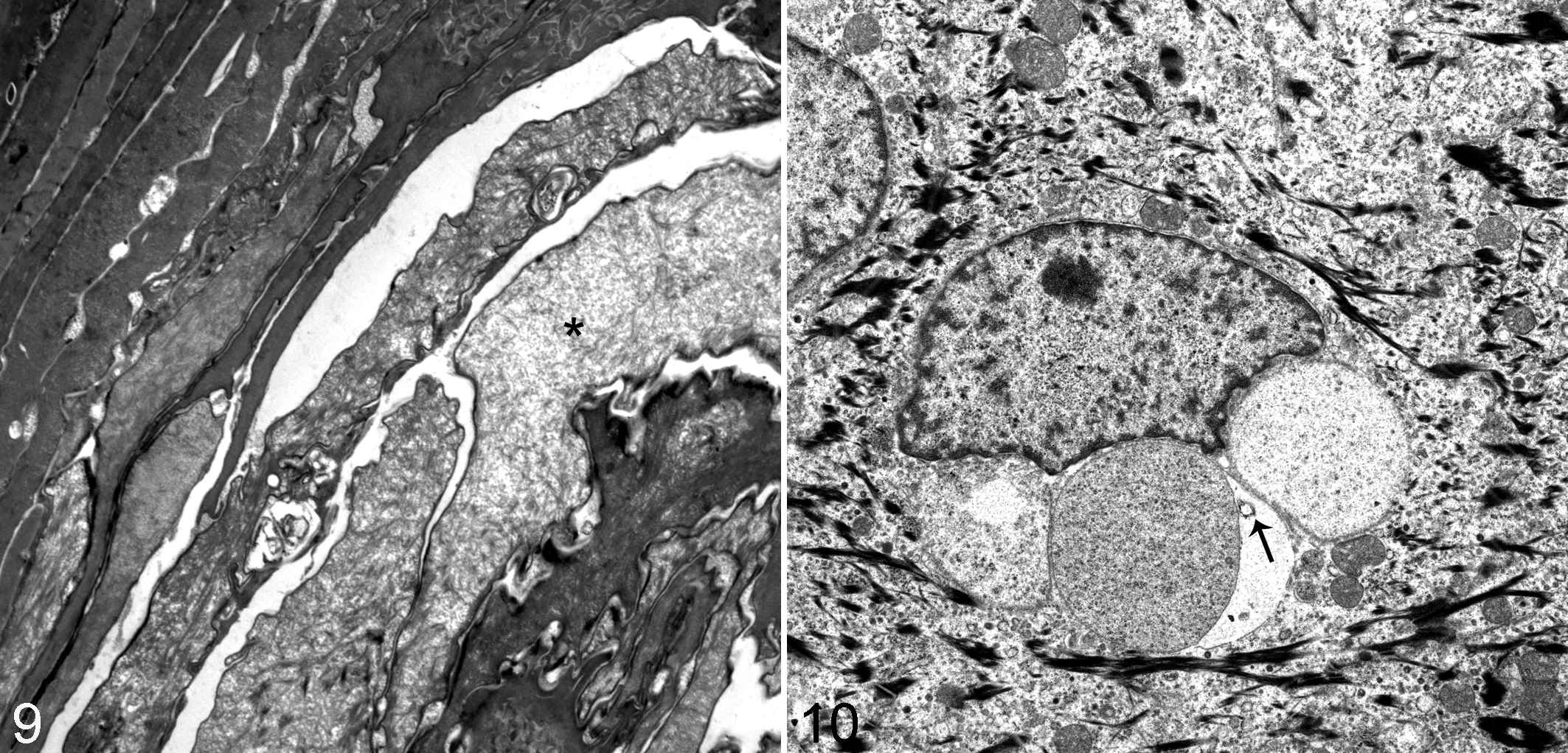
Ultrastructurally, in all 8 affected dogs examined (dog Nos. 1, 2, 8, 9, 10, 12, 13, and 14), marked orthokeratotic epidermal and infundibular hyperkeratosis was present. In the epidermis and infundibulum, the superficial layer of the stratum corneum was structurally normal, while the deeper layers contained large accumulations of a fine granular, amorphous material between the layers (Fig. 9). The thickened epidermis and the hair follicle epithelium had numerous vacuolated keratinocytes, which were mainly located within the stratum granulosum and were also present in the superficial stratum spinosum. The affected keratinocytes had single or multiple, variably-sized, cytoplasmic, membrane-bound vacuoles, which sometimes contained smaller vesicles. The keratinocytic vacuoles often displaced the nucleus and contained varying amounts of an amorphous, fine granular material. The stratum granulosum contained prominent keratohyalin granules varying in size and shape (Fig. 10). The sebaceous glands, besides normal appearing sebocytes, had groups of degenerating and lysed sebocytes, which showed accumulations of an amorphous, electron-lucent material. Such material was also present within dilated hair follicle lumina. In skin samples of the control dog, only few keratinocytes of the epidermis had single, empty, cytoplasmic, perinuclear vacuoles.
Pedigree Analysis
Thirteen dogs of this study were shown to be related (Fig. 11). Dog No. 1 was of unknown parentage and could not be assigned to the pedigree.
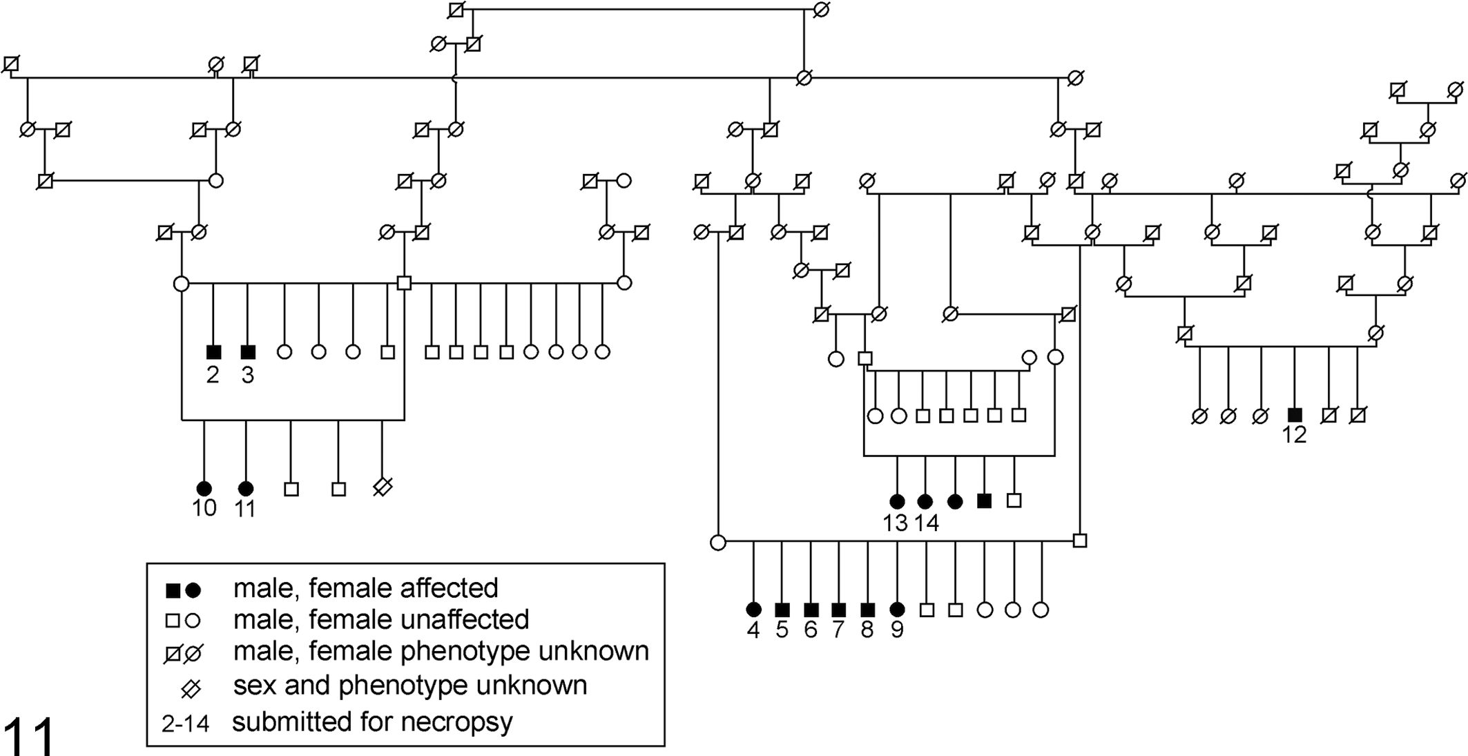
Pedigree, dogs Nos. 2 through 14 showing that 13 dogs are related.
Discussion
The 14 Great Dane puppies examined in this study had identical macroscopic, histopathologic, and ultrastructural skin changes, which based on morphological features are compatible with a primary disorder of cornification. The early onset as well as clinical presentation and histopathologic appearance of the lesions are suggestive of a congenital, non-epidermolytic, lamellar ichthyosis as described in Golden Retrievers.
Macroscopically, all 14 Great Dane puppies had moderate to severe, generalized scaling with fine, dry, and white to yellow scales and a greasy and leathery appearance of the axillary and inguinal skin regions. The histopathologic findings in all dogs were characterized by diffuse, orthokeratotic hyperkeratosis, follicular keratosis, focal keratin plugging, and acanthosis with vacuolization of keratinocytes without epidermolysis. These skin changes are compatible with lamellar ichthyosis as has been described in other purebred dog breeds such as the Golden Retriever, 6,12,13,20 American Bulldog, 19,21 American Pit Bull, 28 and Jack Russell Terriers. 8 Similar findings of abnormal and enlarged keratohyalin granules in canine ichthyosiform dermatoses were present in Norfolk Terriers, a Labrador Retriever, and a mixed-breed dog affected by epidermolytic ichthyosis. 2,5,21 The significance and pathogenesis of the findings in these dogs remain unclear.
Changes in the folliculosebaceous unit containing accumulations of an amorphous material found in Great Danes of this investigation have not been described in other canine ichthyosiform dermatoses. In all cases, prominent sebaceous glands with enlarged glandular lobes were present, in which several sebocytes contained an amorphous, lipid-rich material as revealed by the Sudan red III stain. Such material was also present within dilated hair follicle lumina and on the surface of and within the thickened, hyperkeratotic layers of the epidermal and infundibular stratum corneum. The material within the sebaceous glands and in the infundibula, in addition to lipid staining by Sudan red III, also had alcianophilic staining characteristics. Alcian blue at a pH of 2.5 as used in this study stains all types of glycosaminoglycans including sulfated and carboxylated mucosubstances such as mucin. 24 The lipid-rich and alcianophilic material seen in the folliculosebaceous unit of Great Dane puppies was not digestable with hyaluronate lyase, therefore ruling out the presence of mucinous substances, although the presence of non-sulfated glycosaminoglycans cannot be ruled out. The exact composition of this material is unclear. Further investigations to identify the material are planned. Such findings, to the best of our knowledge, have not yet been described in dogs with congenital ichthyosis. Sebaceous glands produce and release sebum, a complex mixture of lipids, through an excretory duct into the follicular canal, finally being extruded to the skin surface. 25,26 During sebum production, mature holocrine-secreting sebocytes undergo degeneration and lysis and are renewed by division of basal cells. 25,26 The histologic findings in the folliculosebaceous unit of the Great Dane puppies of this study indicate excess production of lipid-rich sebum in hyperplastic sebaceous glands with possible increased turnover of proliferating sebocytes. The ultrastructural findings in 8 of 14 examined dogs were characterized by accumulations of material between the corneocytes of the epidermal and infundibular stratum corneum, perinuclear vacuoles within keratinocytes, as well as the presence of enlarged keratohyalin granules. The material found in the stratum corneum correlated focally with Sudan red III- and AB/PAS-positive reactions, indicating a lipid- and glycosaminoglycan-rich material. Membrane-bound, cytoplasmic vacuoles in keratinocytes were described in other breeds of dogs with different forms of ichthyotic skin disorders in previous studies. 7,18,20,21 In some cases, these vacuoles were accompanied by epidermolysis, which was not seen in the present study on Great Danes. 2,5,7,21 The specificity of perinuclear vacuoles seen in diseased dogs in our study, however, is questionable as vacuoles were also found in skin sections of the control dogs. Furthermore, such vacuoles can often be seen in skin sections as a fixation artefact.
The pedigree analysis of 13 of 14 Great Danes showed that all dogs were related. The Great Dane ichthyosis does not fit in the classification seen in humans where many different forms of autosomal recessive, congenital ichthyoses (ARCI) are classified into subgroups. 12,27 Based on the classification system, most similarities exist with the congenital, autosomal recessive, non-epidermolytic, lamellar ichthyosis described for Golden Retrievers, although differences were present. 12,13,20 Histologic changes not yet observed in ichthyotic dogs include the accumulation of an amorphous material within hair canals and sebocytes found with different staining patterns.
Primary ichthyosis is a non-inflammatory skin disease. 19 Only in 3 dogs of this study were the skin changes accompanied by mild, focal pyogranulomatous or lymphoplasmacytic inflammatory lesions with few intralesional bacteria. These findings indicate that the disease, with advancing age, may lead to secondary infections and inflammatory skin lesions. A predisposition for secondary skin changes such as mycosis and bacterial infection has been described in American Bulldogs with ichthyosis as well as in Shar Pei dogs, possibly due to the wrinkled skin. 19,21
The etiology of the marked wrinkling of the skin seen in all 14 Great Dane puppies of this study remains unknown. This reflects previous reports of ichthyosis seen in American Bulldogs. 19,21 The leathery skin areas could be interpreted as early cutaneous signs for puppies affected by ichthyosis. 13 In the Chinese Shar Pei, skin wrinkles are associated with the presence of dermal accumulations of mucin within the interfollicular dermis, a condition that was suggested to be associated with a mutation in the HAS2 gene. 9,23,29 The alcianophilic material within the interfollicular dermis and subcutis both of Great Dane puppies with skin folds and in control dogs was digestable with hyaluronidase and most likely represents normal amounts of mucin, which is often seen in normal canine skin. 25 Also, the presence of a dermal mucinosis caused by a mutation was ruled out in the puppies of this study.
Ichthyosis is a heterogenous group of congenital disorders of cornification, and the excessive scaling is due to a direct defect in 1 or more steps involved in the formation of the stratum corneum. 19 There are defects that are related to mutations in genes that encode the structural proteins that form the corneocyte, or enzymes involved in lipid formation or lipid transport. 19 In conclusion, this study describes a congenital ichthyosis occurring in Great Dane puppies, which is associated with histologic changes within the folliculosebaceous unit. Based on histopathologic features, we suggest a congenital, non-epidermolytic, lamellar form of ichthyosis with a pathogenesis involving a disturbance in sebum production and/or cellular turnover of sebocytes.
Footnotes
Acknowledgements
We wish to thank Bettina Buck, Klaus-Peter Kuhlmann, and Kerstin Rohn for their excellent technical assistance.
Declaration of Conflicting Interests
The author(s) declared no potential conflicts of interest with respect to the research, authorship, and/or publication of this article.
Funding
The author(s) received no financial support for the research, authorship, and/or publication of this article.