
Abstract
The histologic features of abnormal spectacles in 60 snakes from the 5 families of Boidae, Colubridae, Elapidae, Pythonidae, and Viperidae are described in a retrospective study conducted on specimens submitted to a private diagnostic service during a period of 15 years. Fifty-two snakes had inflammatory reactions in the spectacle. The stroma and outer epithelium of the spectacle were the layers most often involved in inflammatory disease. Lesions of the outer epithelium included edema, hyperkeratosis, and granulocyte infiltration occasionally with bacterial colonies and fungal elements. The stroma had infectious agents and inflammatory reactions in vessels and between the collagen fibrils. The inner epithelium had varying degrees of hyperplasia and hypertrophy, but no infectious agents were seen. Infectious agents in these cases included mites, bacterial disease, fungal disease, or a combination of bacterial and fungal disease. Special stains identified the bacteria most commonly involved to be Gram-positive cocci. Thirteen snakes had dysecdysis of the spectacle. Of these, 5 displayed a concurrent inflammatory reaction of the spectacle, while the remaining 8 snakes had extra keratin layers on a spectacle with an otherwise normal appearance. These keratin layers were attached to serocellular crusts located on the inner surface of the periocular scales. The cause for dyskeratotic lesions of the spectacle was not always apparent, and concurrent acariasis, other forms of dermatitis, trauma, suboptimal husbandry, and visceral disease were considered possible contributing factors. It was notable that only 4% of the submitted cases were found to have spectaculitis and/or spectacular dysecdysis.
Snake eyelids fuse and become transparent during the embryonic state and create the structure known as the spectacle (Fig. 1). The spectacle consists of 3 layers: outer epithelium, stroma, and inner epithelium (Fig. 2). 4 The outer epithelium is composed of basosquamous cells covered with alternating layers of alpha and beta keratin. One layer of alpha keratin and 1 layer of beta keratin together are considered a generation. 8 During the shedding phase, a new keratin generation is created underneath the existing generation, and when complete, the outer generation is shed. 8 The stroma consists of layers of collagen fibrils intertwined with blood vessels and nerves. 4 The inner epithelium is a single layer of flat to very low cuboidal cells with vesicles and microvilli. 4 The spectacle is connected to the periocular scales at the hinge region 9 through a transition zone that lies at the rim of the spectacle proper. 4 Separating the spectacle from the cornea is a fluid-filled subspectacular space. 5
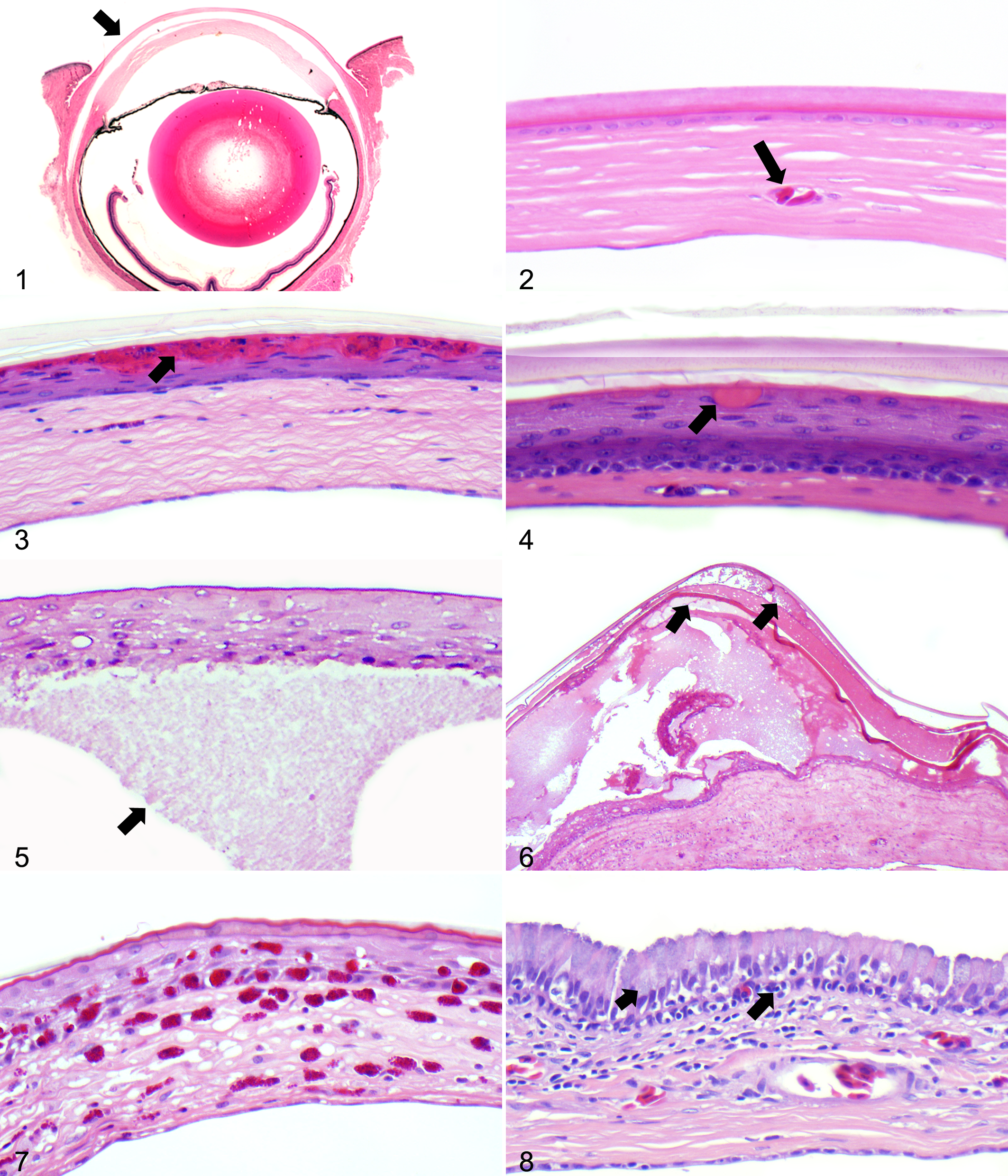
Normal eye; ball python (Python regius). Cross section through the eye of a healthy ball python with the spectacle (arrow) covering the anterior eye globe. Hematoxylin and eosin.
The spectacle is an integumental structure, 11,13 and as pathologic conditions of the skin are common in reptiles, particularly in captivity, 2 spectacle-associated disease is also expected to be a common finding. Also, similar to the skin, the outermost keratin layers of the spectacle may be retained due to various causes. Dysecdysis of the spectacle is mainly observed in captive individuals and may negatively affect the overall condition of the snake. 3 Remarkably though, the spectacle and its response to disease have received very little attention in the literature. This retrospective study presents the first systematic approach to classify histologic lesions in the spectacle of snakes.
Materials and Methods
From 1998 to 2013, approximately 5500 ophidian necropsy cases from several facilities were submitted to a private exotic species pathology service (Northwest ZooPath, Monroe, WA, USA). In approximately 1500 of these cases, the entire head was fixed in 10% neutral buffered formalin before being decalcified in Rapid cal (BBC Scientific, Mt Vernon, WA, USA). The tissues were then processed routinely and embedded in paraffin, sectioned transversely at 5 µm, stained with hematoxylin and eosin, and examined by light microscopy. Archival reports and slides were retrieved, and slides were reexamined by 2 of the authors (M.O.D., M.M.G.), specifically for ocular and/or cutaneous lesions. Eighty-one snakes (5%) were found to have abnormal spectacles. Sixty snakes (4%) had an inflammatory reaction and/or dysecdysis of the spectacle. Specimens from snakes with bacterial infection of the spectacle were stained with Goodpasture’s stain and Fite’s acid fast stain to determine tinctorial and morphologic properties of the bacteria involved. The remaining 21 snakes had lesions outside the scope of this article.
Results
The 60 snakes with pathologic changes of the spectacle seen as an inflammatory reaction and/or as dysecdysis were from the 5 families of Boidae, Colubridae, Elapidae, Pythonidae, and Viperidae.
Inflammation of the Spectacle
Fifty-two snakes (87%) had inflammatory reactions in 1 or multiple layers of the spectacle associated with mites (2 of 52), fungi (8 of 52), or bacteria (30 of 52) either alone or in combination (12 of 52; Table 1). The identity of the agents involved in the lesions was not determined, as cultures were not performed. However, Goodpasture’s stain revealed that Gram-positive cocci were present 3 times more often than Gram-positive (4 of 30) or Gram-negative rods (4 of 30). Only 1 specimen had Gram-negative cocci. None of the specimens had acid-fast bacteria. The outer epithelium was involved in 71% (37 of 52) of the cases, the stroma in 69% (36 of 52), and the inner epithelium in 37% (19 of 52). In 44% of the cases (23 of 52), both the outer epithelium and the stroma were affected; in 37% (19 of 52), both the stroma and the inner epithelium were affected (the outer and inner epithelium were never the only layers to be affected); and all layers were involved in 29% (15 of 52) of cases.
Number of Snakes From Each Family Affected by Bacteria, Fungi, and Mites.a
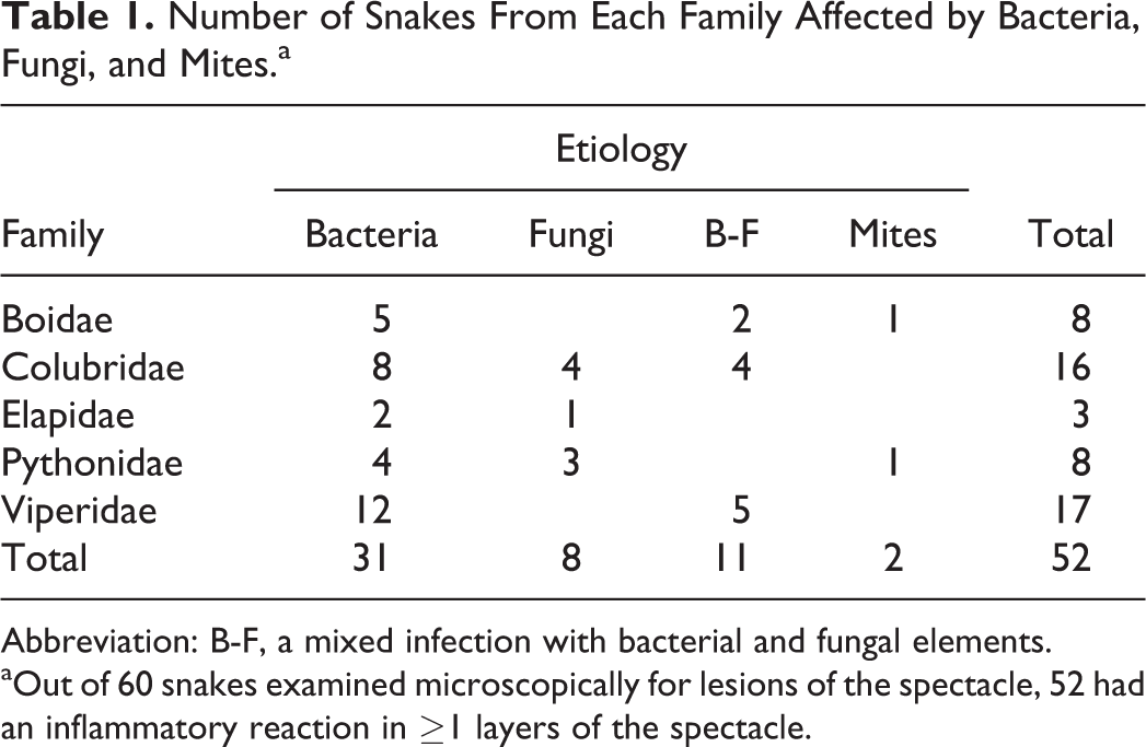
Abbreviation: B-F, a mixed infection with bacterial and fungal elements.
aOut of 60 snakes examined microscopically for lesions of the spectacle, 52 had an inflammatory reaction in ≥1 layers of the spectacle.
All snakes with inflammatory reactions of the spectacle had edema of the spectacle and concurrent dermatitis. Of the cases with fungal infection, 50% had involvement of all layers, in comparison to 26% of the cases with bacterial infection and 27% of the cases with combined bacterial–fungal infection. The snakes were almost equally affected unilaterally (48%) and bilaterally (52%).
Outer Epithelium
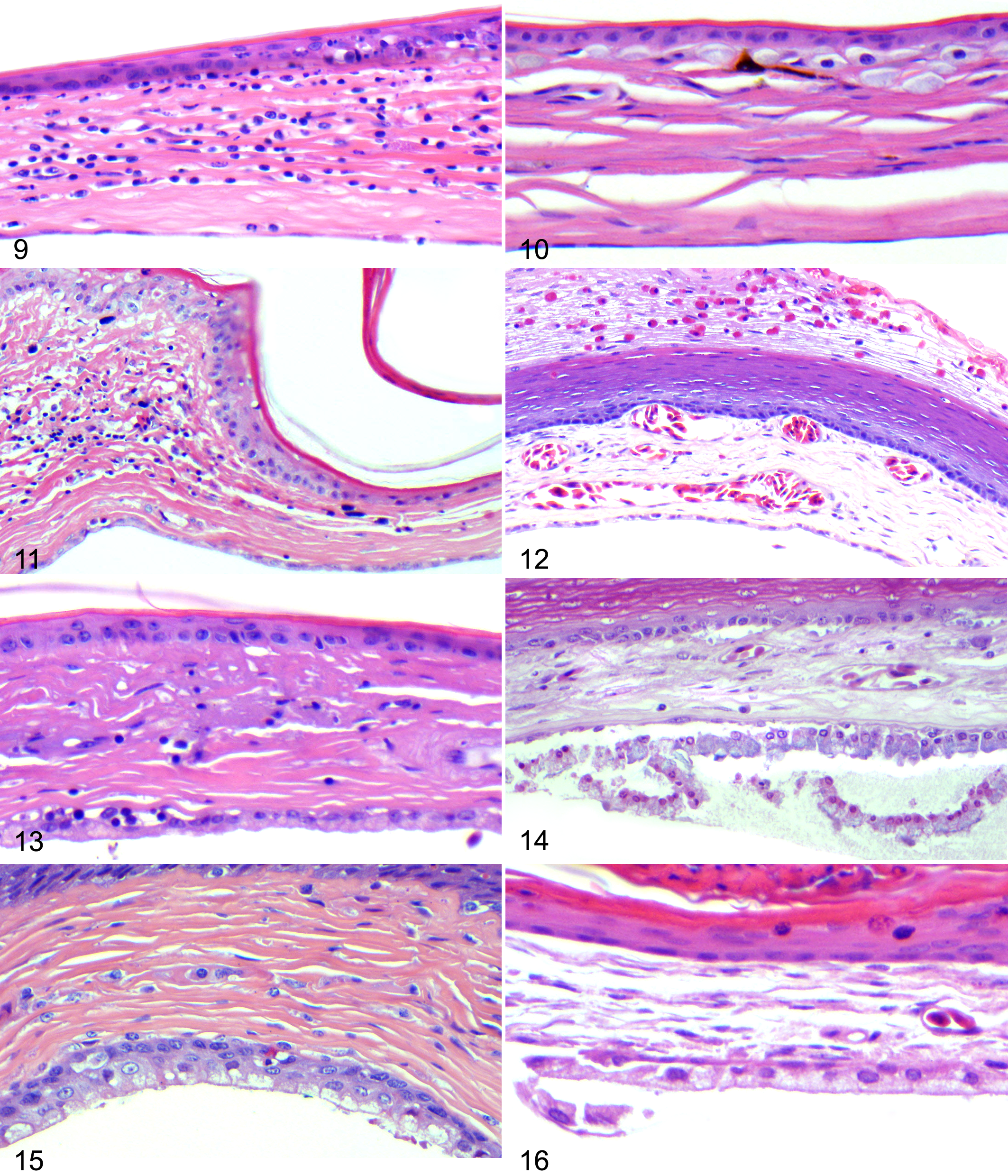
Seventy-one percent of all inflammatory reactions involved the outer epithelium; superficial serocellular crusts comprised focal to diffuse accumulations of serum, fibrin, and necrotic cellular debris (Fig. 3), occasionally colonized by basophilic bacteria and/or nonpigmented fungi. The fungal elements were branching and septate and had parallel walls, although yeasts were also seen in some preparations. The agents were not further classified. In some cases that did not have crust formation, eosinophilic vacuoles were seen in the alpha keratin underneath an intact layer of beta keratin (Fig. 4). The alpha layer showed hyperkeratosis, and the beta layer was intact. Only in severe cases did the crusts involve the beta layer.
Extracellular edema presented as pale staining acellular eosinophilic material and was often seen in the outer epithelium, immediately above the basement membrane, separating the outer epithelium from the stroma (Fig. 5). In more severe cases, edema was also found within the germinal layer and/or the alpha keratin layer (Fig. 6). Edema was never seen in the beta keratin. Intracellular edema was very rarely observed. Acidophilic granulocytes interpreted as heterophils, when present, were located between the basal germinal cells (Fig. 7) or within the alpha keratin layers. They were never present in the beta keratin.
The cells of the outer epithelium of the transition zone reacted differently from those of the spectacle proper (Fig. 8), clearly demarcating this region from the spectacle proper. Lymphocyte migration through the basement membrane was occasionally seen, and the overlying epithelial cells were columnar with ventral nuclear polarity, in comparison to the flat cells of the spectacle proper. The cytoplasm of the columnar cells was filled with microvesicles.
Stroma
Edema was common in the stroma, where amorphous eosinophilic extracellular material separated the collagen fibrils. The edema in the stroma often stained darker than edema in the outer epithelium above the basement membrane. Inflammatory cells were frequently evident in the stroma, as acidophilic granulocytes (Fig. 7), lymphocytes (Fig. 9), or melanomacrophages (Fig. 10). Within the areas of inflammation and within the walls of the blood vessels, colonies of basophilic bacteria and nonpigmented branching, septate fungal elements could be visualized. The stromal inflammation of the transition zone often did not extend into the spectacle proper (Fig. 11).
Spectaculitis;
Neovascularization was observed in the stroma, sometimes to the extent that the blood vessels would expand the stroma and disrupt the regularity of the basement membrane and the overlying basal cells of the outer epithelium (Fig. 12). Collagen degeneration in the stroma was evident as dark eosinophilic and disorganized collagen streams (Fig. 13).
Inner Epithelium
When inner epithelium was involved (37%), this layer displayed cellular hypertrophy and/or hyperplasia of varying degrees (Figs. 14–16), sometimes with concurrent extracellular edema. Sloughed necrotic cells were occasionally present in the subspectacular space (Fig. 14). Fluid-filled vesicles were seen in the cytoplasm of the epithelial cells as basophilic structures (Fig. 14). No inflammation or infectious agents were encountered in the inner epithelial layer. All snakes with changes in the inner epithelium also had involvement of the stroma. Fifteen of them (79%) also had changes in the outer epithelium.
Dysecdysis of the Spectacle
In 13 snakes (22%), retained keratin generations of the spectacle were observed. Nine of these snakes had dermatitis either alone (4 of 9) or in conjunction with inflammatory reaction of the spectacle (5 of 9). The dermatitis was ascribed to bacterial infection (3 of 9), mixed bacterial or fungal infection (3 of 9), mites (1 of 9), and metabolic or environmental causes (2 of 9). The identity of the infectious agents was not determined. Dermatitis included crusts on the inner surface of the periocular scales attached to the keratin layers of the spectacle at the hinge region. The crusts were adhered to 1–3 generations of keratin (Fig. 17). Mites were positively identified in 1 of these snakes (Figs. 18–20) with associated serocellular crust formation of the epidermal keratin layers of the skin, primarily in the scale folds, and with perivascular to diffuse granulocytic, lymphocytic inflammation of the dermis and granulocytic exocytosis (Figs. 18, 19).
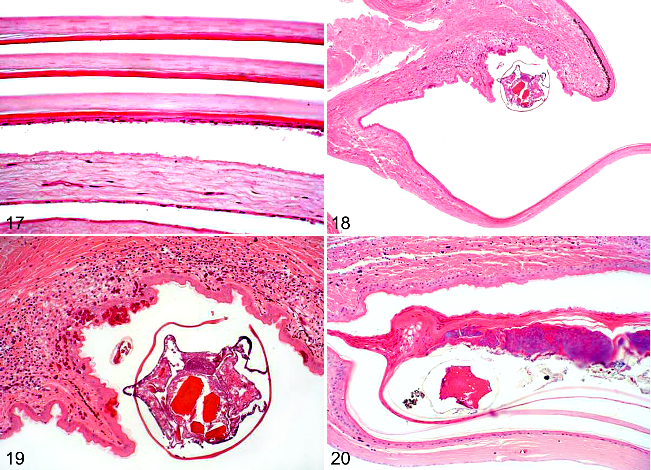
Spectacular dysecdysis; Hematoxylin and eosin.
Of 13 snakes, 4 had retained keratin layers on the spectacle but no further pathologic reaction of the spectacle or skin. One of these snakes was diagnosed with a possible metabolic disorder, and the etiologies of the others were suggested to involve husbandry factors.
Discussion
The majority of snakes with lesions of the spectacle had inflammatory reactions in ≥1 of the spectacle layers—that is, “spectaculitis.” All snakes with spectaculitis had concurrent edema in ≥1 layers and dermatitis, likely indicating a disease process spreading from the skin to the spectacle. This is not surprising, as the spectacle represents modified skin rather than a true ocular structure.4,11,13
The stroma and outer epithelial layers of the spectacle were most often involved in pathologic conditions. The network of stromal blood vessels accounted for distribution of the inflammatory reaction. Stromal edema would often stain darker than edema above the basement membrane, suggesting that transudate is able to pass into the outer epithelium, whereas the more proteinaceous edema of inflammation cannot.
The outer epithelium was often affected underneath the outermost beta keratin layer. Beta keratin is more rigid than the underlying alpha keratin, 1 and the beta keratin layer is thinner toward the transition zone and the hinge region, 4 which could render this area a possible point of entry for infectious agents. In the examined material, infections appeared capable of spread within the alpha keratin layer without affecting the beta layer. The beta layer therefore seemed more resistant to infection than the alpha keratin. The outer epithelium was involved in the lesions of the spectacle with the most severe histologic appearance, indicating that this layer is able to provide some protection against mild infections.
The inner epithelium was the layer least involved in spectacle disease. This normally flat layer demonstrated only 2 ways of reacting; cellular hypertrophy and/or hyperplasia. Affected cells could have edema-filled vesicles. This layer may be the least susceptible to spread of an infectious event. Inflammation in the underlying subspectacular space is often reported in snakes, 10 and future studies are needed to describe histologic findings in the spectacle due to this condition specifically.
Bacteria and fungal hyphae were often encountered in the lesions of the spectacles. Bacteria are reportedly the most common cause of disease in reptiles, 12 with Gram-negative bacteria being the most common bacterial pathogens and with Gram-positive bacteria being infrequently associated with disease. 2,12 In the examined specimens, Gram-positive cocci were most frequently encountered. This could lead us to speculate on the source of infection, as Gram-positives may be more likely to be environmental opportunists secondary to other underlying pathology/immune suppression and treatment should be targeted accordingly. Fungal lesions and clinical signs of fungal disease are often similar to those of bacterial disease; however, mycosis in reptiles is less commonly encountered than bacterial disease, 12 which was also the case in the present study. Fungal disease appears to have a more deleterious effect on the spectacle than that of bacterial disease, as these cases more often showed involvement of all layers of the spectacle. The cases of combined bacterial–fungal infections had a level of involvement of all layers similar to that of the bacterial cases.
Failure to shed the outer keratin layers of the spectacle was seen in 13 snakes. Only the outermost keratin layers of the outer epithelium of the spectacle are involved in the periodic shedding of the skin; therefore, the term retained spectacle is misleading, as the major part of the spectacle is never shed and hence never renewed. Spectacular dysecdysis is a more appropriate term for this diagnosis, as it implies problems with shedding of the keratin layers of the spectacle. Of the 13, 4 had no other abnormalities of the spectacle or the skin. One of these snakes was diagnosed with hypothyroidism, whereas the etiologies of the others were speculated to include environmental factors, such as lack of abrasive material and/or incorrect humidity and heat. Five snakes had spectaculitis and spectacular dysecdysis with concurrent dermatitis, and 4 snakes had spectacular dysecdysis as the only presenting feature of the spectacle that accompanied the dermatitis.
In the examined sections, lesions were found to occur equally unilaterally as bilaterally. Some individuals may have been incorrectly classified, as only 1 section per snake was examined. However, it would be reasonable to deduce that systemic disease would manifest itself bilaterally, while etiologies involving husbandry factors could be unilateral or bilateral.
Artifactual breaks in the keratin layers of the spectacle can occur during preparation for histology, 6 –8 and some specimens may have had additional layers that were lost during preparation.
The snakes examined were not from a single population; therefore, the appropriate prevalence of infectious disease of the spectacle cannot be calculated. However, it must be noted that surprisingly few snakes had histologic lesions of the spectacles. During a period of almost 15 years, out of approximately 1500 cases where eyes were available for histology, only 60 snakes (4%) were found to have spectaculitis or spectacular dysecdysis. The low prevalence could lead to the conclusion that the spectacle is a very resilient structure and seldom involved in disease.
Footnotes
Acknowledgements
We appreciate the assistance of Roy Brown, HT-ASCP (Histology Consultation Services, WA, USA), for preparation of the histopathology specimens and Christie Buie (Northwest ZooPath, WA, USA) for assistance with image layout and photomicrography.
Declaration of Conflicting Interests
The author(s) declared no potential conflicts of interest with respect to the research, authorship, and/or publication of this article.
Funding
The author(s) disclosed receipt of the following financial support for the research, authorship, and/or publication of this article: The research project was partly funded by the Ministry of Higher Education and Science, grant number 10-091595. The Center for Zoo and Wild Animal Health is supported by the Alfred Benzon Foundation.