
Abstract
A 13-year-old cat had a history of seizures for 3 years that resembled temporal lobe epilepsy. Histologic examination of the brain revealed bilateral hippocampal alterations, including hypergyration and broadening of the dentate gyrus associated with hippocampal sclerosis and an intraventricular meningioma near the hippocampal region. The findings in the dentate gyrus were interpreted as a congenital malformation; however, it could not be ruled out that the alterations were induced by the seizures. Similar changes of the dentate gyrus have not been previously described in cats.
Keywords
Malformations of the hippocampus are rare. Two reports exist for dogs: both were associated with epilepsy. 2,5 One of these dogs, a Pekingese, had a unilateral hippocampal cortical hamartoma with concurrent necrotizing meningoencephalitis. 2 The other report describes a neutered mixed-breed dog with dyslamination of the dentate gyrus and concurrent granulomatous meningoencephalitis. 5
One case of developmental hippocampal malformation was mentioned in a magnetic resonance imaging study of brain lesions in dogs and cats, 3 but specific details, including animal species, were not described. A complex developmental anomaly of the central nervous system was reported in a 4-week-old kitten that included a lack of the hippocampal fissure. 4 We believe that the case described herein is the first report of a presumptive hippocampal malformation. The histopathologic changes are similar to those previously reported in dogs. 2,5
A 13-year-old domestic shorthair cat, euthanized due to recurrent otitis and anorexia, was submitted for necropsy. There was a history of epileptic seizures for 3 years. During these episodes, the cat had seeking behavior, anxiety, orofacial automatism, hypersalivation, and occasional generalized convulsive seizures. Necropsy tissues were fixed in 10% neutral buffered formalin, embedded in paraffin wax, transversally sectioned, and stained with hematoxylin and eosin (HE) and Luxol fast blue. Immunohistochemistry—based on a rabbit polyclonal antibody against glial fibrillary acidic protein (GFAP; 1:3000; DakoCytomation, Glostrup, Denmark)—was applied to 3- to 4-μm brain sections through a horseradish peroxidase–polymer system (Ultravision Kit, Thermo Scientific, Fremont, California, USA) and an automated immunostainer (Lab Vision AS 360, Thermo Scientific).
Invading the third ventricle, near the dorsal hippocampus, was a lesion 5 mm in diameter that histologically resembled a psammomatous meningioma (Fig. 1). Despite the size of the tumor, there was no compression or infiltration of adjacent parenchyma or ventricular distension. If this hippocampus were compared with that of an age-matched control cat (Supplemental Fig. 1), it appeared shrunken. Macroscopically and histopathologically, the dentate gyrus was bilateral along its entire length, with a meandering hypergyration of the dorsal parts of the hippocampus (Fig. 2, Supplemental Fig. 2). Neurons of the cornu ammonis (CA) were decreased and, in some areas, completely lost (Fig. 3, Supplemental Fig. 2). Dentate granule cells were dispersed, irregularly distributed, and irregularly shaped and sized with generally enlarged cell diameters (Fig. 4) when compared to a normal dentate gyrus (Supplemental Fig. 3). These irregular granule cells were positive for Nissl staining (Fig. 5) but negative for GFAP. Additionally, some of these neurons had an irregular distribution of Nissl substance, an eccentric nucleus, and swollen cytoplasm reminiscent of balloon cells. Severe astrogliosis, microglialcytosis, and gemistocytosis were present, especially in the CA segment (Fig. 6); this finding was confirmed by GFAP immunohistochemistry (Supplemental Fig. 4). There was edema at the boundaries between the dentate gyrus and CA segments. Additional findings included bilateral otitis externa, mild concentric hypertrophy of the left cardiac ventricle, mild alveolar edema and emphysema, moderate lymphocytic enteritis associated with numerous crypt abscesses, subacute congestion of the liver, mild interstitial nephritis, microurolithiasis, and hydropic degeneration of the renal epithelium.
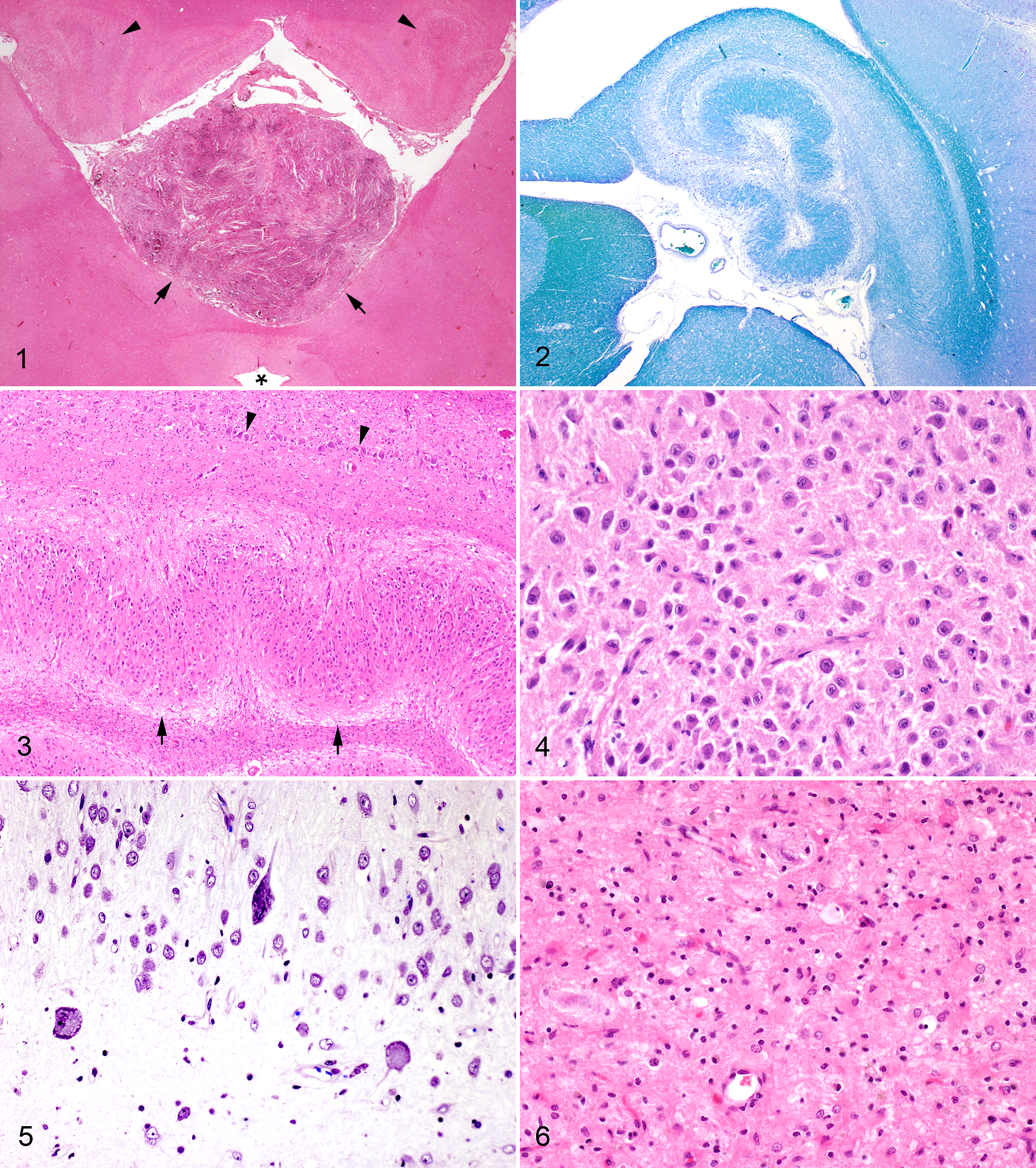
Meningioma, brain, cat. The tumor (arrows) is evident near the dorsal part of the hippocampal region (arrowheads). The asterisk indicates the third ventricle without distension. Hematoxylin and eosin (HE).
In histologic studies of several cats suffering from epilepsy, 6,9 this kind of alteration of the dentate gyrus was not described, nor have these alterations been described in cats in general. Hippocampal malformations described in 2 dogs 2,5 resemble the findings in our cat. In those dogs, hippocampal malformations were also associated with epileptic seizures. Strikingly, in the brain of our cat, an additional pathology—namely, an intraventricular meningioma—was present, similar to the canine cases, which were both associated with encephalitis.
Dysmorphic neurons are described in the hilus of the dentate gyrus in a subset of human patients with temporal lobe epilepsy. 11 Furthermore, studies from rodent models, where the hippocampus was examined after the induction of seizures, have reported such changes as dispersion and structural alterations in dentate gyrus granular cells. 8 In our cat, the exact characterization of the neuronal phenotype in the dentate gyrus requires further immunohistochemical studies.
The lesions in our cat are similar to those of tectonically deformed dentate gyri in a subgroup of human patients with temporal lobe epilepsy associated with bulbous expansions of the CA1 pyramidal cells and subicular layers. 10 Developmental malformation of pyramidal cells and subicular layers may influence the subsequent development of the adjacent dentate gyrus and may render temporal lobe structures hyperexcitable, making patients more vulnerable to seizures, injuries, and subsequent epilepsy. 10
Human cases of hippocampal sclerosis—defined as neuronal cell loss and gliosis at the CA1 segment with relative sparing of other hippocampal regions—can be associated with extra hippocampal lesions. 14 The frequent occurrence of hippocampal sclerosis in epileptic people, particularly in temporal lobe epilepsy, is well known; however, its pathogenesis is not clear. The International League Against Epilepsy has not determined whether hippocampal sclerosis is a nonspecific consequence of a primary epileptogenic lesion or a coincidence. 14 Histologic changes frequently involve more than one brain region, and “dual pathology,” defined as hippocampal sclerosis combined with a second brain lesion, is common. Hippocampal malformations have been postulated to contribute to the development of hippocampal sclerosis in humans. 1 Based on the current classification of focal cortical dysplasia, type IIIa exhibits temporal cortex cortical dyslamination or hypertrophic neurons together with hippocampal sclerosis. 1
Hippocampal sclerosis is a common finding in cats with temporal lobe epilepsy. 6,9,13 Based on case reports, the etiology of hippocampal sclerosis in cats is likely variable and may include hypoxic, inflammatory, toxic, and neoplastic causes. A congenital malformation might be another etiologic factor. In one canine case report, 5 hippocampal sclerosis was present besides malformation of the dentate gyrus.
Meningiomas are the most common primary intracranial and intraspinal tumors of the central nervous system in cats. 12 The tela choroidea of the third ventricle is a fairly common site for meningiomas in cats. 7 Many subtypes of meningiomas are slow-growing benign tumors that can be present for several years and remain relatively small. However, when these tumors reach a large size, they can progressively or suddenly cause neurologic signs. The 3-year history of epilepsy in the cat described herein, with the size of the meningioma, led us to conclude that the meningioma might be causally associated with hippocampal sclerosis, likely through parenchymal compression or vascular compromise; however, coincidence might be possible.
This is the first description of structural change of the dentate gyrus in an epileptic cat. This lesion is associated with hippocampal sclerosis and a concurrent meningioma. Because the hippocampus was not examined before the onset of seizures, it is impossible to prove whether the alterations of the dentate gyrus arose before or after the seizure disorder; however, they do resemble a dysplastic malformation.
Footnotes
Acknowledgements
We thank Ingrid Friedl for skilled technical assistance and Klaus Bittermann for excellent photographic work.
Author Contributions
Conception or design: AK, AP. Data acquisition, analysis, or interpretation: DT, PS, GGK, PH. Drafting the manuscript: AK. All authors participated in critically revising the manuscript, gave final approval, and agree to be accountable for all aspects of work to ensure integrity and accuracy.
Declaration of Conflicting Interests
The author(s) declared no potential conflicts of interest with respect to the research, authorship, and/or publication of this article.
Funding
The author(s) received no financial support for the research, authorship, and/or publication of this article.
References
Supplementary Material
Please find the following supplemental material available below.
For Open Access articles published under a Creative Commons License, all supplemental material carries the same license as the article it is associated with.
For non-Open Access articles published, all supplemental material carries a non-exclusive license, and permission requests for re-use of supplemental material or any part of supplemental material shall be sent directly to the copyright owner as specified in the copyright notice associated with the article.