
Abstract
Collagen type III glomerulopathy, also known as collagenofibrotic glomerulopathy, is a rare renal disease of unknown pathogenesis. The disease occurs in humans and animals and is characterized by massive glomerular accumulations of collagen type III. In the present study, we describe a Drever dog litter affected by an early onset variant of this glomerular disease, where 4 of 9 puppies developed renal failure within 50 days of age. Necropsy specimens of kidney from the 4 affected cases were studied by light microscopy, electron microscopy, and immunohistochemistry, and characteristic lesions compatible with a diagnosis of collagen type III glomerulopathy were found. In addition, 2 cases showed atypical epithelium in the collecting ducts of the medulla, so-called adenomatoid change. Immunohistochemistry of renal specimens from collagen type III glomerulopathy-affected dogs (n = 10) originating from two different dog strains, the Drever dogs and a mixed-breed strain, demonstrated that the deposited glomerular collagen is composed of a mixture of collagen III and collagen V. The distribution of the collagen V corresponded to the localization of collagen III; however, differences in staining intensity showed that collagen type III is the dominating component. Immunohistochemistry for collagen III (n = 9) and a transmission electron microscopic study (n = 1) showed hepatic perisinusoidal collagen type III deposition in affected cases from both dog strains. This is the first report documenting glomerular accumulations of collagen type V and perisinusoidal liver collagen III deposition in canine collagen type III glomerulopathy.
Collagen type III glomerulopathy (Col3GP), also known as collagenofibrotic glomerulopathy, is a rare renal disease with unknown pathogenesis that occurs in humans and animals. In veterinary medicine, the disease is reported to occur in monkeys, 2,5 dogs, 9 –11,21,22 pigs, 25 and a cat. 15 Col3GP is characterized by massive glomerular accumulations of collagen type III. The presence of cross-banded fibrils can be observed by transmission electron microscopy (TEM) and confirmed by immunohistochemistry for collagen III. The morphological phenotype is essentially identical in humans and dogs. 3,7,9,10,22 The accumulated collagen fibrils are located in the mesangium and in the subendothelial space of the glomerular capillary walls and are abnormally curved and frayed compared with normal interstitial collagen. In renal specimens from healthy humans, glomerular collagen type III is not detectable. 29 Various reports have shown concurrent glomerular accumulations of collagen type I in human patients, with a strong 28 or weak 8,16,18,26,27 staining pattern; however, the absence of glomerular collagen type I has been documented in other cases. 6,14 Also, a human case of Col3GP with a widespread glomerular expression of collagen type V has been reported. 14 To our knowledge, the presence of glomerular collagen type I or V has not been reported in Col3GP-affected animals. Previously, we have found evidence of collagen III synthesis by local mesangial cells in an affected mixed-breed dog strain. 22 A precise description of the composition of the pathological collagen could contribute to the understanding of the ongoing pathological processes in the mesangial cells in affected cases.
The pathogenesis of Col3GP is unknown. In dogs, an autosomal recessive inheritance pattern has been shown 22 ; however, the causative mutation has not been identified in humans or animals, and it is not known whether Col3GP is a primary renal disease or a systemic disorder. Two human patients with concurrent pathologic collagen type III fibrils in the liver have been reported. 13,27 Previous case reports in animals have not reported the occurrence of extrarenal abnormal collagen accumulations.
A clinical study of Col3GP-affected mixed-breed dogs showed that the affected dogs developed juvenile chronic renal failure, preceded by nephrotic syndrome and hypertension. 21 The clinical features in dogs are similar to those of affected children. 7 With one exception, 9 all described canine cases 10,11,21,22 developed clinical signs of chronic renal failure within a year of birth.
In the present study, we describe a canine litter affected by an early onset form of Col3GP, where 4 affected puppies developed renal failure within 50 days of age. We also aimed to describe whether glomerular accumulations of collagen type I or V and hepatic collagen type III accumulations could be detected in affected dogs, examining Col3GP-affected cases originating from 2 different dog strains.
Materials and Methods
Clinical Cases
Four puppies from a litter of 9 Drever dogs developed severe disease within 7 weeks of age. All puppies were of normal weight at birth; however, after approximately 4 weeks of age, 3 of the affected puppies (case Nos. 1, 2, and 3, all females) failed to gain weight compared with their siblings. The last affected puppy (case No. 4, a male) showed decreased growth rate from around 6 weeks of age. The puppies presented with growth retardation, anorexia, and respiratory distress. Azotemia was documented in 3 puppies (case Nos. 2–4), and all 4 puppies were euthanized because of poor prognosis and necropsied. Age at euthanasia was 35 (No. 1), 38 (No. 2), 40 (No. 3), and 53 (No. 4) days. Blood samples were collected prior to euthanasia, and serum biochemistry and hematology were available in 2 cases, whereas only serum biochemistry was available in 1 case. The urinary protein/creatinine ratio was available in 1 case. Both parents of the affected puppies were healthy, and there was no history of renal disease in any of the pedigrees. The study was performed with the consent of the owner.
Pathological Methods
Necropsy specimens from the kidneys of the 4 affected Drever dogs were studied by light microscopy (case Nos. 1–4); TEM (case Nos. 3 and 4); conventional immunofluorescence for IgG, IgM, and C3 (case Nos. 3 and 4); and specific immunohistochemical analysis for collagens I (case Nos. 3 and 4), III (case Nos. 1–4), and V (case Nos. 3 and 4); cytokeratin and carbonic anhydrase (cases Nos. 1–4); and immmunoelectron microscopy for collagen III (case No. 4). Liver specimens were studied by light microscopy (case Nos. 1–4), TEM (case No. 4), and immunohistochemical analysis for collagen III (case Nos. 1–4). In addition, necropsy specimens from Col3GP-affected dogs, from another dog strain, 22 including 4 mixed-breed litters with similar origin, underwent immunohistochemical analysis for collagens I and V (renal tissue, case Nos. 5–12) and collagen III (hepatic tissue, case Nos. 5 and 9–12). An overview of the pathological analyses of kidney and liver tissue performed is given in Supplemental Table S1. Specimens from other major tissues were examined by light microscopy and included lung and myocardium (case Nos. 1–4), ventricle and spleen (case Nos. 2–4), and intestines, lymph nodes, pancreas, and thyroid glands (case No. 4).
For light microscopy, slices of tissue were fixed by immersion in 10% buffered formalin and embedded in paraffin. Then, 2- to 3-μm-thick sections of renal tissue were stained with HE, Van Gieson, and periodic acid–Schiff, and 10-μm-thick sections were stained with Congo red; 2- to 3-μm-thick liver sections were stained with Van Gieson, toluidine blue, and von Kossa. Other tissues were stained with HE. The sections were examined under a Nikon Eclipse 50i light microscope, and photomicrographs were captured by a Nikon DS-Fi1 camera using NIS Elements Basic Research Software (Nikon, Tokyo, Japan).
For TEM, small pieces of renal (case Nos. 3 and 4) and liver (case No.4) tissue were prefixed in 2% glutaraldehyde in cacodylate buffer (pH 7.4), postfixed in 1% osmium, dehydrated in ethanol, and embedded in L.R. White (resin). Semi-thin sections were stained with Stevenel’s Blue and examined under the light microscope for selection of glomeruli. Ultrathin sections were stained with uranyl acetate and potassium permanganate. The specimens were observed in a FEI Morgagni 268 transmission electron microscope (FEI, Hillsboro, Oregon), and photographs were captured by an Olympus Veleta CCD camera (Olympus, Münster, Germany).
For immunoelectron microscopy, small pieces of renal tissue (case No. 4) were fixed in 4% paraformaldehyde and 0.1% glutaraldehyde in 0.1 M PHEM buffer, pH 6.9, for 1 hour and transferred to PHEM buffer. Specimens were infiltrated with 2.3 M sucrose, mounted on silver pins, and frozen in liquid nitrogen. Ultrathin cryosections were cut at –110°C (Leica EM FCS ultramicrotome; Leica, Nussloch, Germany) and collected with a 1:1 mixture of 2% methyl cellulose and 2.3 M sucrose. Sections were transferred to formvar-carbon–coated grids and labeled with primary antibodies to collagen type III (Acris Antibodies, San Diego, CA), followed by a bridging secondary antibody (Rockland, Limerick, PA) and 10 nm protein A gold. Sections were observed at 80 kV in a JEOL JEM-1230 electron microscope (JEOL, Tokyo, Japan). Micrographs were recorded with a Morada digital camera (Olympus) using iTEM (SIS) software.
Slices of renal tissue were fast frozen in isopentane cooled in liquid nitrogen and stored at –80°C for immunohistochemical and immunofluorescence studies. Standard immunofluorescence microscopy of renal tissue included staining for C3, IgG, and IgM using indirect immunofluorescence. The following antibodies and dilutions were used for immunofluorescence: goat anti–dog C3 (1:2000; Bethyl, Montgomery, TX), goat anti–dog IgM (1:1000; Bethyl), and goat anti–dog IgG1 (1:1000; Bethyl) and FITC-conjugated anti–goat IgG secondary antibody (1:100; Bethyl). Polymer-based immunohistochemistry was used for collagen types I, III, and V; cytokeratin; and carbonic anhydrase II staining. In addition to the affected puppies from the studied litter, renal and liver specimens from Col3GP-affected dogs from another dog strain 22 were examined; renal tissues were stained with antibodies against collagen V (n = 10) and collagen I (n = 10), and liver specimens were stained with antibodies against collagen III (n = 9) (Suppl. Table S1). For immunohistochemistry, the following primary antibodies and dilutions were used: mouse monoclonal anti–collagen type III (frozen tissue 1:2000, paraffin-embedded tissue 1:500; Acris Antibodies), rabbit polyclonal anti–collagen V (1:50; Abcam, Cambridge, UK), rabbit polyclonal anti–collagen I (1:100; Novus Biologicals, Littleton, CO), rabbit polyclonal anti–carbonic anhydrase II (1:2000; Novus Biologicals), and mouse monoclonal anti-cytokeratin (1:500; DAKO, Glostrup, Denmark). Secondary antibodies used were anti–mouse or anti–rabbit IgG conjugated with a horseradish peroxidase (HRP) labeled polymer, from the EnVision+ System-HRP kit (DAKO). The sections were treated with AEC chromogen solution from the EnVision+ kit and counterstained with hematoxylin. Washing between each of the steps in the procedures was performed with Tris-buffered saline for immunohistochemistry and phosphate-buffered saline for immunofluorescence. The primary antibodies were diluted in a 1% solution of bovine serum albumin in Tris-buffered saline. Negative control staining was performed by replacing the primary antibodies with nonimmunized goat serum, showing no staining. For the immunoglobulins and C3, frozen canine lymph node tissue was used as positive control, whereas for the collagens, connective tissue surrounding the arcuate arteries and the portal triads, in the kidney and the liver, respectively, was used as positive control. The specimens were studied in a Nikon Eclipse 50i microscope, and photomicrographs were captured with a Nikon DS-Fi1 (immunohistochemistry) or an Olympus IX81fluorescence microscope (immunofluorescence).
Renal and hepatic specimens from 3 healthy mixed-breed dogs, 2 puppies (aged 2–3 months), and an adult dog (aged 2 years) were used as control material.
Results
Clinical Findings
All affected puppies showed similar clinical signs characterized by lethargy and dyspnea, increased respiratory rate, and increased respiratory sounds at auscultation. Abdominal distention was observed in all 4 puppies. Case Nos. 1, 2, and 4 had pallor of mucus membranes, and clinical signs of dehydration were observed in case Nos. 1, 2, and 3. All affected puppies had smaller body size than their healthy siblings, weighing 1.2 kg (case No. 1), 1.3 kg (case No. 2), 1.5 kg (case No. 3), and 2.6 kg (case No. 4) at time of euthanasia. Dog No. 3 had a body weight of approximately 1 kg less than healthy siblings at this time point.
Both investigated cases (case Nos. 2 and 3) were anemic (Table 1). Biochemical analyses of serum revealed severely increased serum concentration of urea, normal to moderately increased creatinine, and severely increased phosphate concentrations (case Nos. 2–4). Hypercalcemia was found in case No. 4, whereas case No. 2 was hypocalcemic (Table 1). Large magnitude proteinuria was detected in case No. 4 (Table 1).
Hematologic and Serum Biochemical Values of Puppies Affected by Collagen Type III Glomerulopathy.
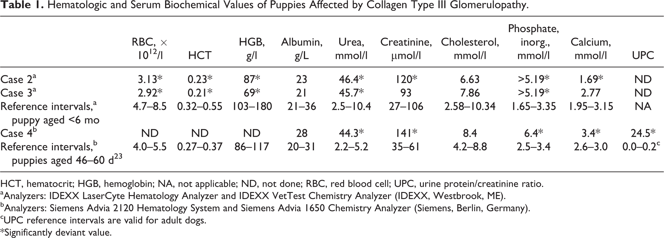
HCT, hematocrit; HGB, hemoglobin; NA, not applicable; ND, not done; RBC, red blood cell; UPC, urine protein/creatinine ratio.
aAnalyzers: IDEXX LaserCyte Hematology Analyzer and IDEXX VetTest Chemistry Analyzer (IDEXX, Westbrook, ME).
bAnalyzers: Siemens Advia 2120 Hematology System and Siemens Advia 1650 Chemistry Analyzer (Siemens, Berlin, Germany).
cUPC reference intervals are valid for adult dogs.
*Significantly deviant value.
Renal Morphology
All affected animals had pale, swollen kidneys with marked tissue hardening (Fig. 1). Essentially identical diffuse glomerular changes were identified in all cases. The lesions were characterized by an eosinophilic material expanding the mesangium and capillary walls of the glomeruli (Fig. 2), and the capillary lumens were frequently narrowed by this deposited material that stained pale pink in periodic acid–Schiff stained sections (Fig. 3) and bright red in sections stained with van Gieson (Fig. 4). Congo red staining did not reveal any red positive staining or green birefringence in polarized light in the glomerular tufts or in other locations, excluding a diagnosis of amyloidosis. Glomerular mesangial and endocapillary hypercellularity was frequently observed and varied from mild to moderate. Glomerular synechiae and parietal epithelial cell hypercellularity and hypertrophy were occasionally observed. Case No. 4 showed glomeruli enlarged in size within dilated Bowman’s capsules. Proximal tubular degeneration was a frequent finding, and dilated distal tubules with eosinophilic or basophilic casts were often observed. Medullary collecting ducts with casts were occasionally observed. Atypical epithelium with adenomatoid proliferation (ie, folded in a ribbon-like conformation in the collecting duct lumens) was observed in case Nos. 1 and 2 (Fig. 5). Increased mitotic activity was not observed in the atypical epithelium. Mineralization of Bowman’s capsules and tubules was present in all cases. Moderate periglomerular and mild interstitial fibrosis was observed.
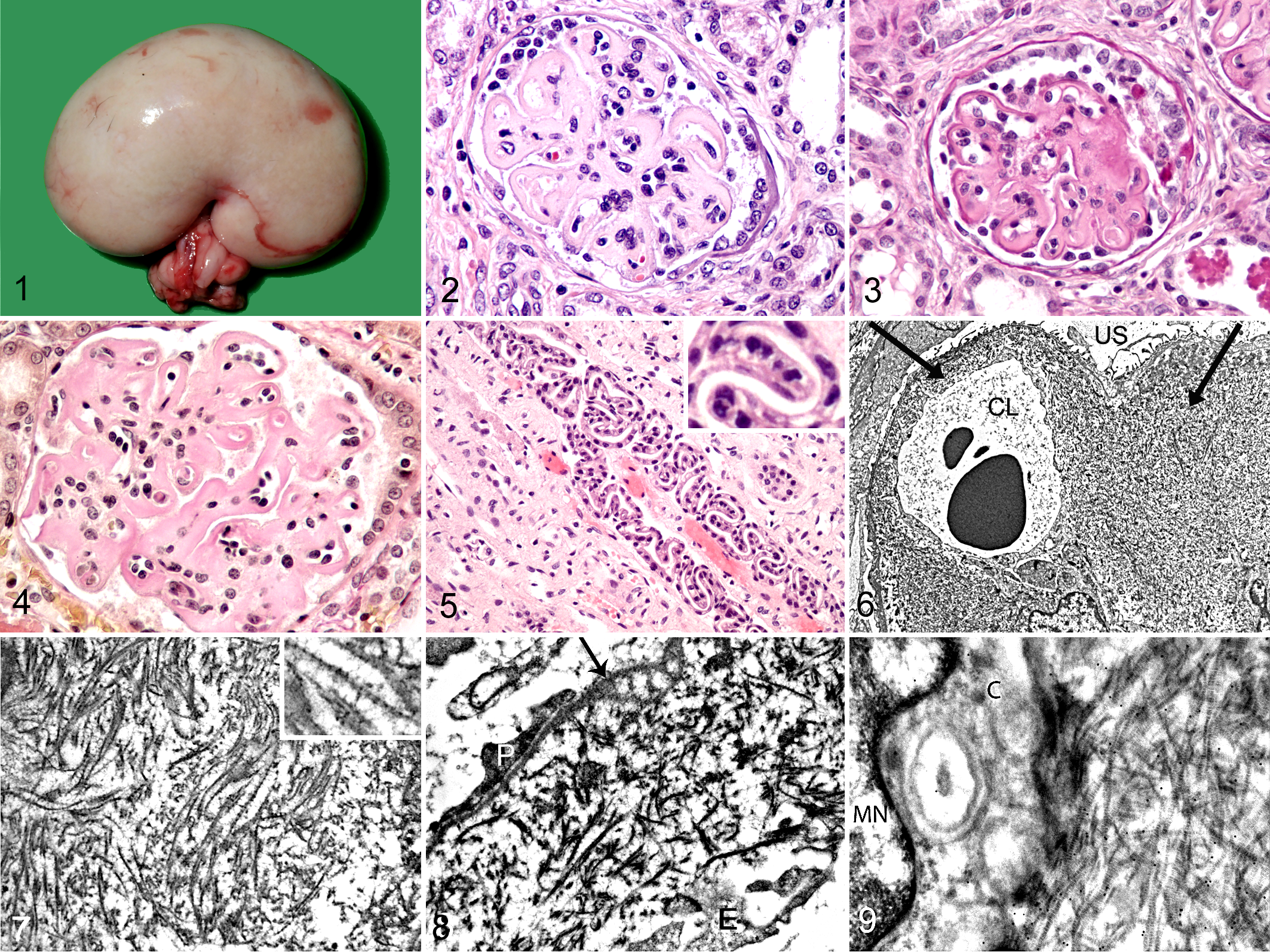
Collagen type III glomerulopathy, kidney, dog.
Both cases studied by TEM (case Nos. 3 and 4) revealed similar ultrastructural glomerular lesions, compatible with a diagnosis of collagen type III glomerulopathy. The mesangial matrix and the subendothelial area of the capillary walls were markedly enlarged by an accumulated fibrillar material (Fig. 6). By the banded structure, the fibrils were identified as fibril-forming collagen. The collagen fibrils were abnormally curved and detached, in an unorganized pattern (Fig. 7). In addition, signs of podocyte injury were frequently found, including multifocal foot process effacement and occasional foot process detachments resulting in denuded glomerular basement membrane areas (Fig. 8). Also, slight thickenings and irregularities of the lamina densa of the glomerular basement membrane were frequently observed (Fig. 8). No evidence of electron-dense immune deposits was found by TEM. Immunoelectron microscopy of cryosections labeled with collagen type III antibody revealed specific immunogold labeling of the cross-banded collagen fibrils accumulated in glomeruli (Fig. 9). The labeling was highly specific, almost exclusively associated with glomerular and interstitial collagen fibrils.
By immunohistochemistry, the glomerular deposits in the mesangium and the capillary walls of all glomeruli in the affected dogs stained strongly positive for collagen III (case Nos. 1–4) (Fig. 10), consistent with a diagnosis of collagen type III glomerulopathy. Furthermore, collagen V was moderately increased in mesangium and capillary walls of all glomeruli in all studied cases (case Nos. 3–12) (Fig. 11). The localization of collagen type V corresponded to the collagen type III deposits. There was also mild segmental positive staining for collagen type I in the capillary walls of some of the glomeruli in all studied cases (case Nos. 3–12) (Fig. 12). In comparison, normal glomeruli from healthy control animals were negative for collagen type III (Fig. 13), showed weak mesangial staining and no staining of capillary walls for collagen type V (Fig. 14), and were negative for collagen type I (Fig. 15). Immunofluorescence studies of affected cases showed linear staining for IgM along the capillary walls and patchy segmental granular staining for C3 in the mesangium and the capillary walls, interpreted as nonspecific staining due to the splotchy and irregular staining pattern. Staining for IgG was negative. The atypical epithelial proliferative cells identified in the tubular lumens stained positive for cytokeratin and showed strong reactivity for carbonic anhydrase antibody (Fig. 16).
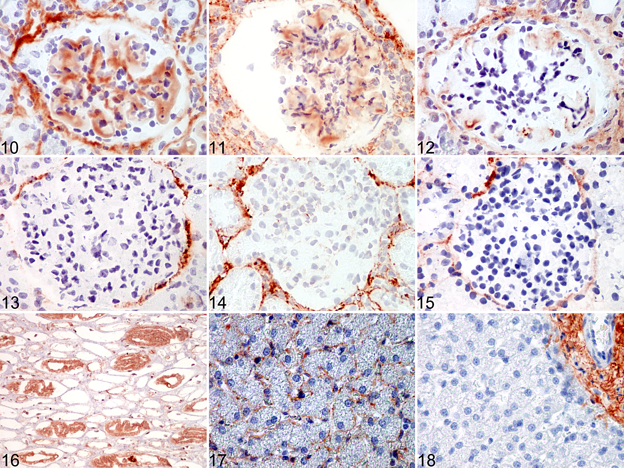
Collagen type III glomerulopathy, kidney, dog.
All affected cases revealed pathologic findings suggestive of secondary uremic pneumonopathy, including calcification of the walls of the alveoli and bronchioles, cellular infiltrations, and edema. Calcification in the myocardium (case Nos. 2 and 3) and liver and stomach (case No. 3) was observed. In addition, case Nos. 2 to 4 had splenic extramedullary hematopoiesis.
Liver Morphology
No gross abnormalities of the liver were observed. Multifocal small granulomas with central variably von Kossa–positive amorphous material were observed in case No. 3. Numerous mast cells identified by numerous intracytoplasmatic granules staining red-purple with toluidine blue (metachromatic staining) were observed along the sinusoids and in the vessel walls in both affected and healthy controls; however, the mast cells of the affected puppies appeared more rounded compared with the spindle-shaped mast cells of the controls. Although no signs of fibrosis were detected by light microscopy of HE- and van Gieson–stained slides, immunohistochemistry for collagen type III was performed, revealing increased collagen type III in the perisinusoidal areas in all investigated Col3GP-affected cases (n = 9) (Fig. 17). Healthy controls showed no collagen type III staining along the sinusoids (Fig. 18). By TEM, the presence of cross-banded collagen fibrils in the perisinusoidal area was confirmed (Fig. 19). The collagen fibrils were straight and well organized in bundles.
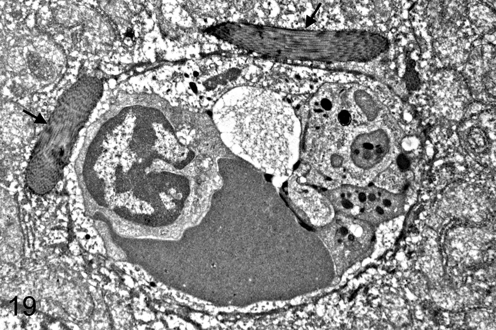
Collagen type III glomerulopathy, liver, dog. Case No. 4. Well-organized bundles of collagen fibrils (arrows) in the liver space of Disse. Transmission electron microscopy.
Discussion
The glomerular lesions observed in the 4 affected Drever puppies in the present study are consistent with previous reports of Col3GP and collagenofibrotic glomerulopathy in dogs and humans. 7,10,22 Both parents of the affected litter were healthy, and among the affected puppies, both sexes were represented, in agreement with an autosomal recessive mode of inheritance that previously has been observed in dogs. 22 The morphologic hallmark of Col3GP is glomerular accumulations of collagen type III, visualized by TEM and confirmed by IHC.
Immunohistochemical examination of glomeruli from affected puppies from 2 different dog strains revealed that the accumulated glomerular collagen is made up of a mixture of collagens III and V, with collagen III dominating. In addition, focal segmental accumulations of collagen type I were observed. To our knowledge, this is the first report investigating the involvement of other types of collagens besides collagen type III in Col3GP-affected animals. In humans, the presence of glomerular collagen I in Col3GP has been documented in some cases, while other cases were negative. To our knowledge, only 1 human case report 14 has examined whether collagen V is present in Col3GP, finding a widespread expression of collagen V, and it has not been clarified whether other Col3GP-affected human cases show the same collagen V expression. The distributions of collagens I, III, and V in affected cases were very similar for the Drever dogs and the mixed-breed dogs. We, therefore, suggest that the phenotypes in the studied Col3GP segregating strains are characterized by a mixture of deposited collagen types III and V. However, it remains to be clarified if morphologically different canine Col3GP phenotypes exist. Studying an affected mixed-breed dog strain, we have previously provided evidence for local mesangial cell collagen production and furthermore excluded the involvement of the Col3A1 gene in this dog strain. 22 While this particular gene might not be the ultimate source of this abnormality in dogs, there are various other genes involved in the posttransitional assembly of collagen fibrils. 12 As a response to injury, mesangial cells can acquire an activated phenotype with excessive production of various substances, including matrix proteins and growth factors 1 ; however, the marked mesangial cell production of collagen type III, characteristic for Col3GP, is not associated with other diseases, including mesangial cell injury.
Hepatic perisinusoidal collagen III deposition was documented in all the studied Col3GP-affected cases. In the normal liver, collagen type III is confined to the connective tissue surrounding the central veins and the portal tracts. Hepatic fibrosis is characterized by the appearance of collagens I and III within the space of Disse (the perisinusoidal areas), as well as an increased amount of collagens in the portal areas and around the central veins. 4 Pathological presence of collagen type III in the perisinusoidal areas in affected cases was shown by immunohistochemistry, and by TEM, the presence of collagen fibrils with cross-striations in the space of Disse was confirmed. The hepatic collagen fibrils were not frayed and curled as the accumulated glomerular collagen; rather, the fibrils appeared as normal straight collagen, well organized in bundles. In comparison, the 2 reported human cases of Col3GP with hepatic fibrosis 13,27 showed curled collagen fibrils in the liver, similar to those of the glomeruli, in 1 case, and less spiraled and frayed in the liver than in the glomeruli in the other case. Liver involvement might suggest the possibility that Col3GP is a systemic disease; however, the observed liver pathology could also be secondary to the clinical progression of the renal failure in the Col3GP-affected cases.
The affected puppies in the studied litter were very young at age of onset compared with other described canine cases. 10,11,21,22 The observed variation indicates that different genetic and environmental factors may be involved in the etiology. A large variation in age of onset is also seen in humans, with the youngest reported case 3 months old 7 and the oldest 78 years old 6 at detection of clinical symptoms.
Atypical renal collecting duct epithelium with adenomatoid proliferation was observed in 2 of the Drever puppies. Such a proliferation of the epithelial cells of collecting ducts in the medulla has previously been described as a primary lesion of canine renal dysplasia. 17,19 Renal dysplasia is defined as disorganized development of renal parenchyma due to abnormal differentiation. In the present study, immunohistochemistry revealed that these cells express cytokeratin and carbonic anhydrase II proteins. Cytokeratins are found in the cytoskeleton of epithelial tissue. Carbonic anhydrase is an important renal enzyme that facilitates renal acidification and is strongly expressed in the intercalated cells of the collecting ducts and weakly expressed in the principal cells of the collecting ducts. 20,24 None of the cases in the present study displayed other signs of renal dysplasia; rather, they were affected by a progressive juvenile glomerulonephropathy (familial renal disease), giving rise to various secondary lesions in the kidney tissue. We speculate that the proliferation of carbonic anhydrase–positive epithelial cells might be compatible with a compensatory mechanism due to the metabolic acidosis caused by the renal failure.
Conclusions
In the present study, we have described a Drever dog litter affected by an early onset version of Col3GP. Assessing tissues from 2 Col3GP-affected dog strains, we found that the deposited glomerular collagen is a mixture of collagens III and V and that focal segmental glomerular collagen I deposits were present. Furthermore, hepatic perisinusoidal collagen III accumulations were demonstrated in all affected cases, indicating that not only the kidney but also the liver is involved in canine Col3GP.
Footnotes
Acknowledgements
We thank Laila Aune, Hilde Kolstad, and Lene Hermansen for invaluable technical assistance.
Declaration of Conflicting Interests
The author(s) declared no potential conflicts of interest with respect to the research, authorship, and/or publication of this article.
Funding
The author(s) received no financial support for the research, authorship, and/or publication of this article.
References
Supplementary Material
Please find the following supplemental material available below.
For Open Access articles published under a Creative Commons License, all supplemental material carries the same license as the article it is associated with.
For non-Open Access articles published, all supplemental material carries a non-exclusive license, and permission requests for re-use of supplemental material or any part of supplemental material shall be sent directly to the copyright owner as specified in the copyright notice associated with the article.