
Abstract
Melanosis coli is a dark discoloration of the colon due to accumulation of pigment-laden macrophages in the lamina propria. Three case submissions were received where rectal discoloration was reported at slaughter in pigs from separate production systems and melanosis coli was confirmed microscopically. Tissues from affected and unaffected cohort pigs were evaluated for evidence of oxidative damage using immunohistochemical staining for 3-nitrotyrosine, 4-hyroxynonenol, and malondialdehyde. Affected colons had significantly greater immunolabeling for all 3 target compounds than unaffected colons (P ≤ .001, all analyses). Hepatic vitamin E levels were low in both affected and unaffected pigs, and there was a trend toward lower values in affected pigs. Given the limited number of slaughter-collected samples available for this investigation, further study is warranted to elucidate the possible association between low vitamin E concentrations and oxidative damage in cases of melanosis coli in pigs.
Keywords
In humans, melanosis coli is characterized by brown or black pigmentation of the colonic mucosa and is commonly associated with chronic use of anthracene cathartics (including cascara, senna, aloes, and rhubarb), which contain the active ingredient anthraquinone. 6,8 Histologically, melanosis coli is characterized by accumulations of pigment-laden macrophages in the lamina propria of the colorectal mucosa. 7 Histochemical staining reactions of the pigment-laden macrophages in the lamina propria of human cases have demonstrated granule composition is not consistent with melanin pigment. 6 As a result, it has been suggested that pseudomelanosis coli may be a more appropriate name; 9 however, others refute this as an erroneous term, which means “false black” or “not black,” and suggest that melanosis coli is appropriate regardless of the pigment type. 5 Accordingly, for consistency, the authors have chosen to use the term melanosis coli to describe the discolored colorectal tissues in swine. Interestingly, a recent investigation has also revealed an intense argentaffin reaction abolished by bleaching in affected human tissues, which is typical of a melanic substance that may originate from anthranoids and/or their metabolites, most of which are brown. 4 A similar reaction was found in the lymph node of Nero Siciliano pigs with acquired pigmentation following acorn ingestion. 10
Melanosis coli is an uncommon condition in swine that has been identified in multiple unrelated production systems. When identified, this condition results in potential financial losses in export niche markets as the discolored tissues are undesirable for human consumption and are therefore devalued or discarded. The etiopathogenesis and potential risk factors associated with the development of melanosis coli in swine are unknown; however, it seems unlikely to be solely breed associated as crossbred pigs predominate in the US pork industry and in these case submissions. A lectin-based histochemical technique demonstrated that the pigment in macrophages in the lamina propria was typical of lipofuscin as well as ceroid in humans. 4 Ceroid accumulation has been documented in multiple species and can be associated with vitamin E deficiency and ingestion of unsaturated fatty acids. 12 Vitamin E is a potent antioxidant, and hypovitaminosis E may lead to lipid peroxidation through lack of free radical scavenging. Recognized markers of oxidative stress include 3-nitrotyrosine (3-NT), 4-hyroxynonenol (4-HNE), and malondialdehyde (MDA). 1,5,13
This report describes multiple case submissions to the Iowa State University Veterinary Diagnostic Laboratory (ISU VDL) from 2 different swine production systems incorporating different farms within each production system where melanosis coli was identified at slaughter and confirmed histologically. The central hypothesis of this investigation is that there is an association between oxidative damage, protein nitration, and hypovitaminosis E in cases of melanosis coli in swine.
Case Description
Three case submissions, including tissues from multiple pigs per submission, were received at the ISU VDL from 2 unrelated production systems and incorporating multiple farms within each production system. In all cases, discolored rectums and colons were observed at slaughter. In case No. 1, an incidence of 8% to 10% was reported at the abattoir from this specific production system; however, this varied by farm within the system where a single farm had an incidence of 73% (509/694). In all cases, pigs were identified as being crossbred, originating from a commercial genetics company and consisting of predominately white breeds (Large White/Landrace females to Large White/Duroc or Duroc terminal line boars), of mixed sex, and 5 to 6 months of age. These pigs, at the time of slaughter, were on a corn/soy/distillers grain diet. Information concerning medications was limited but may have included virginiamycin, tiamulin, lincomycin, and/or ractopamine. As these samples were submitted from the abattoir, clinical history was not provided.
Pathologic Findings
Twenty-six grossly affected distal colons and rectums (case 1, n = 13; case 2, n = 10; case 3, n = 3) and 17 grossly unaffected colons and rectums (case 1, n = 11; case 2, n = 4; case 3, n = 2) were evaluated. Varying degrees of mucosal discoloration and pigmentation were observed in the distal colon and rectum that ranged from severe and diffuse in affected colons to unapparent in grossly unaffected colons in all cases (Figs. 1, 2).
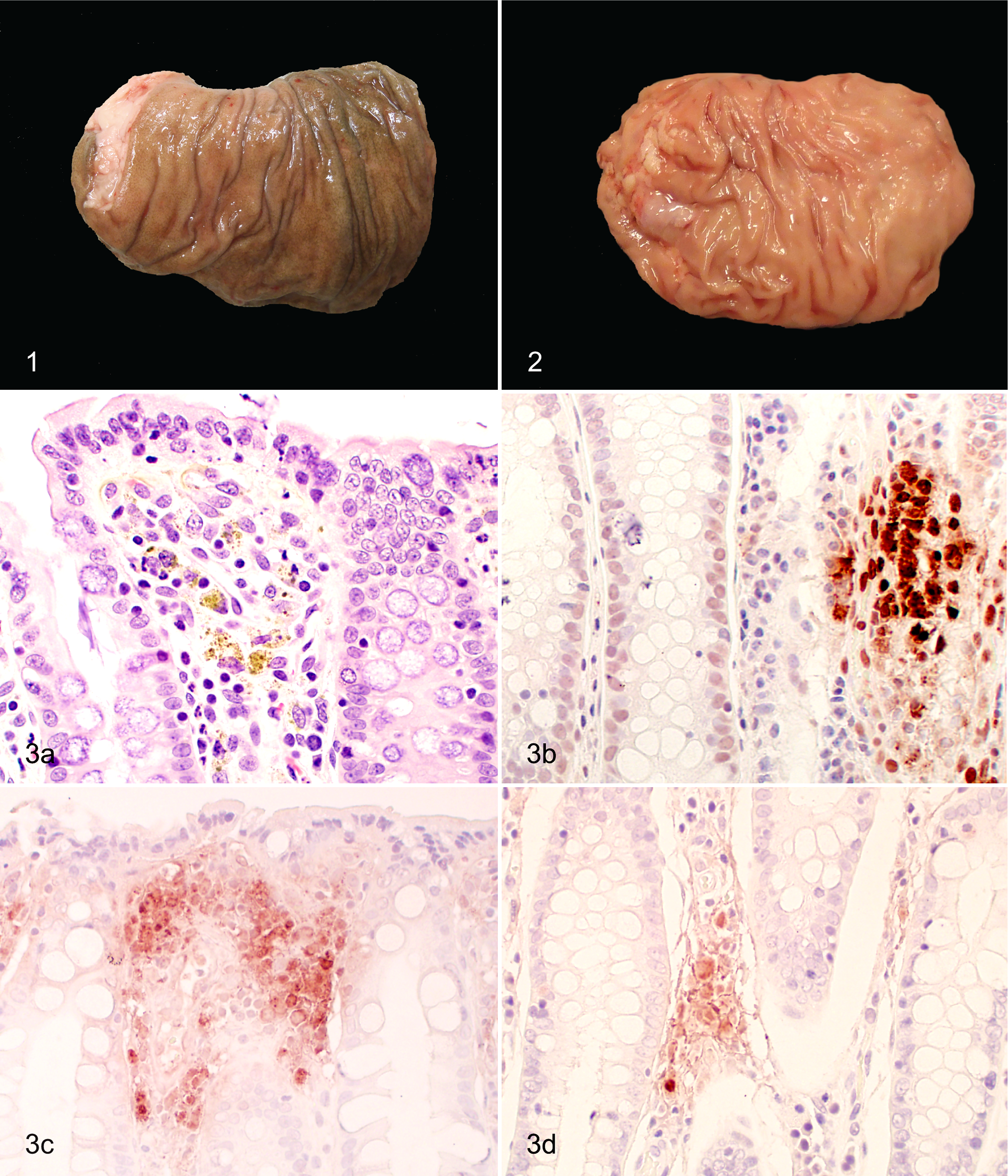
Melanosis coli, rectum; pig; case No. 3. Brown to green mucosal discoloration.
Histologic examination of affected colons demonstrated variable numbers of pigment-laden macrophages in the lamina propria (Fig. 3a), confirming melanosis coli. All sections were evaluated and scored 0 to 3 based on frequency and size of aggregates of pigment-laden macrophages: 0 if there was a lack of pigment-laden macrophages, 1 if small aggregates were present at least every ten 40× high-power field (HPF), 2 if moderately sized aggregates were present at least every five 40× HPF, and 3 if large aggregates that spanned the lamina propria between crypts were present at least every three 40× HPF. Pigment-laden macrophages were rarely observed in the lamina propria of sections from grossly unaffected colons. No pigment-laden macrophages were noted in the submucosa, lymphatics, or musculature of the intestine. Examination of an unstained section of affected colon using a fluorescent microscope revealed autofluorescence of pigment-laden macrophages when viewed by both FITC (fluorescein isothiocyanate)- and TRITC (tetramethylrhodamine)-specific filter sets.
Histochemical stains were used to better characterize the pigment in the aggregates of lamina proprial macrophages. The pigment in the macrophages of all examined colons (1 from case Nos. 1 and 2 and 3 from case No. 3) was negative by Prussian blue staining. Variable but weak acid-fast staining (long Ziehl-Neelsen) was apparent in affected colons from case No. 3, and this pigment was also Luxol fast blue negative, periodic acid–Schiff (PAS) positive but diastase resistant, and Oil-Red-O positive.
Assessment of Oxidative Stress and Protein Nitration
Tissues of 12 pigs with severe (score 3) melanosis coli (case 1, n = 5; case 2, n = 4; case 3, n = 3) and 2 pigs from each case submission without melanosis coli (score 0) were selected for further evaluation using immunohistochemistry followed by quantitative image analysis. See Supplemental Materials for details.
Positive staining for 3-NT, 4-HNE, and MDA was negligible in the lamina propria of colons of unaffected pigs, whereas significant immunolabeling for 3-NT, 4-HNE, and MDA was present in the cytoplasm of pigment-laden macrophages in the lamina propria of colons of all affected pigs (Fig. 3, Suppl. Table S1). Affected colons had significantly more staining for 3-NT, 4-HNE, and MDA than did unaffected colons (P ≤ .001, all analyses) (Fig. 4). When available (case Nos. 2 and 3), vitamin E levels were quantified in the corresponding liver tissue by high-performance liquid chromatography with UV detection. Vitamin E levels were lower than the expected normal range in both affected (mean ± SD, 2.71 ± 0.34 ppm; n = 7) and unaffected pigs (mean ± SD, 3.23 ± 0.52 ppm; n = 4; reference interval: 4.3–11.4 ppm). Compared to immunolabeling, vitamin E levels decreased as positive staining for lipid peroxidation increased (ρ = –0.4368); however, the correlation was not statistically significant (P = .1792).
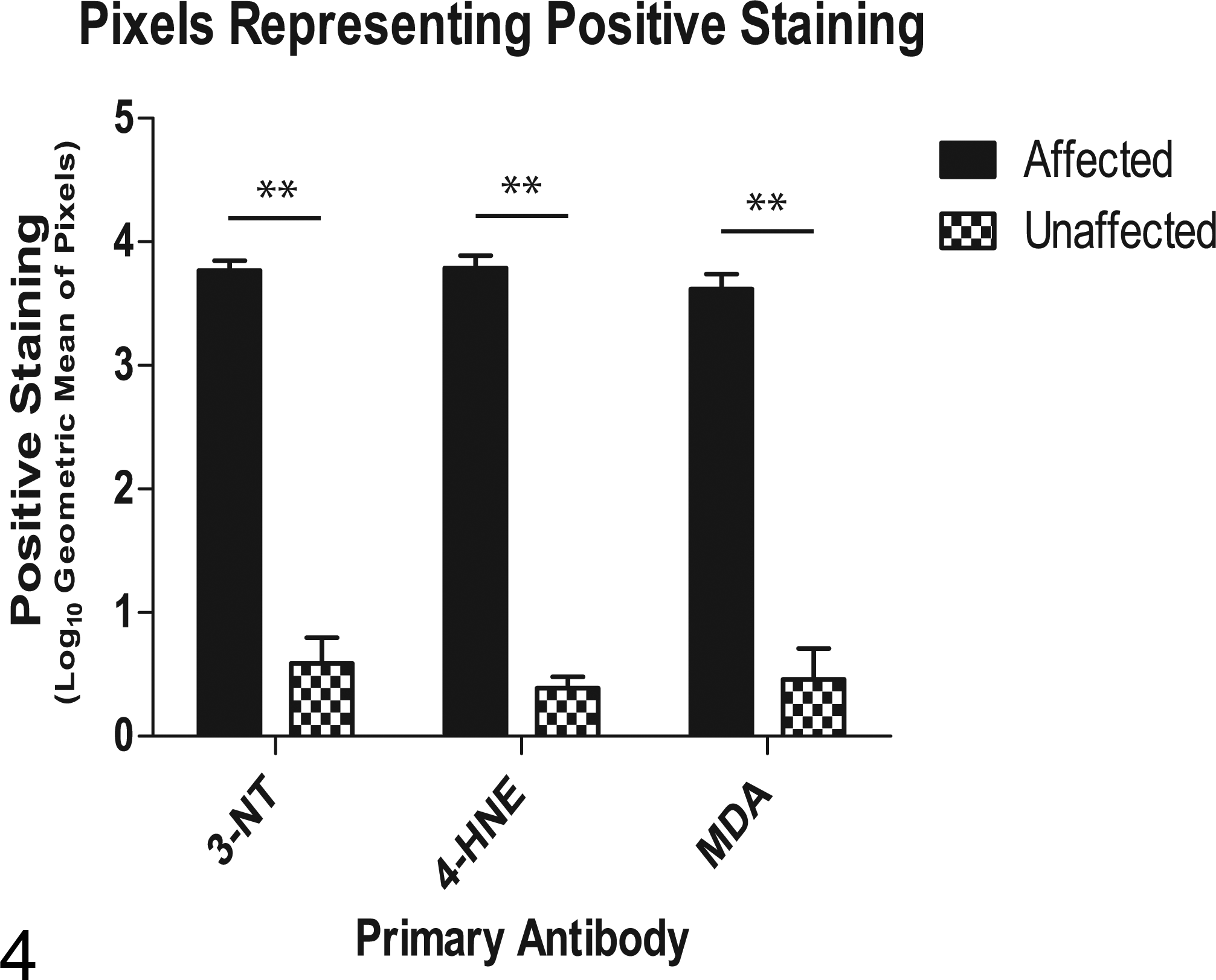
Geometric mean of pixels representing positive immunolabeling of pigment following log transformation of data from pigs with (affected) and without (unaffected) melansosis coli by primary antibody. 3-NT, 3-nitrotyrosine; 4-HNE, 4-hydroxynonenal; MDA, malondialdehyde. **P < .001.
Discussion
To the authors’ knowledge, this is the first detailed description of melanosis coli in swine and the first to investigate the association between oxidative damage, protein nitration, and hypovitaminosis E in cases of melanosis coli. In the present study, the histochemical results and accumulation characteristics of the pigment within macrophages were most consistent with ceroid. 12 Specifically, the location of the pigment in macrophages is similar to the ceroid present in macrophages in feline nutritional panniculitis and suggestive of heterophagy, which is a characteristic of ceroid rather than lipofuscin. The age of the pigs also suggests a rapid rather than very slow accumulation rate.
A statistically significant increase in positive immunolabeling for 3-NT, 4-HNE, and MDA was detected in the pigment-laden macrophages of pigs with melanosis coli, indicating that oxidative damage and protein nitration likely contribute to the pathophysiology of this condition in swine. Alternatively, it cannot be ruled out that these findings were a consequence of macrophage phagocytosis and activation. Lipid peroxidation results from the interaction of free radicals with polyunsaturated fatty acids generating reactive electrophilic aldehydes, the most abundant of which are 4-HNE and MDA. 5,13 Both 4-HNE and MDA have been identified as robust markers of oxidative stress. 5,13 In contrast, 3-NT represents a posttranslational modification of proteins due to a nitrating agent that results in protein tyrosine nitration. 1 The reactive nitrogen species peroyxynitrite is an important nitrating agent in vivo, thus making 3-NT a biomarker for endogenous peroxynitrite activity. 2,16
Lectin-based histochemistry has been used to suggest that the source of the pigment saccharides present in melanosis coli in humans originated from macrophage phagocytosis of apoptotic epithelial cells while also demonstrating that the pigments of melanosis coli have characteristics typical of both lipofuscin and ceroid. 4 Ceroid accumulation has been documented in the small intestine of dogs with intestinal leiomyometaplasia as well as in nutritional panniculitis in cats, mink, foals, and pigs. 12 Insufficient vitamin E, a potent antioxidant embedded in cell membranes, and ingestion of unsaturated fatty acids have been implicated in both conditions. 12 Although hypovitaminosis E was present in the affected pigs of this report, and there was a negative correlation between vitamin E levels and immunolabeling for lipid peroxidation, the correlation was not statistically significant. Since the precise etiopathogenesis of this condition in pigs is unknown, as well as the time course for lesion development and persistence, it is possible that hepatic vitamin E levels at slaughter are an imprecise assessment of the role of vitamin E in disease. In addition, deficiencies in other antioxidant factors may also contribute to disease, and a prospective study is necessary to better elucidate potential disease mechanisms of this condition. Furthermore, the small sample size in the present study may have been an additional limiting factor.
Previous studies have demonstrated a strong association between melanosis coli and chronic laxative use, 3,15 specifically the anthraquinone laxatives in humans, 11 and this condition can be reproduced experimentally in guinea pigs with laxative administration. 14 It remains to be determined if the presence of anthraquinone-containing plants in the diet, ingestion of unsaturated fatty acids, and/or hypovitaminosis E may precipitate the development of melanosis coli in swine. Additional investigations are needed to elucidate such associations with the oxidative damage and protein nitration present in cases of melanosis coli in pigs.
Footnotes
Acknowledgements
We thank Dr Marlin Hoogland, Dr Robert Blomme, and Dr Roger Johnson for their contributions of clinical case materials; Dr Rodger Main for his support of applied research at the ISU VDL; and Deb Moore for her help optimizing the immunohistochemistry procedures.
Author Contribution
Conception or design: BLW, KJS, PCG, ERB. Data acquisition, analysis, or interpretation: BLW, KJS, PCG, CW, ERB. Drafting the manuscript: BLW. All authors participated in critically revising the manuscript, gave final approval, and agree to be accountable for all aspects of work to ensure integrity and accuracy.
Declaration of Conflicting Interests
The author(s) declare no potential conflicts of interest with respect to the research, authorship, and/or publication of this article.
Funding
The author(s) disclosed receipt of the following financial support for the research, authorship, and/or publication of this article: This study was funded in part by the Boehringer Ingelheim Vetmedica Professorship in Food Animal Infectious Diseases.