
Abstract
Seven male Hartley guinea pigs, 3 to 18 months old, died or had to be euthanized because of nonspecific clinical signs unresponsive to supportive treatment. Gross necropsy and histopathology findings in all animals included severe soft tissue calcification affecting the myocardium, kidneys, and occasionally the liver.
History and Findings
Over a period of 2 months, 7 out of 40 male Hartley guinea pigs (Crl:HA, Charles River Laboratories, Houston, TX) aged 3 to 18 months died or had to be euthanized. Six of the animals were used for noninvasive procedural training and 1 was experimentally injected intraperitoneally with ragweed pollen; all procedures were performed with IACUC approval at an AAALAC-accredited research facility. Clinical signs included dyspnea, bruxism, nasal discharge, stiff gait, lethargy, vocalization, rough haircoat, and reduced feed intake/anorexia. These signs varied among animals. Of 4 guinea pigs tested, 2 had hypercalcemia (12.0 and 12.5 mg/dl; reference, 9-11.3 mg/dl), and 4 had hyperphosphatemia (8.0, 8.3, 11.7, and 11.9 mg/dl; reference, 4.2-6.5 mg/dl). Gross necropsy findings included multifocal to diffuse white mineral deposits (mineralization) in cardiac muscle and renal cortices (Fig. 1). Quarterly colony health monitoring indicated animals were seropositive for parainfluenza virus type 3 and seronegative for Sendai virus, Mycoplasma pulmonis, lymphocytic choriomeningitis virus, Encephalitozoon cuniculi, simian virus 5, guinea pig adenovirus, and reovirus. Animals were provided with guinea pig chow and reverse osmosis purified water, corncob or aspen bedding material, and environmental enrichment device in conventional cages. Environmental conditions in housing rooms were 12:12-hour light:dark cycle, 40% to 80% relative humidity, and a temperature of 20°C to 24°C.
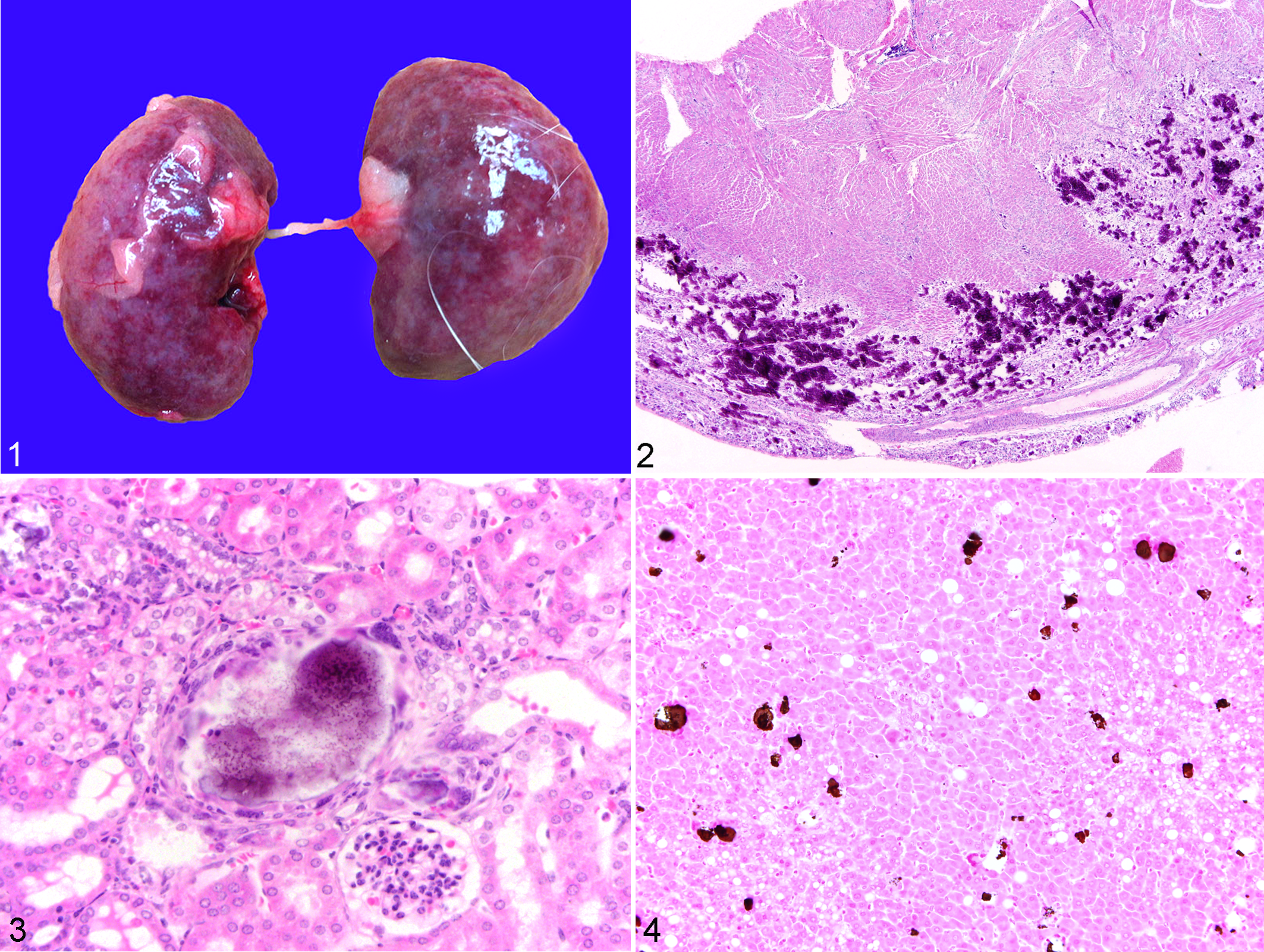
Metastatic calcification, guinea pig. Kidneys. Bilaterally, cortices are irregular with a mottled appearance from the white mineral deposits.
All affected guinea pigs had multifocal to diffuse, moderate to severe biventricular cardiomyocyte mineralization with granulomatous inflammation, cardiomyocyte degeneration, and necrosis (Fig. 2) and mild to moderate multifocal renal predominantly cortical tubular mineralization (Fig. 3). Other variable findings included mild to moderate multifocal and chronic renal cortical infarcts, multifocal hepatocyte mineralization with or without lobular infarcts, and focal to multifocal arterial mineralization within organs. Von Kossa staining of hepatocytes demonstrated abundant mineral deposits (Fig. 4). One guinea pig had mild to moderate multifocal mineralization and foreign-body type inflammation of the lungs, adrenal glands, and pancreas. Another had mild multifocal alveolar histiocytosis with intrahistiocytic hemosiderin pigment (heart failure cells).
Differential Diagnoses and Feed Analyses
The differential diagnoses for metastatic calcification include dietary factors such as low-magnesium (Mg) and high-phosphorus (P) diet and high calcium (Ca) and/or high vitamin D intake, inadvertent cholecalciferol rodenticide poisoning, and renal disease.
Correspondence with feed vendor representatives indicated that there was a feed misformulation affecting the specific batch provided to the animals. A feed sample was sent to Covance Laboratories (Madison, WI) for quantitative analysis of vitamins D and E, selenium, Ca, Mg, and P levels. Results were normal except for the vitamin D3 level, which was 494 IU/g compared to 3.1 IU/g as per product information. 6
Diagnosis and Discussion
Based on signalment, history, temporal pattern of cases, gross and histopathology results, and feed analyses findings, the cause of morbidity and mortality of the animals was hypervitaminosis D–induced metastatic calcification, primarily affecting the heart. Although high-Ca or high-P diets also interfere with Mg metabolism leading to metastatic calcification most often found in guinea pigs over 1 year old, 3 feed analyses indicated that dietary Ca and P contents were normal. Cholecalciferol rodenticide poisoning was ruled out, as this rodenticide was not used in our facility. Renal disease was also ruled out because of its unlikely simultaneous occurrence in the affected guinea pigs, particularly 1 was only 3 months old, and because serum creatinine (range, 0.6-0.9; reference, 0.6-2.2 mg/dl) and urea nitrogen (range, 25-32; reference, 9-31.5 mg/dl) values were normal. More importantly, renal disease typically results in low serum Ca levels. 2 However, it is still important to include renal disease as a differential diagnosis to hypervitaminosis D, especially because both conditions cause polyuria-polydipsia (PU-PD), a condition not reported here but in a subset of guinea pigs presented in a similar outbreak. 5 Hypervitaminosis D may cause PU-PD as the subsequent increased Ca levels may be associated with hyposthenuria caused by reduced antidiuretic hormone efficacy and reduced tubular sodium absorption. 11
One kilogram of guinea pig feed should have approximately 0.04 mg vitamin D if the Ca:P ratio is not adequate. 4 There is very limited literature on vitamin D requirements of guinea pigs. Studies of Ca and P homeostasis in guinea pig maternal-fetal interactions have indicated that dietary Ca and P levels were more important than the dietary vitamin D level. For example, while augmenting Ca and P intake overrides the effects of vitamin D deficiency on fetal development, 9 increasing vitamin D intake does not prevent hypocalcemia in dams provided with restricted dietary Ca and phosphate. 10 Based on product literature, the properly formulated feed meets the nutritional requirements for Ca and P and has additional vitamin D content. 6 While the properly formulated feed has not caused any toxicity, the extremely high vitamin D level in the misformulated feed caused tissue mineralization in vital visceral organs. In our cases, cardiomyocyte mineralization caused weak and stiff myocardium, leading to reduced filling and contraction and ultimately to heart failure as evident by heart failure cells in 1 case.
The role of vitamin D in mineralization is complex. The active form of vitamin D3 (1,25-dihydroxycholecalciferol) increases the active transcellular transport of Ca and P in the small intestinal mucosa 7 and may facilitate parathyroid hormone action on bones. 2 Another mechanism is the formation of fetuin-A mineral complexes consisting of a calcium phosphate mineral phase and fetuin and matrix Gla proteins in association with a decrease in free serum levels of fetuin-A, a calcification inhibitor. 8 Although the biochemical linkage is not yet clear, high levels of these complexes are predictive of artery and soft tissue calcification in rats treated with toxic doses of vitamin D. 8
Serum Ca levels of 2 out of our 4 animals tested were normal. This was not surprising as serum total Ca may not parallel ionized Ca, 11 which is a more important variable as it is the major active form in the body. Thus, the serum ionized Ca levels of our guinea pigs presumably were increased relative to the increased P levels. However, it is important to note that it is not necessary to have increased blood Ca to produce tissue calcification. 1 Besides serum testing for ionized Ca, a more direct antemortem test for hypervitaminosis D includes serum level determination of 25-hydroxycholecalciferol and 1,25-dihydroxycholecalciferol.
Footnotes
Acknowledgments
We thank Mrs. Marita Husted and the rest of the administrative, veterinary, and husbandry support staff of the Animal Resources Center at the University of Texas Medical Branch, Galveston, TX.
Declaration of Conflicting Interests
The authors declared no potential conflicts of interest with respect to the research, authorship, and/or publication of this article.
Funding
The author(s) disclosed receipt of the following financial support for the research, authorship, and/or publication of this article: The authors received financial support from the Land O’Lakes Purina Feed for the research of this article.