
Abstract
Cattle grazing turnips or other brassica forage crops occasionally develop hepatogenous photosensitization. In New Zealand, cases of bovine photosensitization associated with such crops frequently occur during late summer and fall, and this coincides with the facial eczema (sporidesmin toxicosis) “season.” Clinical chemistry findings in acute photosensitization cases associated with both brassica and facial eczema include marked serum elevations in γ-glutamyl transferase and glutamate dehydrogenase activities. Distinctive bile duct lesions of “subacute” turnip photosensitization in 2 cows, comprising microscopic cholangiectasis with concentric periductal fibrosis of small bile ducts, and a spectrum of changes from bile duct necrosis progressing to obliterative sclerosis are described. These bile duct lesions are compared with those in 3 cases of “subacute” facial eczema in adult cows, where medium-sized and larger ducts tend to be involved and bile duct hyperplasia and portal fibrosis are more prominent, often leading to bridging between neighboring portal triads.
Fast-growing forage crops, comprising cultivars of turnip (Brassica rapa ssp rapa), rape (Brassica napus ssp biennis), swede (rutabaga) (B. napus ssp napobrassica), and kale (Brassica oleracea ssp acephala), and interspecies hybrids fulfill an important niche in the provision of high-quality, easily digestible feed during dry months of the year in many countries worldwide. On the North Island of New Zealand, brassica are considered “safe” crops during late summer and autumn (fall) when facial eczema risk is high. 9 Daily access by dairy cattle to such crops is normally restricted according to time and/or intake per cow (such as with break feeding). Cattle grazing brassica crops occasionally develop a variety of disease signs of which photosensitization is the most prevalent. 7
Facial eczema (FE), a hepatogenous (secondary) photosensitization of farmed ruminants, is a disease of major economic importance in New Zealand. It is caused by the epipolythiodioxopiperazine mycotoxin sporidesmin found in conidia (spores) of the saprophytic fungus Pithomyces chartarum growing on dead pasture litter. Serum biochemistry findings in acute clinical FE cases are characterized by markedly elevated activities of γ-glutamyl transferase (GGT) and glutamate dehydrogenase (GDH), as well as raised concentrations of phytoporphyrin (phylloerythrin). In cows with FE, GGT activities frequently range from 500 to 2000 U/L and can be more than 4000 U/L (normal range, 0–36 U/L), while GDH activities range from within the normal range to 2000 or more U/L (normal range, 8–41 U/L).
Unfortunately, from a diagnostic point of view, clinical chemistry is of little value in distinguishing FE from turnip photosensitization, since GGT and GDH activities in the latter are usually of a strikingly similar magnitude. 1 Both have raised phytoporphyrin concentrations, confirming that the photosensitizing mechanism is hepatogenous. 1,2 Another confounding feature is that the 2 diseases frequently occur concurrently during late summer and autumn (fall) in New Zealand. Liver biopsies taken from cows showing acute turnip photosensitization have, to date, been disappointing in that no consistent biliary or parenchymal lesions have been identified.
Since cases of turnip photosensitization occur sporadically and, at this stage, cannot be predicted, and because the hepatotoxic phytochemical in brassica is unknown, 1 there is no way to satisfactorily reproduce the disease. In this communication, prominent bile duct lesions, occurring in 2 cows (Nos. 1 and 2) that were killed 1 and 2 weeks after the appearance of acute turnip photosensitization, are described. These lesions are compared with those in 3 confirmed naturally acquired cases of facial eczema (cow Nos. 6, 7, and 8). The respective bile duct lesions in both diseases partly explain some of the clinical chemistry findings.
History and Clinical Signs
Cow No. 1 was a 5-year-old Friesian that was 1 of 480 cows on a farm near Palmerston North, in the Manawatu district of the North Island. It developed severe photosensitization on nonpigmented skin of all 4 legs, the udder, and teats on January 21, 2011, after having been break fed on a turnip (Barkant cultivar) crop for just 4 days. The estimated intake of the crop was 4 kg/cow/d. The attending veterinarian collected venous blood into a Vacutainer containing a clot activator for serum separation. Despite the usual treatment, the cow did not improve and was eventually euthanized as a “downer cow” on January 28 (day 11 after the first day of access to the crop). A venous blood sample for serum separation was collected immediately prior to death, and a complete necropsy was performed within 1 hour. Samples of affected skin, liver (dorsal and ventral lobes), kidney, and a number of other organs and tissues were fixed in 10% buffered formalin. The histopathological lesions, including distinctive bile duct changes, have been described. 1 This farm is in a region where P. chartarum spore counts can reach dangerous levels on an annual basis.
Cow No. 2 was an adult Ayrshire, one of a herd of 330 dairy cows. The farm is located near Richmond, in the Tasman district of the South Island. The farmer had fed turnip crops to his cows for the past 16 years without any cases of photosensitivity occurring. Facial eczema has never been recorded on the property. Cows had been break fed for a maximum of 2.5 hours daily on a turnip (Barkant cultivar) crop until February 10, 2013 (in an attempt to estimate the amount of time that liver lesions—described below—could develop, this is taken as day 0). They were then moved onto a chicory crop until February 20 (day 10), before being break fed on a mature, good-quality crop of turnips (Envy cultivar) starting on February 21 (day 11). Each cow was offered approximately 4 kg crop per day. A few cows were noticed to be irritable within a day of going onto the second turnip crop. The following day, a small number of cows were noticeably ill with signs of early photosensitization. A veterinarian was called on the afternoon of February 24 (day 14) when 6 cows were severely agitated, and some were recumbent and showed subcutaneous edema of nonpigmented skin of the legs (and occasionally of the submandibular skin), purplish discoloration of the muzzle mucosa, as well as skin of the teats and around the vulva. Some were febrile with temperatures up to 41.5°C. Venous blood samples from 4 of these cows (Nos. 2–5) were collected into Vacutainers for serum separation. The worst was cow No. 2; another one, cow No. 4, died shortly afterward. Unfortunately, no necropsy was performed on this animal. At that time, there were about a dozen cows that were less severely photosensitized. Affected animals were provided with shade and analgesics were administered. The break feeding of the turnip crop was reduced, and supplementary silage was provided as well as greater access to pasture. The farmer monitored the remaining severely affected cows until day 30, when a veterinarian was called since cow No. 2 was extremely distressed. Blood was collected into a Vacutainer immediately before euthanasia and a necropsy was performed. Samples of fresh liver from the dorsal and ventral liver lobes were fixed in 10% buffered formalin. The farmer shot another 4 severely affected cows the following day; unfortunately, no samples were taken. Two neighboring farmers subsequently reported that a number of their cows grazing turnips (Envy cultivar) had also developed photosensitivity.
For comparative purposes, the serum biochemistry results and the histopathology of liver (dorsal and ventral lobe) lesions in 3 adult lactating Friesian-cross dairy cows (Nos. 6, 7, and 8) that developed naturally acquired facial eczema are presented. Cow No. 6 had been moved from its birth farm in the Southland district of the South Island (where facial eczema is not known to occur) to a dairy farm near Palmerston North on April 2, 2012. Cow Nos. 7 and 8 had been resident on this farm for longer than a year. None of them had grazed a brassica crop for at least 6 months prior. During this period, P. chartarum spore counts in the district were at dangerous levels (>100 000/g). Through an oversight, no zinc prophylaxis was administered to these cows. As part of another trial, all were blood sampled on the day of arrival of cow No. 6 (day 0). By day 16, all 3 cows had developed photosensitivity, and a veterinarian was consulted. Blood samples were collected into Vacutainer tubes for serum biochemistry. Only the ventral udder and teats (the only nonpigmented skin) of cow No. 6 were affected. Skin lesions of the other 2 cows were extremely severe and the decision to euthanize was made. Samples of dorsal and ventral liver lobes from both cases were collected and fixed in formalin. Cow No. 6 was dried off and provided with shade. It appeared to be recovering well until severe mastitis developed in one quarter. The cow was blood sampled before being euthanized on day 30; fixed dorsal and ventral liver lobe samples were collected at necropsy.
Serum Biochemistry
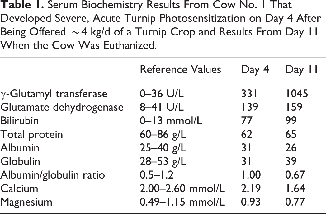
The results from cow No. 1 for day 4 (first appearance of photosensitization) and day 11 (day it was euthanized) are given in Table 1. Those from cow Nos. 2 to 5, including the cow that died, on day 14 (acute photosensitization), as well as those for cow No. 2 on day 30 (when it was euthanized), are provided in Table 2.
Serum Biochemistry Results From Cow No. 1 That Developed Severe, Acute Turnip Photosensitization on Day 4 After Being Offered ∼4 kg/d of a Turnip Crop and Results From Day 11 When the Cow Was Euthanized.
Serum Biochemistry for Cow Nos. 2 to 5 With Severe, Acute Turnip Photosensitization Collected on Day 14 Following Last Access to a Barkant Turnip Crop and That of Cow No. 2 on the Day of Euthanasia on Day 30.
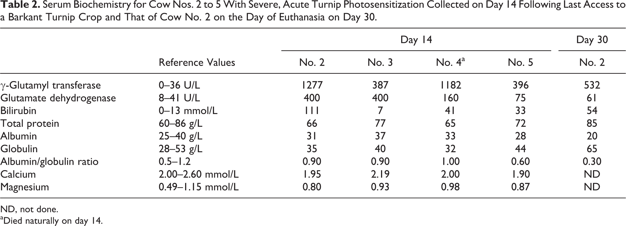
ND, not done.
aDied naturally on day 14.
The serum biochemistry findings of cow Nos. 6 to 8 that were sampled on day 0 and that had developed clinical facial eczema on day 16 (the day that cow Nos. 7 and 8 were euthanized), as well as those for cow No. 6 that was euthanized on day 30, are given in Table 3. As can be seen, cow Nos. 7 and 8 had increased activities of both GGT and GDH on day 0, probably indicating that sporidesmin liver damage was already under way.
Serum Chemistry Results of Cow Nos. 6 to 8 That Developed Clinical Facial Eczema on Day 16 and Those for Cow No. 6 on Day 30.
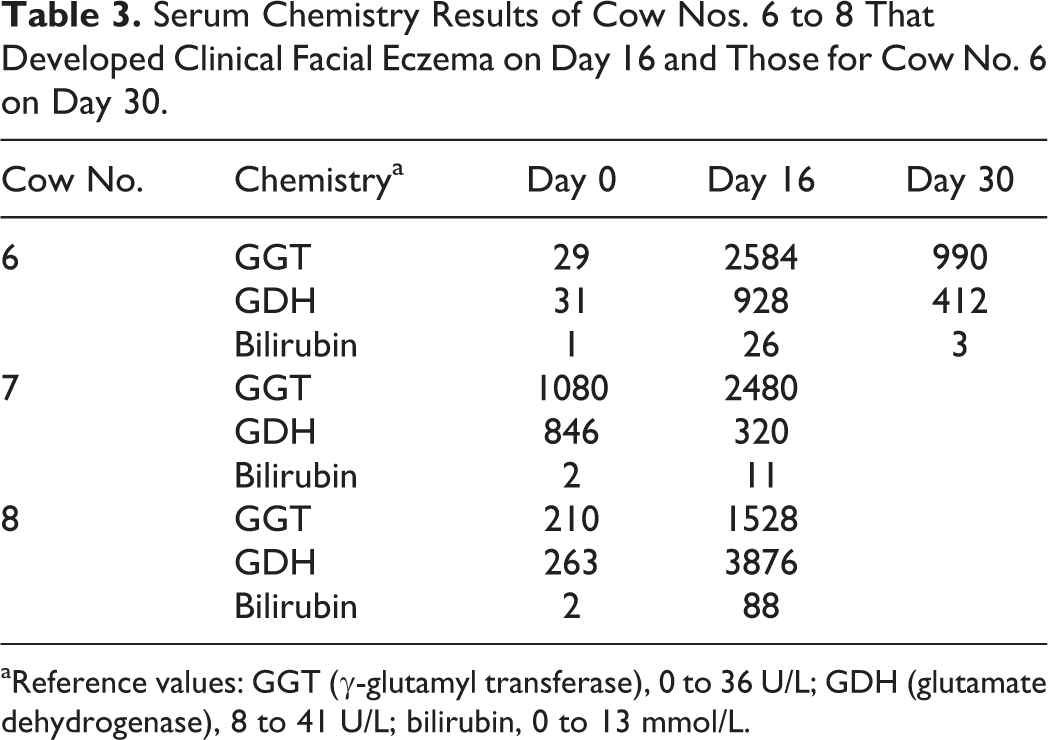
aReference values: GGT (γ-glutamyl transferase), 0 to 36 U/L; GDH (glutamate dehydrogenase), 8 to 41 U/L; bilirubin, 0 to 13 mmol/L.
Histopathology
Fixed liver samples from cow Nos. 1, 2, and 6 to 8 were routinely processed for histology and embedded in paraffin, and 3-μm sections were stained with hematoxylin and eosin (HE) before being examined with a light microscope.
Liver lesions in cow Nos. 1 and 2 were very similar. A conspicuous feature in both was the number of small interlobular bile ducts in the dorsal and ventral lobes that were dilated and lined by attenuated (atrophic or squamous) epithelium, comprising flattened nuclei and hyalinized cytoplasm (microscopic cholangiectasis) and surrounded by thin bands of concentric (“onion-skin”) fibrosis (Fig. 1). Other small bile ducts showed varying degrees of sclerosis (Fig. 2) culminating in fibrotic scars (Fig. 3). Some small interlobular bile ducts were necrotic and almost unrecognizable. There was sometimes a spectrum of changes in small bile ducts in single portal areas, ranging from biliary epithelium with cells showing pycnosis and hypereosinophilia, indicating necrosis, to periductal fibrosis and asymmetrical epithelial regeneration (Fig. 4). Some medium-sized bile ducts contained plugs of neutrophils. Also conspicuous were ducts lined by regenerating epithelial cells that appeared to be “piling up” in places and that were surrounded by loosely arranged (edematous) concentric fibrosis with a mild to moderate infiltration of mononuclear inflammatory cells further expanding the portal areas (Fig. 5). Occasional bile plugs were evident within canaliculi. There were rare inflammatory foci that usually surrounded proliferating bile ductules and that sometimes contained free bile (“bile infarct”) (Fig. 6). In cow No. 2, some portal tracts showed tangled proliferations of small bile ductules, in the presence of minimal fibrosis, that stretched toward adjacent tracts. On the whole, portal tracts in both cows were only mildly expanded by fibrosis. Lesions in the hepatic parenchyma included cell swelling, variation in staining intensity, increased mitotic rate, some fatty change, and occasional foci of lytic necrosis.
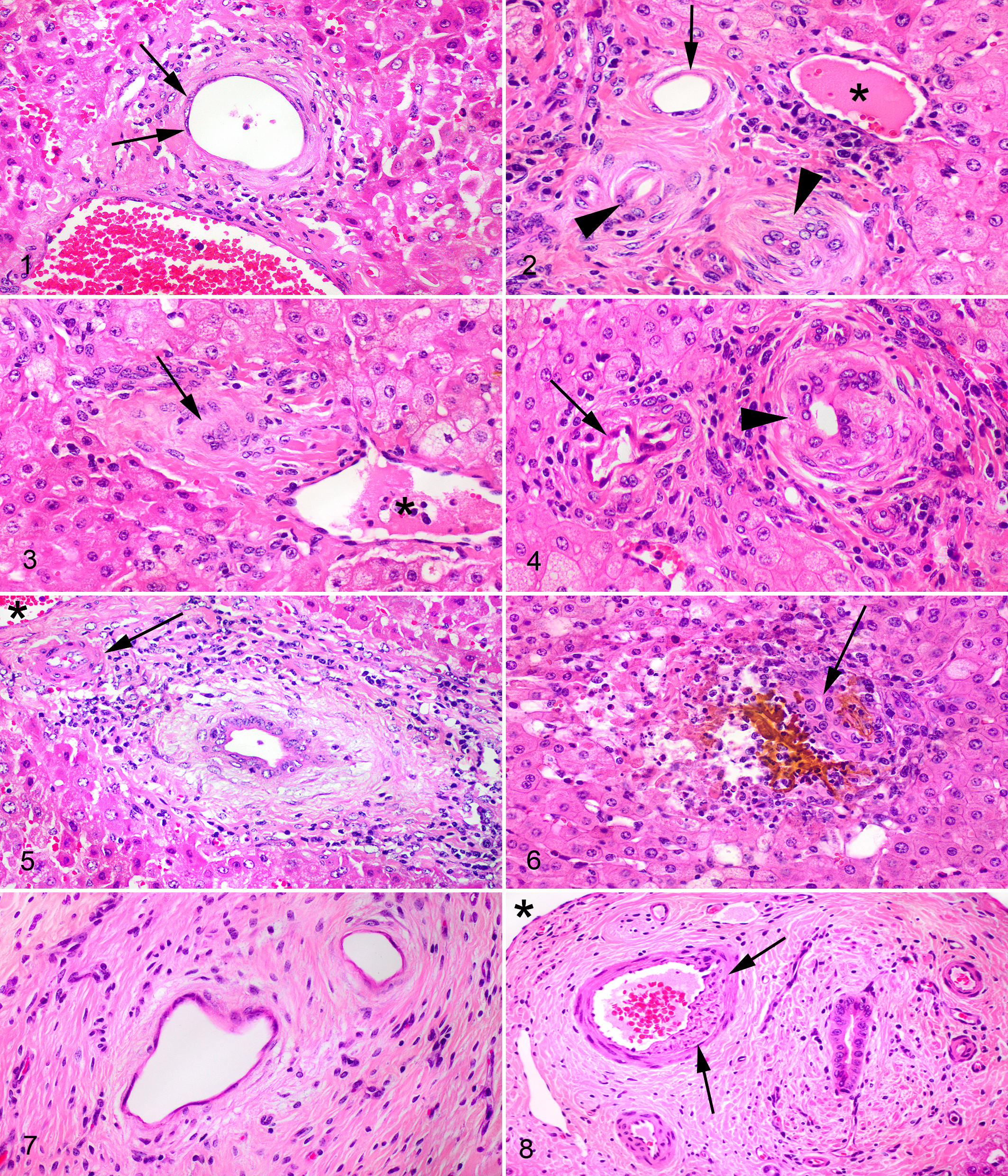
Typical lesions of subacute sporidesmin toxicosis (facial eczema) were present to a similar degree in both regions of the liver in cow Nos. 6 to 8 but were more extensive in cow Nos. 7 and 8. These lesions comprised moderate to severe expansion of the portal tracts with fibrous tissue, proliferated bile ductules, arteriolar hyperplasia, some mononuclear inflammatory cells, and occasional interlobular fibroductular bridges. Small- to medium-sized bile ducts were frequently surrounded by characteristic loose concentric rings of fibrosis, very similar to that in Fig. 5, except that their epithelium tended to be more regular cuboidal. A rare finding was cholangiectasis of small bile ducts with attenuated epithelium and concentric fibrosis, similar to the lesions seen in cow Nos. 1 and 2, except that these ducts were embedded in thick scar tissue (Fig. 7). In the latter, occasional scars that obliterated preexisting bile ducts were also encountered. A rare lesion in larger hepatic arterioles in portal tracts of cow No. 6 was the eccentric subintimal fibroblastic proliferation on the side adjacent to a bile duct (Fig. 8). In cow No. 8, some of the medium-sized bile ducts contained neutrophil plugs. The lamina propria of large hilar bile ducts occasionally showed ulceration of the epithelium and suppurative exudation, while the lamina propria contained granulation and scar tissue. Parenchymal lesions were minor in cow No. 6. In cow No. 7, fatty vacuoles were prominent in centrilobular hepatocytes. There were foci of centrilobular coagulative necrosis and randomly scattered small foci of suppurative inflammation in the liver of cow No. 8.
Discussion
Cows affected by turnip photosensitization or facial eczema exhibit many common features, such as the (usually nonpigmented) skin lesions typical of photosensitization, the elevation of GGT and GDH enzyme activities, and bilirubin and phytoporphyrin concentrations, typical of a hepatogenous photosensitization. In FE, the mechanism of toxicity involves the autoxidation of sporidesmin that initiates a free radical chain reaction in biliary epithelium, 8 which would explain the widely recognized and diagnostically utilized GGT activity elevations. In the case of turnips, similar GGT elevations would indicate some sort of mechanism causing injury to bile ducts. Without knowing the cause, however, this mechanism remains unknown.
Although the number of cases is too small for statistical comparison, in the cows described here, some features would appear to differentiate bile duct lesions in “subacute” turnip photosensitization from those seen in subacute facial eczema. In turnip photosensitization, the following changes to bile ducts appear distinctive: lesions appear to be restricted to small interlobular ducts, microscopic cholangiectasis with attenuation of biliary epithelium in some ducts, irregular regeneration of biliary epithelium in others, obliteration of other small ducts by unrecognizable debris or sclerosis, and lack of excessive portal tract fibrosis or bile duct hyperplasia. Bile duct lesions ranged from acute necrosis to sclerosis, possibly indicating an ongoing cholangiotoxic process.
In contrast, facial eczema lesions tend to involve medium-sized and larger bile ducts, 2,4,6 and portal tract fibrosis with bile duct hyperplasia is frequently markedly excessive, often leading to bridging between neighboring portal tracts. The latter lesions in FE frequently become relentlessly progressive, leading to fibrotic atrophy of the ventral lobe and eventually culminating in hepatogenous photosensitization at any time of the year, or ill-thrift, liver failure, or death. The more extensive liver lesions seen in cow Nos. 7 and 8 probably resulted from a longer period of exposure to high P. chartarum spore counts and, consequently, greater cumulative sporidesmin intake. The pathognomonic vascular-occlusive lesion, sometimes seen in portal veins and hepatic arterioles of sheep and goats with facial eczema, characterized by eccentric subintimal fibroblastic proliferation on the side adjacent to affected bile duct, 3,4,10 was present (although very rare) in arterioles of cow No. 6 but was not seen in any of the turnip cases.
The fact that signs of clinical photosensitization in the herd that comprised cow No. 2 started within 24 hours of access to grazing on the second crop of Envy turnips may have been coincidental and related to the increased availability of chlorophyll. However, another possibility is that prior exposure to the Barkant crop may have led to greater sensitivity to the putative cholangiotoxic turnip metabolite when the cows began grazing Envy. This phenomenon, potentiation, has been demonstrated for sporidesmin in sheep. 5
In conclusion, the spectrum of bile duct lesions described above could be diagnostically useful in cases of suspected turnip photosensitization in cattle. It would appear that turnips and probably other forage plants or weeds belonging to the Brassicaceae contain secondary compounds that, under certain unknown circumstances of metabolism, produce cholangiotoxic derivatives.
Footnotes
Acknowledgements
I thank Kevin Lawrence, Mike Hogan, Mark Gilmour, Roger Bay, Danny Hajdu, Mark Freeman, John Munday, Evelyn Lupton, Saritha Gils, and the staff of New Zealand Veterinary Pathology, Palmerston North.
Declaration of Conflicting Interests
The author(s) declared no potential conflicts of interest with respect to the research, authorship, and/or publication of this article.
Funding
The author(s) disclosed receipt of the following financial support for the research, authorship, and/or publication of this article: This work was supported by funding from the McGeorge Fund.