
Abstract
The veterinary literature contains scattered reports of primary tumors of the urinary tract of fish, dating back to 1906. Many of the more recent reports have been described in association with the Registry of Tumors in Lower Animals, and most of the spontaneous neoplasms of the kidney and urinary bladder are single case reports. In rare instances, such as described in nephroblastomas of Japanese eels and tubular adenomas/adenocarcinomas of Oscars, there is suggestion of a genetic predisposition of certain populations to specific renal neoplasms, environmental carcinogenesis, or potentially an unknown infectious etiology acting as a promoter. Hematopoeitic neoplasms have been infrequently described as primary to the kidney of a variety of fish species, and therefore those case reports of renal lymphoma and plasmacytic leukemia are addressed within the context of this review.
Between 30 000 and 40 000 species of fish have been reported in the literature and included in a variety of taxonomic classes falling under the infraphylum Gnathostomata or jawed vertebrates, which further consists of the extant class Chondrichthyes or cartilaginous fish and superclass Osteichthyes or the bony fish. 37 Within that enormous number of species, there is an extraordinary variety of structure and physiology, with species ranging in size from the smallest recorded fish (Paedocypris progenetica), measuring a mere 7.9 mm, 48 to the whale shark (Rhincodon typus), measuring up to 20 m in length. 15 It is therefore reasonable to expect that the pathology of fish encompasses an immense domain of entities and pathogeneses, representing a boundless field of potential discovery in veterinary pathology, particularly concerning spontaneous neoplastic disorders. The current paucity of information is due in large part to the environment in which the subjects live, the lack of our ability to track individuals throughout their range, and the fact that autolysis and predation affect the vast majority of samples, leaving the pathologist with a limited caseload of spontaneous neoplasms within wild populations. On the other hand, zoo and aquarium populations, private tropical and marine aquaria, and farmed fish are providing veterinary pathologists with a steady flow of novel entities and new interactions between host species and disease.
The following review of neoplastic disease in the urinary system of bony and cartilaginous fishes relies heavily on the body of knowledge accumulated by the National Cancer Institute’s Registry of Tumors in Lower Animals (RTLA) (Experimental Pathology Laboratories, Inc, Sterling, VA), which were donated from myriad sources as well as from the diligence of those researchers who have published their findings in the international literature. In addition, other sources were consulted as part of this review to include the Armed Forces Institute of Pathology (AFIP) archives, the Smithsonian National Zoo, department of pathology, and through the generous inclusion of selected syngnathid tumors from the Toronto Zoo.
The histopathology of most of these exemplars from the RTLA and the AFIP were reexamined, and photomicrographs from selected cases are included in this article. The RTLA alone consists of a collection of more than 7500 pathologic specimens of cold-blooded vertebrates and invertebrates with historical data and pertinent literature. 19 In addition, this review carefully examined both current and historical literature dating back more than 100 years and scoured all references to neoplastic lesions within the urinary tract of fish.
The veterinary literature contains scattered reports of primary tumors of the urinary tract of fish dating back to 1906. Many of the more recent reports have been described in association with the RTLA, and the vast majority of the spontaneous neoplasms of the kidney and urinary bladder are single case reports. In rare instances, such as described in nephroblastomas of Japanese eels (Anguilla japonica) and tubular adenomas/adenocarcinomas of Oscars (Astronotus ocellatus), there is suggestion of a genetic predisposition of certain populations to specific renal neoplasms, environmental carcinogenesis, or potentially an unknown infectious etiology acting as a promoter. Hematopoeitic neoplasms have been infrequently described as primary to the kidney of a variety of fish species, and therefore those case reports of renal lymphoma and plasmacytic leukemia will be addressed within the context of this review.
Primary renal neoplasia in higher vertebrates is predominantly split into the 2 most common forms, being nephroblastomas and some variant of adenocarcinomas. This appears to be statistically true within fish as well, with the vast majority of both spontaneous and experimental renal neoplasms falling into one or the other diagnostic group. The important distinction lies in the prevalence of primary renal lymphoma in fish due to the particularities of renal anatomy and physiology in those affected species. The most common renal tumor in domestic species is lymphoma, but these are almost all metastatic processes with the potential exception of chickens affected with Gallid herpesvirus type 2 or avian leucosis virus. 60
Few classical spontaneous renal neoplasms have been unequivocally diagnosed in fish. Of these, nephroblastomas far exceed other tumors in prevalence, with 83 cases having been either catalogued within the RTLA archives or described within the literature. Japanese eels and, to a lesser extent, rainbow trout (Oncorhynchus mykiss) represent greater than 50% of all individual cases, but 16 distinct species of fish have been reported with nephroblastomas. The second largest group of spontaneous renal neoplasms consists of the epithelial tumors comprising various subtypes of benign adenomas and approximately equal numbers of adenocarcinomas. All told, these have been described in 17 diverse species and are typically believed to arise from terminally differentiated tubular epithelium.
A variety of chemical etiological causes have been attributed to piscine carcinogenesis in the veterinary literature, including methylazoxymethanol acetate (MAM), N-methyl-N′-nitro- N-nitrosoguanidine (MNNG), dihydroepiandrosterone (DHEA), multiple aflatoxins, nitrosamines, and polynuclear aromatic hydrocarbon (PAH), and while these compounds have been primarily reported to induce hepatic tumors, there are several reports of nephroblastomas in association with a confirmed carcinogen. 13 Other environmental studies examining epizootics of hepatocellular and biliary carcinomas in Brown bullhead catfish (Ameiurus nebulosus), 6,7,70 lake whitefish (Coregonus clupeaformis), 61 and English sole (Pleuronectes vetulus) 54,57 and exocrine pancreatic tumors in mummichog (Fundulus heteroclitus) 18 have also shown a direct correlation between water contaminants and neoplastic transformation in various target tissues.
Retroviruses have been associated with tumor formation in an ever expanding cohort of species, as have reports of DNA viruses such as herpesvirus, papovavirus, and adenovirus. 1,12,45,46 One of the most comprehensively researched species-specific epizootics of piscine tumors are the damselfish neurofibromatosis and chromatophoromatosis, which have been associated with retroviruses. 13,76 Examples of similar outbreaks associated with retroviral etiologies include lymphomas in muskellunge (Esox masquinongy), 81 northern pike (Esox lucius), 85 and madai (Pagrus major), 63 as well as epidermal tumors in white sucker (Catostomus commersoni), 82 walleye (Sander vitreus), 87 and smelt (Osmerus mordax). 1 While none of these are associated with renal-specific targeting for carcinogenesis, there have been several reports of primary renal and metastatic lymphomas associated with retroviral neoplastic transformation. Salmonid herpesvirus type 2, colloquially known under a variety of names, such as Yamame tumor virus, Coho salmon herpesvirus, rainbow trout kidney virus, and Nerka tumor virus, among others, has been reported to form renal epithelial tumors in masu salmon (Oncorhynchus masou), sockeye salmon (Oncorhynchus nerka), chum salmon (Oncorhynchus keta), Coho salmon (Oncorhynchus kisutch), and rainbow trout, but the primary tumors are typically found within cutaneous tissues. 45 The virus causes age- and isolate-correlated high rates of mortality, with experimentally infected rainbow trout having rates ranging from 34% to 77% mortality. In surviving salmonids, tumors may develop in up to 100% of infected fish within a population. 20 Additional tumor-associated herpesviruses that result in systemic disease include Herpesvirus cyprini, herpesvirus of Japanese flounder, and epizootic epitheliotropic disease virus of lake trout (Salvelinus namaycush). 2
Familial or breed-specific renal tumors have been reported in humans, German Shepherd dogs, and Eker rats, associated in humans with molecular change within the von-Hippel Lindau (VHL) tumor suppressor gene, while the Eker rat is an animal model for hereditary renal carcinogenesis due to the development of renal carcinomas associated with mutation of the Tsc-2 tumor suppressor gene. In German Shepherd dogs, tumor induction is associated with a missense mutation in the Birt-Hogg-Dubé (BHD) gene, which is a tumor suppressor gene located on chromosome 5. Although the genetic interplay between several of the retroviral-induced tumors and their hosts’ genome has been closely examined, 73 to date the spontaneous renal tumors described in fish have not been shown to have a familial predisposition with the exception of Japanese eels and the development of nephroblastomas, which appear to show multiple nucleic acid changes associated with the Wilms tumor 1 gene. 64
Renal Neoplasms
Nephroblastomas
Nephroblasma is traditionally described as a tumor composed of neoplastic cells originating from the embryonic kidney, specifically in higher vertebrates such as humans, from the metanephric blastema. In these cases, the tumors develop from shared stem cells during embryogenesis or from embryonic nephrogenic rests and undergo transformation into neoplastic blastema and stromal cells. The tumor is a relatively common disease in humans, in whom it is termed Wilms tumor, but has been described also in most domesticated veterinary species, 29 laboratory animals, 30 and some exotic species. 30,80 In pigs and chickens, it is considered the most common primary renal tumor and the second most common in cats and dogs. 60 In humans, familial forms of the disease exist in which the Wilms tumor gene, located on chromosome 11, is correlated to tumorigenesis. WT-1 is implicated in approximately 22% of human cases, while WTX and CTNNB1 genes, both involved in the WNT/beta-catenin signaling pathway, account for an additional 20% to 30% of human nephroblastomas with minimal overlap between the genes involvement. 55,75 Furthermore, rat models have been developed using strains of Sprague-Dawley and Noble Hooded rats, which have demonstrated nephroblastoma development as a spontaneous process 31,59 as well as through experimental induction using chemical carcinogenesis. 32,79
With the exception of lymphoma, nephroblastomas represent the most common primary renal neoplasm in fish, having been reported in both spontaneous and carcinogen-induced instances and affecting a wide variety of fish species. Epizootics of spontaneous nephroblastoma have been reported in over 60 Japanese eels, 56,64 while individual cases have been reported in European eels (Anguilla argentina), 21 striped bass (Morone saxatilis), 38 koi (Cyprinus carpio), 84 Crucian carp (Carassius carassius), 10 Japanese dace (Tribolodon hakonensis), 58 smelt, 42 Siamese fighting fish (Betta splendens), 52 sockeye salmon (RTLA), rose bitterling (Rhodeus ocellatus), 34 banded cichlid (Heros severus), 24 and rainbow trout. 22,25,66,74 While a definitive correlation to a specific etiology resulting in the high incidence of nephroblastomas in the Japanese eel has not been proven, the authors of the various articles suggest that a water vector or carcinogen cannot be ruled out, and 1 unpublished report suggested the presence of virions in correlation with an eel affected with a concurrent nephroblastoma. 62 The carcinogen theory is consistent with extensive research conducted in rainbow trout, which have been demonstrated to undergo neoplastic transformation to nephroblastoma when experimentally exposed to both dimethyl nitrosamine (DMN) and N-methyl-N′-nitro-N-nitroguanidine (MNNG). 5 Furthermore, studies examining the environmental impact of MAM have produced neoplastic transformation in several visceral organs in a variety of species, including guppies (Poecilia reticulata), zebrafish (Danio rerio), and medaka (Oryzias latipes). The predominant populations of neoplasms were concentrated within the liver of each individual species, but medaka produced rare nephroblastomas when exposed to the carcinogen. 23
While the clinical presentation in reported cases varies, several examples in the literature displayed some form of vertebral pathology, such as lordosis, combined with coelomic enlargement. The gross presentation also typically includes organ displacement and the presence of a large, firm, pale, frequently unencapsulated mass. Nephroblastomas vary in size from small protuberances within the kidney tissue of less than half a centimeter in diameter to large growths, attached to the dorsal body wall, displacing the abdominal viscera. The biggest nephroblastoma in the RTLA collection occurred in a 32-cm-long rainbow trout and measured 7 × 8.5 cm. Most descriptions indicate the surface of trout nephroblastoma to be smooth, sometimes lobulated, and grayish to darkly pigmented. On cut section, the neoplasms are frequently white to pale beige and either firm and often granular or cystic in texture. Tissue of origin also varies between species, with all incidents in Japanese eels arising exclusively from the posterior aspect of the kidney. The same is reported in the case of koi, while the spontaneous reports in Siamese fighting fish, rainbow trout, striped bass, and Japanese dace appear to have obliterated the majority of both the anterior and posterior kidneys by the time of presentation, thereby negating the ability to definitively determine an anatomical point of origin (Fig. 1). Considering the variety in anatomic structure of fish kidneys, the embryological development of the anterior pronephros and the posterior opisthonephros, and the differences in glomerular vs aglomerular fish, it is unlikely that nephroblastomas are strictly limited to the genesis within the posterior kidney in all fish, but it stands to reason that those species with an anterior kidney predominantly composed of hematopoetic and endocrine tissues are overwhelmingly reported to have nephroblastomas developing in the posterior kidney. Predominantly, nephroblastomas in fish appear to be locally aggressive, expansile, and frequently cystic but rarely metastasize to distant sites. However, the reports in Crucian carp, Japanese dace, rainbow trout, and Japanese eels have described metastases to distant organs within affected animals. 50
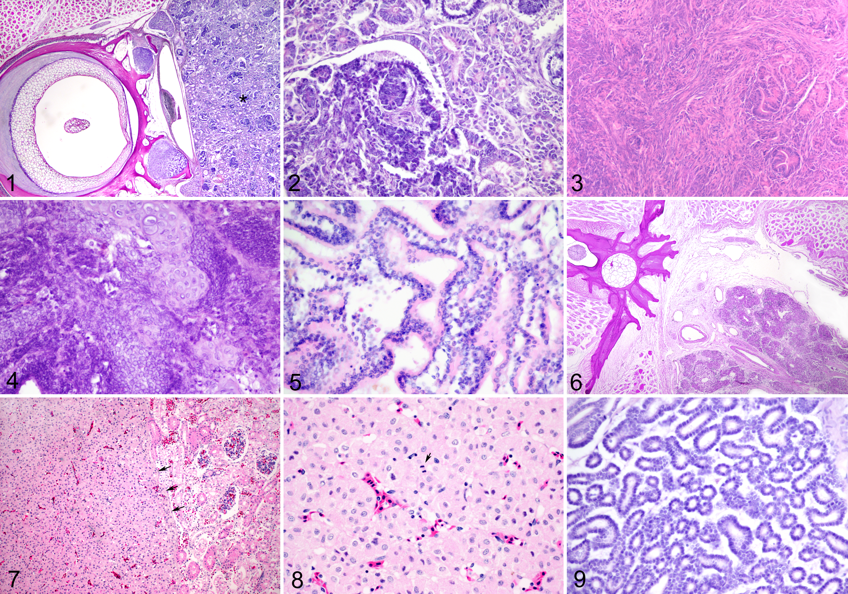
Microscopically, fish nephroblastomas appear to be consistent with the traditional triphasic histopathological appearance of the tumors in humans. The 3 diagnostic criteria include some combination of blastemal, epithelial, and mesenchymal elements, with the presence of embryonic or abortive glomerular structures considered as the most critical. 60 Those exemplars of piscine nephroblastomas described in the literature to date include tumors that predominantly present as large masses that efface the preexisting renal parenchyma. The majority are described as being unencapsulated and poorly demarcated, with infiltration into the renal interstitium, separating, surrounding, and replacing the renal tubules and glomeruli (Suppl. Fig. S5). The neoplasms are composed of the 3 distinct cellular populations in varying degrees of development and in various ratios. These include the epithelial component frequently arranged in disorganized tubules lined by cuboidal to columnar cells and tufts of epithelium with the appearance of embryonic or primitive glomeruli that invaginate into a luminal space lined by flattened cells similar to the parietal epithelium of Bowman’s capsule (Fig. 2). The blastemal population is characterized by small polygonal cells with minimal cytoplasm and dense nuclei with indistinct nucleoli, arranged in large swaths or smaller nodules (Suppl. Fig. S6 and Fig. 3). The final element is the primitive mesenchyme, composed of loose streams and whorls of spindle to stellate cells with scant cytoplasm and elongate nuclei. Within the mesenchymal population, there are frequent reports of terminal differentiation of the cells into striated muscle, cartilage, or even bone and, as such, form irregular islands of these tissue types. The mitotic rate in each population is extremely variable between reports.
The RTLA archive contained single cases of nephroblastoma in 4 other species of the salmonid family besides the rainbow trout—namely, brown trout (Salmo trutta), Coho salmon, Chinook salmon (Oncorhynchus tshawytscha), and chum salmon. In the former 3 species, the tumors histologically fell within the range expressed by the rainbow trout nephroblastomas, with primitive glomerulus formation in the brown trout specimen, loose sheets of blastema with minimal epithelial differentiation in the Chinook salmon, and squamoid differentiation within blastemal islands in the Coho salmon. The latter tumor had invaded skeletal muscle of the dorsal body wall.
Two nephroblastomas encountered in striped bass, one in a Siamese fighting fish and one in a brown bullhead, exhibited a feature not described in the salmonid renal neoplasms. In addition to the pathognomonic presence of blast cell aggregates and differentiation into primitive tubules, cartilage was a prominent feature of the bass and the Betta neoplasms. The cartilage was distributed histologically as either disorganized solid sheets or small islands of mature chondrocytes often separated and surrounded by contiguous areas of blastema and tubules (Fig. 4). Derivation of the cartilage by direct differentiation from blastema appeared probable but is not proven.
A series of tumors diagnosed as nephroblastoma in eels presented an important departure from those seen in the rainbow trout. Thirty-nine tumors are held in the RTLA files, 38 of them from Japanese eels and one from an American eel (Anguilla rostrata). A single case has also been recorded in the silver eel (Anguilla anguilla). 21 Collectively, these tumors displayed a histological spectrum more reminiscent of human Wilms tumors.
As a group, the Japanese eel tumors presented a histologic pattern that was, for the most part, similar to that of salmonid nephroblastomas, including diffuse sheets of basophilic blast cells with some fascicular disposition, irregular tubular structures, squamoid differentiation, and primitive glomerulus formation. However, most of the eel tumors, including that in the American eel, were also admixed with atypical striated muscle ranging from misshapen or round cells to bundles of more typical elongated myotubes. The silver eel tumor is described as having areas of cartilage and osteoid but no striated muscle fibers.
Consistent with published descriptions and illustrations, 49,56 some of the nephroblastomas in Japanese eels displayed a striking admixture of poorly to well-formed striated muscle cells with diffuse blastema and tubular and glomeruloid structures. The islands of muscle cells were sometimes in intimate juxtaposition with the undifferentiated blast cells, suggesting transitional stages between the blasts and the rhabdomyoblasts. In this series of neoplasms in eels, it became unambiguously evident that striated muscle can be one product of differentiation of neoplastic renal blastema, as is sometimes the case in human nephroblastomas.
Viewed as a series, these tumors in eels represent a spectrum ranging from purely epithelial nephroblastoma to embryonal renal tumors with increasing degrees of differentiation into striated muscle, with all examples retaining to some extent a diffuse distribution of blast-like cells and some distinct tubule formation. Human Wilms tumor, which predominantly affects children, also comprises a spectrum of histological variants ranging from purely blastemal and epithelial forms (the majority of human nephroblastomas) to neoplasms consisting of blastema, differentiated into tubule profiles, and neoplastic secondary mesenchyme in the form of sarcoma cells and striated muscle. It is this potential for biphasic differentiation into neoplastic elements of secondary mesenchyme as well as into epithelium that is connoted by the term Wilms tumor in humans. Anguilla spp is the only fish genus in which renal neoplasms resemble human Wilms tumor with respect to a capacity for rhabdoid differentiation.
As discussed earlier, nephroblastomas have been experimentally induced through administration of 2 different nitroso compounds. In the first of these experiments, rainbow trout were fed doses of dimethylnitrosamine ranging from 7 to 1920 mg/100-g dry diet in their daily ration for 12 to 20 months. 3 These various exposures to dimethylnitrosamine resulted in a 1% incidence of nephroblastoma in the survivors. Another study found that juvenile rainbow trout injected with either single or repeated intraperitoneal doses of 100 μCi of Iodine 131 had an increased incidence of nephroblastoma formation. 3,4
MNNG-induced nephroblastomas have been experimentally associated through treating both embryonic and near-adult stages of rainbow trout. In 1 study, MNNG was administered by stomach tube to 12- to 15-month-old fish, and nephroblastomas of macroscopic size were observed within 12 months. 44 In another study, rainbow trout embryos were incubated in water containing 10 ppm MNNG for 24 hours at 10°C. In this instance, a nephroblastoma measuring approximately 4.5 cm was observed at 9 months following carcinogen exposure. The cumulative frequency of nephroblastoma in an effective group of 122 survivors was 7.5% with the peak of tumor incidence occurring at 15 months postexposure. 45 Further investigations have induced nephroblastomas in embryos after a single hour of MNNG exposure. 39 The histopathology of these experimentally induced nephroblastomas in salmonids and their variations with respect to the predominance of certain histological features are the same as in the spontaneously occurring neoplasms described in the literature in other fish species.
Epithelial Tumors in Fish
There are only rare reports of spontaneous renal tubular cell tumors in the literature occurring in fish, and many of those that do appear range from a century ago. Equally, the RTLA archives contain scarce submissions of either adenomas or carcinomas in fish. The few descriptions that do arise are often mentioned without a histopathological description and as such are of limited use in drawing population or interspecies correlates. In 1911 and again in 1924, cystic adenocarcinomas were described in silver eels. 72,78 Other early reports include a benign renal tumor in a catfish composed of neoplastic cells forming acini and papilliferous projections into cystic spaces, suggestive of a renal papillary cystadenoma 77 and a similar spontaneous tumor that was described in the mesonephric duct epithelium of a Chinook salmon. 50 Single case reports can also be found in goldfish, catfish, 77 Mozambique tilapia (Oreochromis mossambicus), 24 and yellow or spotted seahorses (Hippocampus kuda). 51 Furthermore, a review of the RTLA archives revealed submission of a renal papillary cystadenoma in a winter flounder (Pleuronectes americanus), as well as references to similar tumors in a Tiger barb (Barbus tetrazona) and in a penguin tetra (Thayeria obliqua). 47
Renal Cystadenoma/Renal Tubular Adenoma
Oscars appear to have a predisposition for some variant of renal adenomas, which have recently been determined to be of proximal tubular origin. There has been some debate on the cell of origin of these neoplasms, with the first having been described in 1996 but without proffering a definitive diagnosis at the time. In that case, the mass, which was composed of mildly pleomorphic epithelial cells forming papillary projections into cystic regions, significantly effaced the renal parenchyma (Fig. 5). While determined to be locally expansile and potentially regionally aggressive, there was no evidence of malignancy, and the authors proffered a more benign categorization of the mass, with a favored diagnosis of an epithelial papilloma of mesonephric duct origin. 67 Similar tumors of the posterior kidney were observed in 3 other Oscars submitted to the RTLA archives. In these instances, the masses were also predominantly expansile and cystic, with minimal pleomorphism, and a low mitotic index, but 1 of the 3 appeared to be invading the adjacent preexisting renal tissue and all 3 had some degree of necrosis, as well as granulomatous inflammation and in 1 case an accumulation of birefringent crystals. More recently, an additional 6 animals have been described in the literature with tumors described as renal cystadenomas. These are similarly described as being expansile neoplasms, composed of disorganized cystic tubular structures or endophytic papillary projections with cuboidal to columnar epithelium supported by a fine fibrovascular stroma (Suppl. Fig. S7). In all cases, the neoplastic cells are noted to have minimal pleomorphism and a low mitotic index. 40
Exclusive of those described in Oscars, there are individual case reports of renal adenomas scattered in the literature, including examples in a tilapia (Sarotherodon spilurus). 28 Chinook salmon, 50,53 brown bullhead, 77 rainbow trout, 50 and northern pike 65 with experimentally induced adenomas in zebrafish and medaka. For the most part, these are described as renal cystadenomas or papillary cystadenomas; however, the RTLA archives include examples of diagnoses of papillomas in various levels of the urinary tract. These will be described in the section covering the urinary bladder.
A population of Crucian carp was affected by an epizootic of renomegaly. Upon necropsy, several of the animals were found to have renal adenomas as well as a high percentage of polycystic kidneys. The authors of that particular study suggested that while polycystic kidneys may be a precursor to adenoma, in Crucian carp and goldfish, there is a correlation between the myxozoan Sphaerospora dykovae (previously Sphaerospora renicola) infection and both hematopoietic cell and renal epithelial hyperplasia. 41
Another group of interest was brought to light in a recent study of syngnathid mortality at the Toronto Zoo, which determined a 4.1% incidence of neoplasia, and of those, approximately 50% were associated with the kidney. The study evaluated the postmortem findings of 172 deaths in captive spotted or yellow seahorses, potbellied seahorses (Hippocampus abdominalis), and weedy sea dragons (Phyllopteryx taeniolatus). A total of 7 neoplasms and 2 lesions described as neoplastic-like were identified, to include a renal adenocarcinoma, a renal adenoma, and 2 renal round cell tumors interpreted to be lymphoma. The tumor diagnosed as a renal adenoma was associated with a 2-year-old male yellow seahorse that had an expansile mass occupying approximately half of the coelomic cavity (Fig. 6). Histopathological examination of the tissue noted a renal neoplasm composed of variably sized tubules often supported by a dense fibrovascular stroma. The authors described minimal atypia but acknowledged that there was moderate autolysis of the tissue, which confounded a more detailed description. 51
Recent necropsy of a polka dot sting ray (Potamotrygon leopoldi) in Thailand revealed a firm, pale, well-circumscribed, expansile renal mass measuring 4 × 3.5 cm. Microscopic examination of the mass noted a densely cellular polygonal cell neoplasm (Fig. 7). Occasionally, the neoplastic cells formed vague tubules. The neoplastic cells had abundant eosinophilic, finely granular cytoplasm and oval nuclei with stippled chromatin (Fig. 8). There was approximately 1 mitotic figure per high-powered field and mild anisokaryosis. This neoplasm was diagnosed as a renal cell adenoma and represents the first report of a renal tumor in an elasmobranch and only the second in a condrichthyan.
Experimentally Induced Renal Adenomas
Renal cystadenomas have been reported in rainbow trout subsequent to exposure to MNNG. These tumors occurred with a 40% incidence rate 32 months after experimental intoxication and occasionally occurred simultaneously with nephroblastomas. 50
A renal tubular adenoma was also described in a zebrafish as an extrahepatic tumor resulting from a MAM-ac exposure in a carcinogenesis study. While the authors directly correlate the tumor to the experimental intoxication, the tumor represents a single incidence. 86
Adenocarcinomas/Renal Cell Carcinoma
Adenocarcinomas arising from well-differentiated (but not postmitotic) tubule cells have occurred spontaneously in an elasmobranch as well as in several teleost species. They were single tumors in each case. Like nephroblastomas, adenocarcinomas also have been induced experimentally by chemicals in small aquarium species.
The RTLA archives contain few examples of spontaneous renal adenocarcinoma, including individual descriptions in the spiny dogfish (Squalus acanthias), banded cichlid, Mozambique tilapia (Oreochromis mossambicus), winter flounder, and Chinook salmon. Each of these 5 tumors consisted of a population of epithelial cells forming tubules, without the aggregates of blastemal cells that characterize nephroblastoma.
The example in a spiny dogfish is the only renal neoplasm that has been found to date in the order of Squaliformes. The tumor was attached to the dorsal body wall in the location of the kidney. It consisted of well-formed tubules, often irregular in shape, lined by a single layer of cuboidal to columnar epithelium usually having eosinophilic cytoplasm. The cells were polarized, with basally disposed vesicular nuclei, each containing a single prominent nucleolus. In some neoplastic tubules, there was papillary proliferation of the lining cells. Tubules were surrounded by a desmoplastic stromal reaction that was scirrhous in parts of the tumor where epithelial structures were limited to islands and cords. Some epithelial cells in scirrhous areas were more anaplastic, having increased cytoplasmic basophilia and larger nuclei. Necrosis of epithelial lobules or of intraluminal cells was prominent. The tumor was partially enclosed by a fibrous envelope, and invasion of normal parenchyma was evident. Mitotic figures were found particularly at the invasive periphery of the tumor.
The tumor in the banded cichlid was similar in appearance to that described in the adenocarcinoma of the spiny dogfish. Well-formed basophilic tubules were supported by a loose, hypocellular stroma (Fig. 9). At the periphery of the tumor, there was no evidence of a fibrous pseudocapsular reaction. Mitotic figures were infrequent and nucleoli faintly staining.
The variant described in the Mozambique tilapia is of an unencapsulated, multiloculated neoplasm composed of neoplastic polygonal cells arranged in multiple layers of cells lining tubules and papillary invaginations into the larger cystic areas. The cells are supported by a fibrovascular to myxoid stroma (Suppl. Fig. S8 and Fig. 10). The neoplasm was restricted to the kidney, and while the unencapsulated edge of the mass abutted the skeletal muscle of the body wall, no invasion was noted. 24
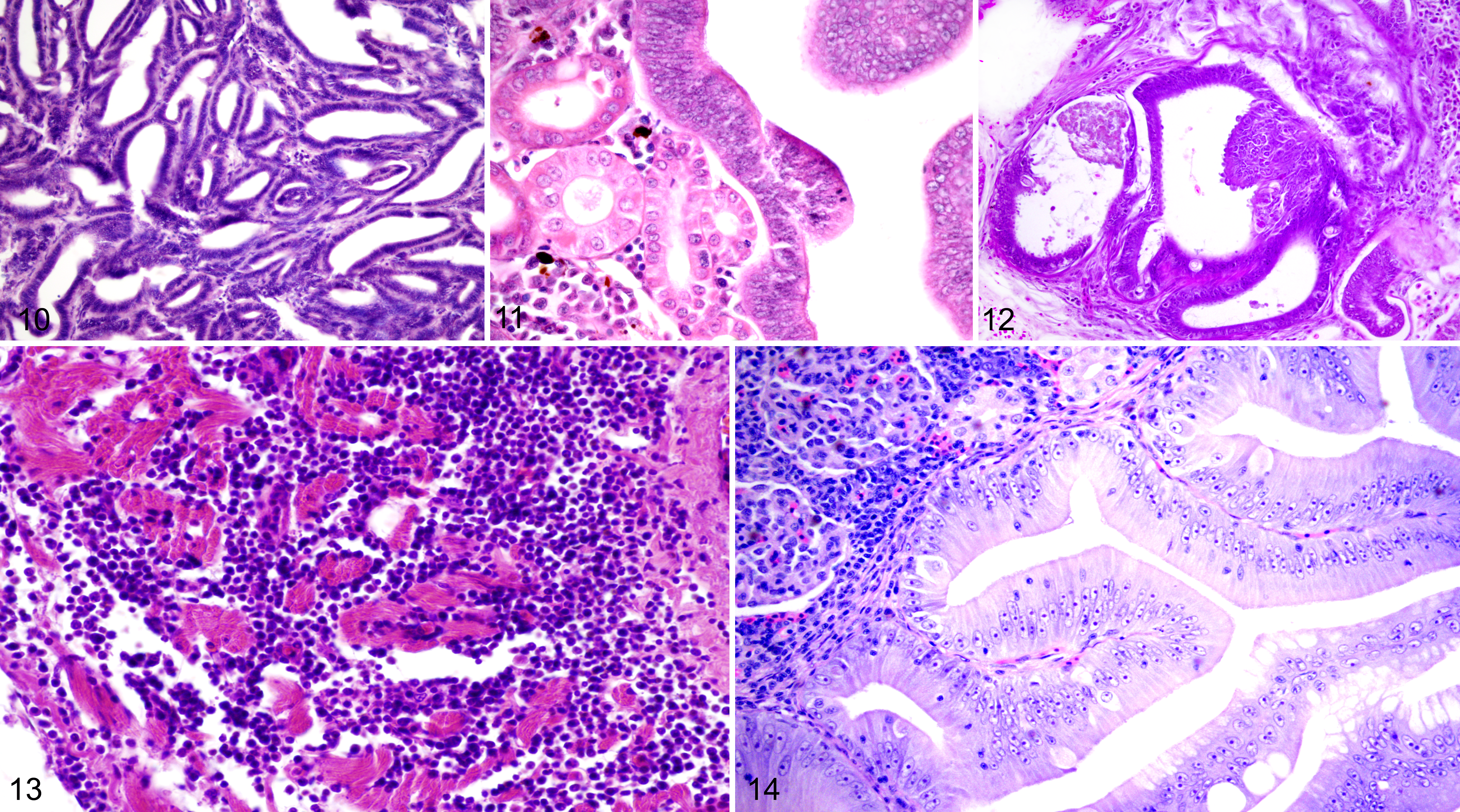
The renal adenocarcinoma in the Chinook salmon presented with a more microcystic and occasionally papillary pattern in which the tubules formed convoluted and anastomosing spaces. The lining epithelium was frequently of a pseudostratified or multilayered columnar type, with densely crowded nuclei and some mitotic activity (Fig. 11). The stroma consisted mostly of narrow vascularized strands of connective tissue running between the plicated, dilated tubule-like structures, but there were also large stromal tracts containing hematopoietic tissue and normal proximal tubules, representing entrapped preexisting renal tissue.
The winter flounder was noted on necropsy to have a large retroperitoneal mass that interdigitated with the intervertebral depressions and compressed the adjacent skeletal muscle while not invading either the musculature or the spinal column (Suppl. Fig. S9). The renal architecture was replaced by myriad coalescing cysts ranging up to 1 cm in diameter. Histopathologically, these were lined by disorganized simple to lightly stratified layers of neoplastic columnar epithelium that often formed papillary projections into the cyst lumen (Fig. 12).
Within the literature is a recent report of a captive bred adult male yellow seahorse that at necropsy was noted to have an expansile renal mass contiguous with the posterior kidney and occluding a large portion of the coelomic cavity. Microscopically, the mass was described as being a poorly encapsulated neoplasm that effaced and replaced the preexisting renal parenchyma. Neoplastic cells were polygonal, separated by a fine fibrovascular stroma and arranged in irregular cords, nests, and rare acini or tubules. Neoplastic cells had indistinct cell borders, a moderate amount of fibrillar or microvacuolated eosinophilic cytoplasm, and large centralized nuclei with minimal anisocytosis and anisokaryosis (Suppl. Fig. S10). The mitotic rate averaged approximately 2 per high-powered field, and there was frequent single-cell necrosis with no significant inflammation. A well-demarcated portion of the neoplasm consisted of a marked desmoplastic response admixed with high numbers of melanomacrophages. 51
Experimentally Induced Renal Adenocarcinomas
In carcinogenesis studies, the guppy and medaka are reported to have developed papillary renal adenocarcinomas secondary to experimental exposure to polycyclic aromatic hydrocarbons. 35 The chemical was administered in the ambient water at varying concentrations for a brief period of exposure in posthatched fish. Three guppies and 1 medaka that had been examined between 36 and 50 weeks posttreatment were submitted to the RTLA. They had renal lesions ranging from hyperplasia of the proximal tubules to frank renal adenocarcinoma. The latter was identical in form to a tumor described in Chinook salmon, consisting of a convoluted pattern of anastomosing tubules lined by mitotically active and densely crowded, basophilic columnar cells. Of interest are renal neoplasms described as having occurred within laboratory settings in zebrafish and demonstrated a notable difference in response to exposure to treatment with MNNG in comparison with trout. Zebrafish exposed during development to MNNG have developed both renal adenomas and adenocarcinomas but do not develop the typical nephroblastomas noted in trout fry with incidence rates upward of 50%. 83
Lymphoma/Leukemia
Hematopoietic tumors of the kidneys of fish are described with some frequency in comparison with other types of neoplasm, and as opposed to mammals and birds, there is an anatomical basis for a lymphoma to be primary to the kidney. Lymphomas have been described in up to 22 species of fish, with the thymus and kidney being statistically the most common primary sites for neoplastic transformation. 8,9,16,17,68 Historically, the species variation and histological anatomy of the fish kidney may have resulted in inaccurate diagnoses of lymphoma in the kidney. The normal presence of hematopoietic tissue within the anterior kidney of many fish species may have resulted in exaggerated descriptions of both mononuclear cell nephritis and lymphoma being maintained within the earlier published literature. In reviewing reports from a century ago, cases have been evaluated based on both gross findings of a mass effect as well as the histopathological appearance of the kidney. An example of this is illustrated in the description of a lymphoma in the kidney of a conger eel (Conger oceanicus) published in 1931. In this case, the neoplastic cells are characterized as lymphocytes widely separating entrapped uriniferous tubules and compressing the preexisting renal tissue. Combined with a macroscopic description of a 16 × 9 × 7 cm mass expanding the anterior kidney, causing a bulging of ventral body wall, there is little doubt of the mass effect and the presence of the described tumor; however, the author himself cautions the reader of the fact that “the kidney of teleosts is an extremely lymphocytic structure.” 88
Typically, the histopathological descriptions of lymphoma in the fish kidney are similar to those in higher vertebrates. Reports consist of highly invasive round cell neoplasms that infiltrate, replace, and efface the renal architecture, forming sheets of distinct lymphoblastic cells separated by preexisting tissue. These cells often have a high nuclear to cytoplasmic ratio and a moderately high mitotic index with mild anisocytosis and anisokaryosis. Frequently, these tumors are noted to have metastasized widely throughout the viscera (Fig. 13). 27,36
Lymphoreticular tumors in northern pike from Ireland and in muskellunge from Canada have been shown to have a retroviral etiology, and unlike other forms of spontaneous lymphoma, both of these species-specific tumor types have a seasonal incidence and have been described as beginning within the cutaneous tissues and metastasizing to the viscera, including the kidney. Histopathologically, both lymphomas consist of uniform populations of round cells with a blastoid appearance. The northern pike variant has a tendency to have a “starry-sky” appearance, which is reported not to occur in the muskellunge counterpart. 33
An epizootic in pen-raised Chinook salmon resulted in significant mortality in multiple production sites in British Columbia in the late 1980s and 1990s. The disease was colloquially known as marine anemia and was characterized by antemortem gill pallor and bilateral exophthalmos. Necropsy of selected animals revealed renal and splenic enlargement with the kidneys being uniformly enlarged and pale. Histopathologically, the affected tissues were severely infiltrated by innumerable neoplastic cells described by the authors as plasmablasts. The renal interstitium was markedly expanded by the neoplasm, and frequently there was evidence of a glomerulopathy consisting of expansion of the glomeruli by leukocytes with concurrent thickened basement membranes and hyperplasia of the Bowman’s capsule parietal cells. In one epizootic, the renal lesions in 50% of the fish were complicated by the presence of granulomatous inflammation with intracytoplasmic bacilli suggestive of Renibacterium salmoninarum. The same neoplastic cells were noted within the splenic vasculature, the heart, the intestinal tract, hepatic sinusoids, and the choroid gland. The neoplastic cells were characterized histopathologically and ultrastructurally as round cells with a large, clefted nucleus and with distinct nucleoli. Electron microscopy noted an abundant, regimented rough endoplasmic reticulum with occasionally dilated cisternae suggestive of Russel bodies. On the basis of these findings, the authors proffered a diagnosis of plasmacytoid leukemia. Plasmacytomas have been described in several fish species, and a single case of plasma cell leukemia has been described in a brown bullhead catfish. The reports in Chinook salmon proved critical as the tumors were found to be experimentally transmissible with cell-free filtrates, and reverse transcriptase activity was noted, suggesting a retroviral etiology. 43
A variety of case reports have described lymphoma in the kidneys of fish to include brook trout (Salvelinus fontinalis) 16 and several animals culled during a mass mortality in lake trout in New England. In those cases, the renal architecture was effaced by a clonal proliferation of atypical lymphocytes characterized by sheets of round cells with moderate pleomorphism, hyperchromatic nuclei, and a markedly increased mitotic index. These cases were interesting in that they were animals having concurrent severe mixed bacterial infections and necrotizing cellulitis due to the combined presence of the neoplasm and the bacteria. This particular study was not able to definitively prove a viral etiology; however, based on the high mortality in multiple locations, it is highly suggestive of an oncogenic and potentially immunosuppressive virus resulting in the transmission of disease and the high incidence of concurrent bacterial infections. 17 Another interesting series involves the previously described round cell neoplasms in 2 adult male seahorses. In the first animal, 90% of the coelomic cavity was filled with a large pale mass, composed microscopically of sheets of round cells compressing preexisting parenchyma and rare uriniferous tubules (Suppl. Fig. S11). The neoplastic cells had indistinct cell borders, a high nuclear to cytoplasmic ratio, large oval nuclei with roughly clumped chromatin, and a distinct central nucleolus. The cells displayed mild to moderate anisocytosis and anisokaryosis and a high mitotic ratio. The neoplastic cells were noted within the vasculature and had metastasized to the intestinal wall. The second case was essentially identical, with increased pleomorphism and a greater degree of tissue invasion and necrosis (Suppl. Fig. S12). While immunohistochemistry was not pursued in these cases, the morphology strongly favored a diagnosis of lymphoma. 51
Urinary Bladder and Collecting Duct Tumors
As previously discussed, there appears to be a predilection in Oscars for epithelial neoplasms of the urinary tract, with 10 cases having been described in the literature and some having been observed but not published. 26,40,69 Four neoplasms in the RTLA archive were originally described as likely having originated from the transitional epithelium lining the renal collecting ducts and urinary bladder. All 4 cases were discovered in Oscars, although the individual specimens were separated by both time and geographic location. Grossly, the 4 tumors were huge nodular masses, up to approximately 8 cm in diameter, in the caudoventral region of the abdomen with 1 protruding from the anus. They appeared to originate in the transitional epithelium lining the collecting duct urinary tract system. However, the exact point of origin, whether in the collecting ducts or the bladder, was not determined. Histologically, the masses formed cysts with neoplastic cells that were characterized as a cuboidal to columnar epithelium forming papillary projections supported by a fine fibrovascular stroma (Suppl. Fig. S13). These projections often invaginated into the lumina of the cystic spaces. Some cysts contained acidophilic material resembling proteinaceous casts. Also consistent with a renal origin, 2 of the tumors contained birefringent urate crystals with the characteristic radiating pattern. As such, these tumors were interpreted to be urothelial adenopapillomas of the urinary bladder/urinary collecting duct system. A second study in Oscars collected samples from 5 animals from various locales in Europe and compared them microscopically and ultrastructurally with the previously described US cases. Histopathologically, all neoplasms were determined to be morphologically similar, characterized by partially encapsulated, densely cellular masses that replaced a significant portion of the renal parenchyma. Neoplastic cells were polygonal, arranged in variably sized tubules, which occasionally were markedly dilated and cystic. Multifocally, the neoplastic cells formed similar papillary projections to those previously described. Neoplastic cells had indistinct cell borders, a moderate amount of eosinophilic cytoplasm, basilar or central nuclei with granular or finely stippled chromatin, 1 to 3 distinct nucleoli, and a low mitotic index (Suppl. Fig. S14). Occasionally, the cells were covered in cilia. Only 1 of the 5 cases in this second study was thought to have progressed to the point of malignancy and was noted to be invading the adjacent renal tissue. Based on the concomitant presence of the cilia and ultrastructurally of microvilli in these 5 cases, as well as those RTLA cases examined in conjunction with this report, the authors suggested that these tumors originated from the proximal tubules rather than the urinary bladder, collecting duct, or the mesonephric duct. 40
In the literature, 2 mesonephric duct lesions have been reported in rainbow trout that were exposed as fry to MNNG. 50 These were cystic papillary growths of basophilic anaplastic cells that projected into dilated luminal spaces. At the point of attachment, they compressed the duct wall and periductular connective tissue but did not breach the basal lamina. While they were diagnosed as adenomas of the mesonephric duct, the possibility of these lesions representing a hyperplastic or metaplastic response to an obstructive stimulus was also considered based on their small size (approximately 1 mm) and their proximity to a compressing renal cell cystadenoma in 1 case and a nephrocalcinotic lesion in the other.
Several tumors involving the urinary bladder of fish have been recorded in the literature over the past century as individual case reports. The earliest description, which was published in 1909, appears to be that of a carcinoma involving the neck of the urinary bladder in a goldfish. 71,72 That particular tumor, which obstructed the animal’s distended bladder, was reported as being composed of epithelial cells in an alveolar arrangement. Another instance was diagnosed as a squamous epithelioma associated with crystal formation in the bladder wall of a cod (Gadus morhua). 89 Furthermore, 3 similar tumors from specimens collected in the Benares district of India were described as being large rounded masses arising from the epithelial lining of the bladder wall in a climbing gouramie (Ophiocephalus gachua), a spiny eel (Mastacembelus armatus), and a ghost knife fish (Notopterus notopterus). In all 3 cases, it was suggested that the lumen of the urinary bladder was significantly compromised by the mass. Unfortunately, there is little microscopic detail in the descriptions other than line drawings within the text. 14
A more detailed description of a urinary bladder neoplasm is noted macroscopically as a nodular lesion within the coelomic cavity of a yellow perch (Perca flavescens). It was classified as carcinoma of the bladder involving the urogenital sinus, urinary bladder, and vas deferens. 11 The description demonstrates an invasive, papillary tumor with both squamous and transitional cell components, a feature that is encountered in urinary bladder neoplasms in humans and other mammals. Because both the bladder and the urogenital sinus were occupied by the tumor, the exact site of origin could not be ascertained. In view of the very close developmental origins of the urinary bladder and the urogenital sinus (both being lined by endodermal urothelium), there is little reason to suppose that epithelial neoplasms at these sites should differ from each other. In humans and other mammals, the neoplasms originating from urothelium of the urinary outflow tract generally include the same rather narrow range of histological variants, regardless of whether they are located in renal pelvis, ureter, bladder, or urethra. This particular case may very well typify urothelial cancers of the lower urinary tract in fish, but significantly more specimens would be required before this can be considered as established.
The RTLA archive contains a single case of a transitional cell papilloma from a brown bullhead catfish, which is theorized to have arisen from the renal collecting duct epithelium. The neoplasm is composed of thick arborizing papillary projections lined by tall columnar cells with indistinct cell borders, supported by a fine fibrovascular stroma. The neoplastic cells have abundant eosinophilic cytoplasm, basilar oval nuclei, finely stippled chromatin, and 1 to 2 distinct nucleoli. Frequently, these cells appear to undergo maturation to form goblet cells and contain a large clear apical vacuole (Fig. 14).
Conclusions
Review of fish renal lesions underscores the need to accurately discriminate between infectious processes, hyperplasia, and neoplasia. Tumors of the urinary tract of fish are uncommon, and those reported in the literature, be they spontaneous or experimental examples, are predominantly individual case reports. Of the many neoplasms and variants described in the kidneys of higher vertebrates, only a select few have been reviewed in fish. This is likely due to the greater ease of seeing disease in terrestrial species as well as the greater scrutiny that domestic species have received. Considering the significant superiority in numbers and variety of fish species, there is a vast body of knowledge waiting to be discovered. The pathobiology of renal tumors of fishes has not been extensively studied. There are only few reports addressing the ultrastructure of fish neoplasms and equally few cell culture studies.
With the exception of nephroblastomas in Japanese eels and proximal tubular epithelial neoplasms in Oscars, there does not appear to be specificity or predilection for any particular tumor type. Nephroblastomas and kidney primary lymphomas dominate the field, although as each year passes, additional reports of adenocarcinomas and adenomas appear in the literature, augmenting the robustness of our understanding and increasing the depth of our knowledge base. Publication of new findings is critical to this endeavor.
Footnotes
Acknowledgements
We offer our sincere gratitude to Dr Jeff Wolf for his support in accessing the RTLA archives; Dr Veronique LePage and her colleagues at the Fish Pathology Laboratory, Department of Pathobiology, University of Guelph for allowing access to the materials from cases in syngnathids; Dr Wes Baumgartner for examples of disease in catfish and Gulf killifish; Dr Tabitha Viner and Dr Timothy Walsh for access to the Smithsonian Zoo’s pathology archives; and to the myriad dedicated fish pathologists and clinicians who have donated their material to the RTLA for the advancement of knowledge and science.
Declaration of Conflicting Interests
The author(s) declared a potential conflict of interest (e.g. a financial relationship with the commercial organizations or products discussed in this article) as follows: E. D. Lombardini is a Lieutenant Colonel in the US Army. The opinions or assertions herein are those of the authors and do not necessarily reflect the view of the Department of the Army or the Department of Defense.
Funding
The author(s) received no financial support for the research, authorship, and/or publication of this article.
References
Supplementary Material
Please find the following supplemental material available below.
For Open Access articles published under a Creative Commons License, all supplemental material carries the same license as the article it is associated with.
For non-Open Access articles published, all supplemental material carries a non-exclusive license, and permission requests for re-use of supplemental material or any part of supplemental material shall be sent directly to the copyright owner as specified in the copyright notice associated with the article.