
Abstract
The identification, application, and qualification of safety biomarkers are becoming increasingly critical to successful drug discovery and development as companies are striving to develop drugs for difficult targets and for novel disease indications in a risk-adverse environment. Translational safety biomarkers that are minimally invasive and monitor drug-induced toxicity during human clinical trials are urgently needed to assess whether toxicities observed in preclinical toxicology studies are relevant to humans at therapeutic doses. The interpretation of data during the biomarker qualification phase should include careful consideration of the analytic method used, the biology, pharmacokinetic and pharmacodynamic properties of the biomarker, and the pathophysiology of the process studied. The purpose of this review is to summarize commonly employed technologies in the development of fluid- and tissue-based safety biomarkers in drug discovery and development and to highlight areas of ongoing novel assay development.
A biological marker or biomarker has been defined as a defined physical sign or laboratory measurement that is objectively measured and evaluated as an indicator of normal biological processes, pathogenic processes, or pharmacologic responses to a therapeutic intervention. 4,44,96 Ideally, these measurements should be standardized/traceable (ie, metrological traceability to a primary reference material and definitive measurement method), 15 but often reference material is not available or, if present, is not standardized. Although biomarkers are commonly developed to measure pharmacodynamics, they are also useful in the assessment of safety, which will be the focus of this review. In general, to be of use in safety assessment, the quintessential safety biomarker will need to be a specific marker of early clinical injury. In the past, the pharmaceutical industry was not very enthusiastic about the identification of safety biomarkers during preclinical development due to the lack of direct and well-established translational corroboration in determining early clinical injury. 73 However, it has become apparent that the identification, application, and biological qualification of preclinical safety biomarkers that have “fit-for-purpose” translational specificity and sensitivity are becoming increasingly critical to successful drug discovery and development as companies are striving to develop drugs for difficult targets (ie, those with known narrow therapeutic indices) and for novel disease indications in a risk-adverse environment.
The quest for potential safety biomarkers should be an integral component during the earliest stages of drug discovery. This should start with the compilation of a list of putative safety concerns during drug target identification. A thorough knowledge of the biology of the intended target and full characterization of genetically modified mouse models sets the foundation for early safety biomarker exploration. The predictability and utility of these selected safety biomarkers should be established as part of pharmacology and early safety studies supporting lead optimization. In fact, many of these safety biomarkers can be the same as those biomarkers used as efficacy/pharmacodynamics (PD) markers. This is particularly the case with the development of biopharmaceuticals. As part of ongoing pharmacology studies, adequate knowledge of the biology of the target and detailed pharmacokinetics (PK) of both the test article and safety biomarker should be incorporated into study design to ensure proper sampling following administration of the test article. The PK of the test article, the pathophysiology of the toxicity/tissue injury, and duration of pharmacologic response and serum/plasma/tissue half-life of the safety biomarker will inform the exploratory studies and aid in the characterization of the safety biomarker. During lead optimization, the applicability of these putative safety biomarkers should be assessed for human relevance. As compounds progress into early preclinical safety testing, these biomarkers need to be qualified for use in preclinical species and in the clinic.
Translational safety biomarkers that are minimally invasive and are specific and sensitive markers of early clinical injury are urgently needed to assess whether toxicities observed in preclinical toxicology studies are relevant to humans at therapeutic doses. 9 However, despite the discovery of numerous novel safety biomarkers during preclinical research, only rarely are they applied in clinical trials. 96 Evaluation criteria for safety biomarkers, including adequate effect-associated specificity, reliability, sensitivity, and standardization/traceability, are key hurdles to overcome before clinical application. 96 Moreover, interlaboratory differences in methodologies and lack of independent validation of assays hinder broad industry acceptance of safety biomarkers.
To address this issue, multiple consortia have been formed with the objective of qualifying safety biomarkers in preclinical (eg, the Predictive Safety Testing Consortium [PSTC] and International Life Science Institute/Health and Environmental Sciences Institute [ILSI/HESI]) and clinical (the Safer and Faster Evidence-based Translation consortium [SAFE-T]) studies. For example, the PSTC has tackled this hurdle by bringing together representatives of pharmaceutical companies to qualify preclinical safety biomarkers under the advisement of the Food and Drug Administration (FDA), the European Medicines Agency (EMA), and the Japanese Pharmaceutical and Medical Devices Agency (PMDA). Currently, there are 6 independent PSTC working groups covering liver, kidney, cardiac, testicular, skeletal muscle, and vascular toxicity. It is through such consortia that novel safety biomarkers will be discovered and qualified.
The utility of efficacy/PD biomarkers to the successful development of appropriate animal models of human disease and determination of safety margins is undeniable, but the benefit of a subset of biomarkers that can monitor specific drug-induced toxicity during human clinical trials is critical to expedient clinical development. The purpose of this review is to summarize commonly employed technologies in the development of fluid- and tissue-based safety biomarkers in drug discovery and development and to highlight areas of ongoing novel assay development and acceptance by regulatory agencies.
Considerations for Biomarker Development
Method validation is the process of assessing the assay for measurement performance characteristics (ie, the range of conditions under which an assay will give reproducible and accurate data), whereas qualification is the process of linking a biomarker with biological processes and clinical end points. 94 With respect to biomarker qualification, the interpretation of biomarker data should not be done in isolation of other biomarker parameters and reduced to a simple yes or no answer. The results depend on not only the analytic method used but also the biology of the biomarker and the cell(s) of origin, the physiology of the associated organ(s) and of the animal (including circadian rhythms), the pathophysiology of the process studied, the drug used, the mechanism of action of the drug, its PK and PD (efficacy) properties, and the PK and PD properties of the safety biomarker itself. An example that appropriately highlights the importance of knowing the comparative physiology of the organ and the PK/PD of the biomarker is with serum amylase and lipase and pancreatic injury. Both serum amylase and lipase are commonly used traditional diagnostic parameters to assess pancreatic injury. However, little is known regarding the mechanism of release of these enzymes. Under physiologic conditions, such as feeding, amylase and lipase are released from the apical side of the pancreatic acinar cells and are transported to the intestine via the pancreatic ducts. 17 During normal feeding, the enzymatic activity is also increased in the serum of rats, partly due to increased salivary gland amylase. The mechanism of this increase is still controversial and could include either reabsorption at the basolateral membrane of acinar cells or reabsoption of the enzyme via the enterocytes to the enterohepatic recirculation. During pancreatic injury, the increases of these enzymes in the serum might be due to reabsorption of those enzyme zymogens at the acinar/ductular region or via recirculation at the basolateral membrane of the acinar cell or just release in blood vessels adjacent to the area of injury. Over time, the injured pancreas will also be depleted of the enzyme-producing cells, and consequently chronic injury might result in subnormal values. In addition, the half-life of the enzyme in rodents is very short (17 and 18 minutes for amylase and lipase, respectively), while it is much longer in humans (6–8 hours). 37 Consequently, when sampling in a toxicology study is limited to a single time point (eg, 12–24 hours after last dose), a negative result may be misleading as an inappropriate time point was measured (past the peak). If injury is observed microscopically, it is not necessarily indicative that the biomarker is not useful but that the sampling protocol might not have been adequate. Knowing the PK of the drug used, the half-life of the active enzyme, and the effect of the drug on appetite should allow a better assessment of the sampling window. Conversely, an increase in measured enzyme activity must also be interpreted with caution as it might indicate necrosis, but it may also be to an effect of the drug on feeding (in rats) or an effect of the drug on the release by the acinar cells without causing necrosis. In addition, in such a case, relying on a single enzymatic assay might also be misleading since during the necrotic process, the enzymatic activity might be affected and consequently give a false-negative result, while the inactivated protein could have been present at a high level in the peripheral blood and detected with an immunoassay. Thus, the interdependency between various biomarkers and various approaches should be effectively used to increase predictability.
In conclusion, this example emphasizes that the assessment of safety biomarker data in toxicology studies is complex and relies on the knowledge of biology, physiology, and pathophysiology of the biomarker in the context of the species and toxicity evaluated. The complexity of such processes becomes exponential as the ultimate goal is to translate those biomarkers to other species and ultimately to human since the biology of the process, the biomarker, and the PK/PD properties of the drug might be different.
Technologies
Fluid Based
Routine clinical pathology
Routine clinical pathology includes hematology, coagulation profiling, clinical chemistry, and urinalysis. Despite guidelines that have been produced from various sources, substantial differences between companies persist as to which specific end points/core tests from each of these routine clinical pathology assays should be analyzed. However, the core tests recommended for toxicity studies are outlined in the publication from Weingand et al, 97 and while those tests represent the minimal battery, others might be added based on the study objectives, target, or known toxicity of the compound. Although the use of serum is standard practice in Good Laboratory Practice (GLP) preclinical studies (and in the clinic) for a full chemistry panel, plasma may be used during research. Caution should be taken to harmonize the sample collection across studies for a similar compound or class of compound. Factors to consider include the following: the feeding status of the animal (fasted vs fed), time and frequency of collection, goal and duration of the study, mechanism of action of the drug, and the species selected. The future evolution in standard clinical pathology probably lies in the ability to measure the current parameters with smaller sample volumes, allowing more frequent sampling and the utilization of assays adaptable to chemistry analyzers that cross-react with nonclinical species (eg, C-reactive protein [CRP], high-density lipoprotein [HDL], low-density lipoprotein [LDL], HbA1c, CH50, cystatin C), which will expand the standard panel.
Immunoassays
An immunoassay measures and quantifies the binding between an antigen (ie, enzyme or hormone) and its homologous antibody. Various immunoassay formats (eg, enzyme-linked immunosorbent assay [ELISA], radioimmunoassay) are used to complement the standard battery of clinical pathology parameters. The choice of using an immunoassay over an enzymatic, mass spectroscopy (MS), or other assay can be driven by sensitivity, specificity, and availability of reagents. Traditionally, creatine kinase (CK) and lactate dehydrogenase (LDH) have been used to detect cardiac injury in preclinical species using enzymatic reaction. However, cardiac troponin (cTnT or cTnI), detected using troponin immunoassays, is now becoming accepted as a standard biomarker of cardiac myocyte injury. 53,66 First developed commercially for humans, this biomarker represents a good example of reverse translation from clinical use to preclinical safety (http://www.fda.gov/downloads/Drugs/DevelopmentApprovalProcess/DrugDevelopmentToolsQualificationProgram/UCM294644.pdf). The qualification and acceptance of this biomarker have greatly benefited from the formation of consortia such as the HESI cardiac troponin biomarker working group. 7,56,66 With the advent of new high-sensitivity assays, the limits of detection of this biomarker are now reaching nanograms/liter. 2 For example, the commercially available high-throughput assay for cTnT (hs-cTnT assay from Roche, Basel, Switzerland), which has not yet obtained FDA clearance but is used clinically worldwide outside the United States, is in this range as well as other investigative high-sensitivity cTnI assays recently applied to rat cardiotoxicity studies. 2,3,67 These unapproved investigative high-sensitivity cTnI assays allow the detection of baseline concentrations of cTnI in people and animals, as well as in myocardial injury that is below detectable limits using the common FDA-approved assay. 67 Other commercially available assays are available that show adequate cross-reactivity for rats, dogs, and nonhuman primates. 57 However, the half-life of cTnT and cTnI might provide some challenges in detecting myocardial changes. For instance, in chemically or surgically induced myocardial damage in the preclinical or clinical setting, respectively, cTnT can often peak within minutes of the initial cardiac injury and return to baseline within a few hours. 16,46,67 Consequently, a good understanding of the PK/PD of the drug, time course/pathogenesis of the cardiac lesions, and study design is essential to making an accurate interpretation of a negative or positive signal.
Urinary biomarkers represent another good example of immunoassay utility and where reverse translation and the consortia such as the PSTC (including industry, biotech, regulatory agencies such as the FDA and EMA, and academia) helped expedite the qualification of safety biomarkers in preclinical species (see special edition of Nat Biotechnol. 2010;28:399–528). Historically, most of the renal monitoring in preclinical species relied mostly on measurements of serum/blood urea nitrogen (BUN) and serum creatinine. Urea is a metabolite (primarily produced in liver) of protein catabolism and metabolism of dietary protein that is excreted by the kidney. Serum creatinine, the product of muscle creatine catabolism, is secreted by the kidneys and is rather specific for kidney damage, although it can be elevated with ongoing profound muscle catabolism. However, neither BUN nor creatinine is a clinically sensitive end point as renal reserve allows for significant injury before those biomarkers are increased. Nowadays, multiple other renal injury biomarkers are being evaluated, with the most studied being urinary kidney injury molecule 1 (KIM-1), clusterin (CLU), albumin, total protein, β2-microglobulin, cystatin C, and trefoil factor 3 (TFF3). Some of these have shown to be specific (organ and/or nephron segment specific) and sensitive biomarkers of renal injury. For instance, increases in urinary KIM-1, CLU, and albumin reflect renal tubular alteration in rats, whereas total increases in urinary protein, β2-microglobulin, and cystatin C may indicate acute glomerular damage leading to impairment of renal tubular reabsorption. 20
Flow cytometry
Flow cytometry enables the identification, enumeration, and characterization of numerous cell types by immunofluorescence and cell morphology. Flow cytometry of peripheral blood and lymphoid tissues may be a component of toxicology studies, particularly those evaluating test articles with known immunomodulatory mechanism of action or if immune system abnormalities have been observed in previous studies. At a minimum, changes in lymphocyte subsets, including T cells, B cells, and natural killer (NK) cells, are assayed prior to dosing and at predetermined times during and after cessation of dosing. Detailed reviews of these assays (methods and interpretation) in rats 74 and nonhuman primates 30 exist. Currently, on a more limited basis, a targeted assessment for PD and/or safety biomarkers is employed with specific subsets of circulating immune cells or progenitor cells assayed. The literature contains numerous references to putative biomarkers investigated clinically by flow cytometry. For instance, regulatory T-cell counts in the peripheral blood of lung transplant recipients were associated with an increased risk of rejection, 75 and the expression of mHLA-G, a candidate biomarker in patients with early rheumatoid arthritis, can be evaluated in peripheral blood CD14-positive cells by flow cytometry. 68 Quantifying circulating CD34+/KDR+/CD45dim endothelial progenitor cells by flow cytometry as biomarkers in various cardiovascular diseases has been described, 91 and various circulating immune effector cells (T, B, NK, and NK-T cells), myeloid-derived suppressor cells, circulating plasmacytoid dendritic cells, and melanoma-associated antigen-specific T cells have shown promise as predictive biomarkers of efficacy in patients with metastatic melanoma treated with ipilimumab. 86,87
More recently, advanced techniques in flow cytometry have been applied to the detection of microvesicles/microparticles, which are small membrane-bound vesicles released from activated and/or apoptotic cells and enter the biofluids displaying proinflammatory and prothrombotic activities. 63 As biological fluids can contain various types and amounts of proteins that can aggregate and share biophysical parameters with microparticles and confound assessment, analysis has benefited by the application of modified flow cytometry methods, including preparation of platelet-free fluid and differential detergent analysis. 13,32,72 These microvesicles/microparticles are showing promise as novel biomarkers for numerous clinical conditions, including rheumatic disease, 32,63,79 venous thrombosis, 59,103 systemic lupus erythematosis, 55 acute liver injury, 79 and atherosclerosis. 6 The recent development of semiconductor nanocrystals, also known as quantum dots (QD), has significantly affected flow cytometry, providing researchers with a new class of fluorescent labels with the ability to have better resolution of dimly staining markers in multiplexed assays. 14,19 QDs are derived from semiconductor materials, such as cadmium, selenium, and tellurium, that assemble into nanometer-scale crystals that can absorb and emit light very efficiently, which permits highly sensitive detection. As opposed to organic dyes, the fluorescence emission spectra of QDs are related to particle size, and a single wavelength can be used for the simultaneous excitation of all the different-sized QDs. 14,19
Circulating RNAs
Recently, many publications have highlighted the use of circulating microRNA (miRNA) or messenger RNA (mRNA) as biomarkers of disease or injury. 50,99 Currently, these biomarkers are solely used as investigative end points. There are multiple advantages to using an RNA biomarker strategy, including the large number of known sequences across species (which facilitate the rapid development of cross-species assays), the potential to amplify the signal via rounds of reverse transcription polymerase chain reaction (RT-PCR) amplification to increase sensitivity, and the ready access to information regarding the unique location and amount of specific miRNA or mRNA biomarkers in tissues or cells within tissues, allowing the rapid identification of potential novel biomarkers of injury. However, as this technology is also novel, there are still some hurdles such as gaps in understanding of the exact mechanism of release of those biomarkers (free, argonaute complexes, in particles/vesicles), the half-life, and their variation in disease state. Nevertheless, more evidence is mounting for miRNA or mRNA biomarkers to be part of a future battery of biomarkers. For liver injury, miR122 and albumin mRNA have been used successfully to assess injury in preclinical species and are currently included in the panel of biomarkers being evaluated by the clinical SAFE-T consortia for clinical use. 82,95 miRNA 133a is also a potentially specific and sensitive biomarker of cardiac injury. 12,18,28,42,95 While RNA biomarkers are still in their infancy, and despite some of the gaps in having a clear understanding of the exact mechanism of release of these biomarkers, the future seems bright due to the ease of selection and amplification of such markers.
Gas chromatography– and liquid chromatography–mass spectrometry
As mentioned above, the choice of using an immunoassay over other assay formats such as MS is primarily driven by the required level of sensitivity and specificity, as well as availability of reagents. Mass spectrometry is a process that determines the mass of a molecule by measuring the mass-to-charge ratio (m/z) of its ion. 29 Mass spectrometry is enhanced by using it in tandem with chromatographic separation techniques such as gas chromatography (GC) and liquid chromatography (LC), which separates compounds by gas or liquid, respectively, before they are introduced to the mass spectrometer. 29,43
GC or LC coupled with MS (GC-MS, LC-MS, or LC-MS/MS) offers significant advantages over immunoassays by being more specific in distinguishing similar heterogeneous analytes. For example, steroid analysis is routinely performed by immunoassays since they are sensitive, high throughput, and relatively inexpensive, but at times MS is used. Circulating steroids occur in a wide variety of biologically active forms due to extensive phase I and phase II metabolism, and their blood concentrations have been used as biomarkers for many pathological conditions. 25 Analytical strategies for the detection of steroid metabolites by GC-MS have been augmented by the use of LC-MS/MS, allowing for the detection of previously unreported steroid metabolites and for the direct detection of phase II metabolites. 25 LC-MS has been used to measure hormones in urine and fecal samples from nonhuman primates and rats and is a useful tool for investigative toxicology and putative biomarker identification. 64,84,98 However, the disadvantages of LC-MS/MS are the requirement for separate and distinct technical expertise to establish assay methods and to operate analyzers. 100,101
Tissue Based
Histology, immunohistochemistry, and biomarker qualification
Clinical and anatomic pathology remain the gold standard for preclinical safety assessment, and thus it is a natural progression that the biomarker qualification process relies heavily on these 2 disciplines. Prior to the use of any new biomarker to support safety-related decisions during drug development, a substantial data set that critically assesses the analytical and biological performance of that biomarker is required. 81 Histopathological and clinical pathology evaluations are significant components in the assessment of biomarker performance. For example, the PSTC Nephrotoxicity Working Group used histopathological examination of target tissues from animal toxicology studies as the basis for the biomarker response to qualify safety biomarkers for nephrotoxicity that could outperform BUN and serum creatinine. 81 This work culminated in the qualification of 7 renal safety biomarkers (total protein, albumin, cystatin C, TFF3, β2-microglobulin, KIM-1, and CLU) for use in nonclinical rat safety assessment. 22 This list was expanded to 8 after the ILSI/HESI Committee on Biomarkers of Nephrotoxicity evaluated a panel of renal safety biomarkers in 2 strains of male rat that would be specific for renal papillary necrosis and added renal papillary antigen 1 to the list. 33 Renal papillary antigen 1 is specifically expressed in collecting ducts of rat kidney and was superior to other reference biomarkers in the diagnosis of injury to the collecting ducts in the rat model tested. 33,65 For a review of the use of these 8 qualified renal biomarkers in preclinical safety, see Xie et al. 102 Thus, histopathologic scoring of lesions as the reference standard comparator remains a key element for determining the predictive or diagnostic value of novel nonclinical safety biomarkers during the qualification process. 10 A set of best practices for the conduct of histopathology review for safety biomarker qualification for nonclinical studies was endorsed by the Society of Toxicologic Pathology (STP) in 2011. 10 The key tenet of the best practices is that a generalized blinded evaluation of histological slides should be avoided, and a tailored approach is employed as follows: (a) the pathologist is “unblinded” or has access to study data to support the meta-analyses of these data, and (b) the blinded evaluation is applied only after well-defined criteria for specific histopathologic findings are identified. 10
A natural offshoot of anatomic pathology is immunohistochemistry (IHC), which is a common technique for biomarker evaluation. In a review by Dunstan et al, 23 the authors state that for IHC to be considered a “top-tier” biomarker assay, it will need to evolve from a discipline that is descriptive to one that is quantitative by providing data comparable to nonmorphologic assays. This will require reference standards around tissue collection, fixation, processing, and staining, along with morphologic criteria for slide assessment, digitization of images, and image analysis. 23 In December 2011, an FDA Draft Guidance for Industry on the use of Histology in Biomarker Qualification Studies (http://www.fda.gov/Drugs/GuidanceComplianceRegulatoryInformation/Guidances/default.htm) was released. The guidance focuses on the conduct biomarker qualification studies in which histology is used as a reference standard.
Tissue microarrays
Originally described by Kononen et al, 40 tissue mircroarray (TMA) blocks can be generated containing full sets of tissues representing all distinct microanatomical regions in 60 or more tissue cores from humans or preclinical species, including animal models and toxicology species. 41,62 TMAs, although not generally within the scope of developing translational safety biomarkers, are an essential tool in the discovery and qualification of efficacy biomarkers, particularly within the oncology area. 35 However, TMAs are useful in determining differentially expressed targets (efficacy or safety) in preclinical species vs humans and as such are useful in overall safety assessment. TMAs are created by taking tissue cores ranging in size from 0.6 to 2 mm from multiple paraffin-embedded tissue blocks from multiple donors of normal or diseased tissues or neoplasms that are then reinserted into a single paraffin block. 62 Automated or manual arrayers (eg, Beecher Instruments, Sun Prairie, WI) are helpful for in-house use, and there are multiple commercial sources for TMAs (US Biomax, Rockville, MD; Asterand, Detroit, MI). Smaller TMAs consisting of 12 to 72 frozen tissues can be generated and be extremely helpful with interpretation of IHC to some antigens that are poorly detectable in paraffin-embedded tissues and when antibodies directed against human epitopes are being used on preclinical species. TMAs enable rapid and efficient analysis of many samples in a more cost-effective manner than examination of whole-tissue sections for any individual marker (for an excellent review on design and use of TMAs, see Pinder et al 62 ). Once established, TMAs are used to complement in-house and commercially available gene expression databases (see Table 1) conducted across various normal and diseased tissues. Expression patterns for each target are subsequently evaluated for concordance/discordance with the human expression profile. In addition, IHC and/or in situ hybridization on TMAs provide important subtopographic and cellular signal resolution complementary to gene expression data. If commercially available antibodies recognizing target in any of the relevant preclinical species are available, IHC screening in the intended species can be performed and staining methodology optimized. If suitable antibodies are not available, in situ hybridization can be used. Once targets are identified, TMAs can be a tool used in translational research and clinical trials, allowing the efficient use and high-throughput profiling of large numbers of tissues, particularly tumors. 62
Genome Online Sources.
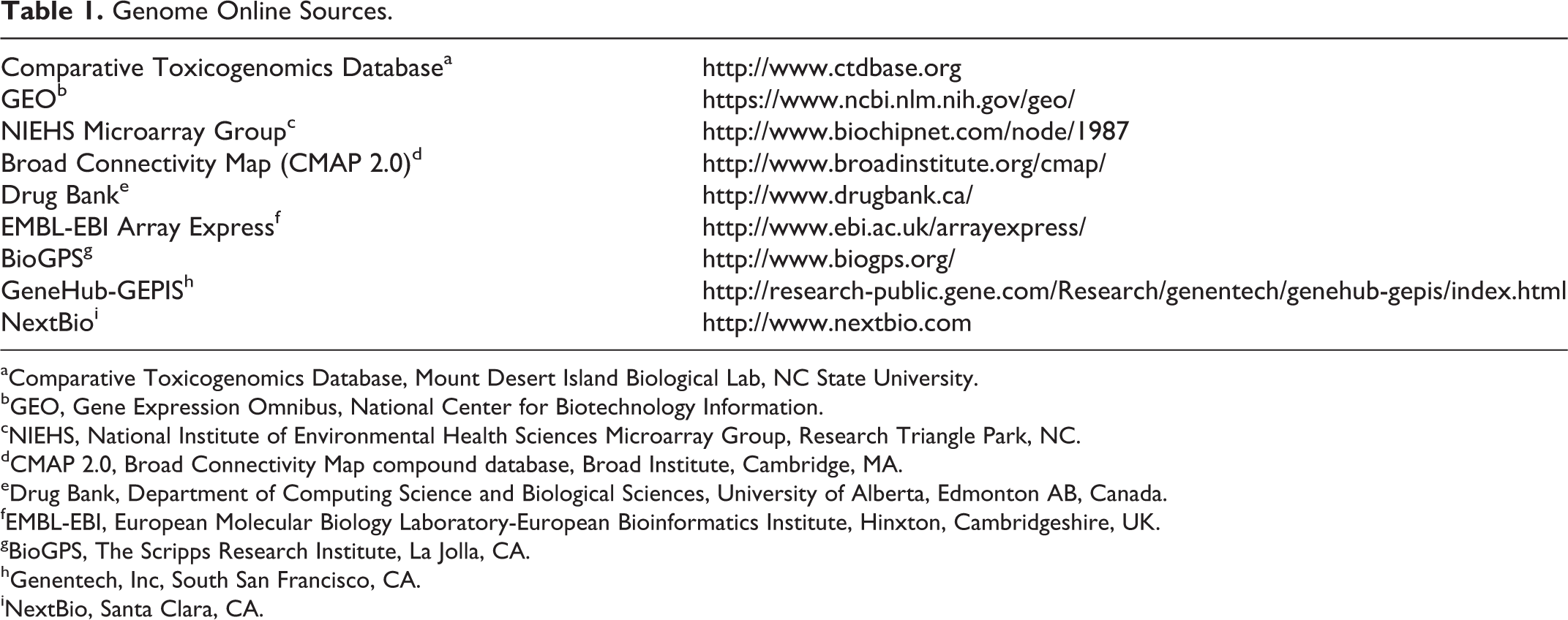
aComparative Toxicogenomics Database, Mount Desert Island Biological Lab, NC State University.
bGEO, Gene Expression Omnibus, National Center for Biotechnology Information.
cNIEHS, National Institute of Environmental Health Sciences Microarray Group, Research Triangle Park, NC.
dCMAP 2.0, Broad Connectivity Map compound database, Broad Institute, Cambridge, MA.
eDrug Bank, Department of Computing Science and Biological Sciences, University of Alberta, Edmonton AB, Canada.
fEMBL-EBI, European Molecular Biology Laboratory-European Bioinformatics Institute, Hinxton, Cambridgeshire, UK.
gBioGPS, The Scripps Research Institute, La Jolla, CA.
hGenentech, Inc, South San Francisco, CA.
iNextBio, Santa Clara, CA.
Quantitative laser scanning cytometry
An efficient and cost-effective method for evaluating and quantifying multiple targets in a large number of tissue samples is accomplished by using routine IHC on TMAs coupled with laser scanning cytometry (LSC). Laser scanning cytometry is a technology that combines the strengths of flow cytometry with tissue architecture retention. 60 Tissue sections are processed by routine IHC methods, and chromatically or fluorescently labeled proteins are simultaneously measured by automated imaging systems (eg, iCyte Automated Imaging Cytometer; CompuCyte Corporation, Westwood, MA). The data acquired for each fluorescent or chromagenic label are assembled into a set of images and quantified based on expression levels for each parameter coupled with cell characteristics such as cell number and size. 41 Krull and Peterson 41 demonstrated the use of this technology for quantification of biomarkers in TMAs derived from Zucker diabetic fatty rats, an animal model of human diabetes, and to quantify tissue co-localization of therapeutic antibody.
Transcriptomics, Metabonomics/Metabolomics, and Proteomics
“Omic” technologies is a term used to define the 3 platforms of transcriptomics, metabonomics/metabolomics, and proteomics. These data-driven platforms have helped transform drug safety from being a subjective and observation-based discipline to one capable of defining the biologic basis of adverse events at the cellular, molecular, and biochemical levels. 89 The combination of “omics” data sets can provide a more thorough and efficient means to obtain insight into mechanisms of disease/toxicity, leading to the identification of novel disease biomarkers. 49 When “omics” are used either alone or better yet in conjunction with other technologies (systems biology approach), they can rapidly fill the gap in the development of new minimally invasive and easily accessible biomarkers in preclinical and clinical studies. 24,48,85 Systems biology is the combination of the “omics” platforms, bioinformatics, and other biologic data, which focuses on interactions within and between the mechanisms that combine to give rise to the function and behavior of a biological system. 34,89 Deciphering complex, highly networked, and pleiotropic pathways that give rise to adverse events aids in the identification of specific biomarkers, which ultimately helps us understand the patient risks and improves the overall safety testing process. 34,89 The leveraging of systems biology technologies may make preclinical safety testing more efficient and accurate, leading to better decisions on patient risk/benefit. 89
Transcriptomics
The use of transcriptional profiling in drug discovery has provided early biomarker and mechanistic toxicology information, but its impact on reducing drug attrition has been limited, in part due to diversity of approaches, differences in sponsors’ commitment to incorporate and evaluate the technology, and available study data. 27 Moreover, although both genome-wide and differential expression of select gene sets has been applied at all stages of drug discovery and development from early in vitro assays through nonclinical pharmacology and toxicology studies and clinical trials, the lack of qualified markers and validated methods, as well as accessible tissues (eg, usually limited to peripheral blood cells) in clinical trials, severely limits its use in studies to support regulatory filings. 76 However, transcriptomic data can still make useful contributions toward characterization of the safety of a compound, particularly when used in combination with other “omics” technologies and with traditional toxicology end points. The InnoMed PredTox project is a European collaborative effort of pharmaceutical companies, service providers, and universities investigating mechanisms of toxicity and identifying safety biomarker candidates by evaluating a combination of results from transcriptomics, metabolomics, and proteomics in relation to routine clinical chemistry and histopathology. 24,85 With this systems biology approach, this collaborative and others have demonstrated that for liver, renal, and other diseases, transcriptomics data are becoming more pertinent in contextualizing the molecular basis of a disease process and aiding in qualifying safety biomarkers. 24,36 Notably, another limitation with the use of transciptomics in the clinic is inaccessibility of these target organs; however, peripheral blood cells are commonly used as a substitute. 5 The limitations on tissue access and the focus on single or groups of transcripts are largely overcome with the use of metabonomics/metabolomics.
Metabonomics/metabolomics
Metabonomics is a biochemical profile reflecting the temporal state of an organism through its endogenous small molecules (sometimes referred to as metabolites). 70 Metabolites in this context are amino acids, carbohydrates, nucleic acids, or fatty acids from biochemical pathways. 47 There can be multiple levels of data interpretation ranging from a spectral result of the overall changes in metabolite concentration without identifying the actual components that are changing (fingerprint) to a quantitative list of metabolites linked to specific pathways. 70,72 The latter mechanistic approach has sometimes been referred to as a subset of metabonomics, termed metabolomics or metabolic profiling. 26 Others have defined metabolomics as the unbiased global survey of the small molecules in a biofluid, tissue, organ, or organism, with the term metabolite profiling used for a more restricted or targeted assessment of these molecules. 8 However, the terms metabonomics, metabolomics, and molecular profiling for the most part have been and will continue to be used interchangeably. Metabonomics/metabolomics offers the researcher a sensitive tool using biofluids that can be collected during the course of the study, allowing temporal changes to be captured. Metabonomics/metabolomics requires analytical methods based on nuclear magnetic resonance (NMR) and MS to generate reproducible spectral patterns used to identify metabolites within the various types of preclinical and clinical samples. 8 In addition to or in combination with NMR and MS, various other technologies are used, including LC-MS, GC-MS, ultraperformance liquid chromatography–electrospray ionization quadrupole time-of-flight mass spectrometry (UPLC–ESIQTOFMS), electrostatic axially harmonic orbital trapping (orbitrap-MS), Fourier transform ion cyclotron resonance MS, and tandem quadrupole MS. 1,21,69,71,78,80,92 Despite continued technological advances in chromatographic separations and detection platforms, no single platform can capture all metabolites, and a combination of the approaches mentioned above is necessary to increase coverage of the metabolome. 51,52 For a review of the extraction of analytes from biofluids, data capture, alignment and normalization, and multivariate data analysis, see Montanez et al. 51 In a recent forward to a special focus issue on metabolomics in the journal Bioanalysis, Nicholls 54 provided a list of databases that encompasses information on the biological involvement of components of the metabolome molecules, sources of analytical data for identification of components, and repositories for the sharing of metabolomics experimental data (Table 2).
Metabolome Online Sources.
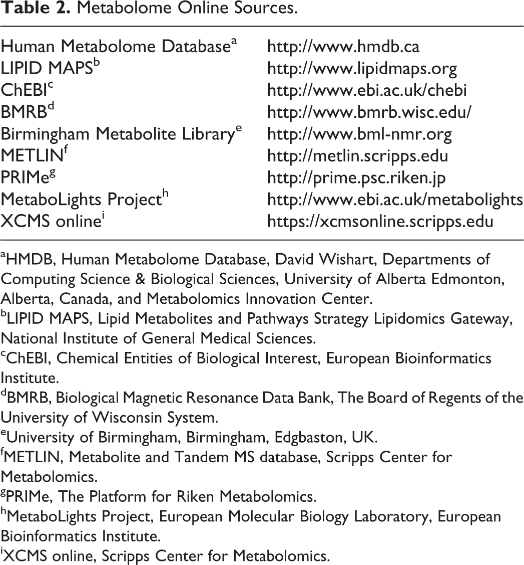
aHMDB, Human Metabolome Database, David Wishart, Departments of Computing Science & Biological Sciences, University of Alberta Edmonton, Alberta, Canada, and Metabolomics Innovation Center.
bLIPID MAPS, Lipid Metabolites and Pathways Strategy Lipidomics Gateway, National Institute of General Medical Sciences.
cChEBI, Chemical Entities of Biological Interest, European Bioinformatics Institute.
dBMRB, Biological Magnetic Resonance Data Bank, The Board of Regents of the University of Wisconsin System.
eUniversity of Birmingham, Birmingham, Edgbaston, UK.
fMETLIN, Metabolite and Tandem MS database, Scripps Center for Metabolomics.
gPRIMe, The Platform for Riken Metabolomics.
hMetaboLights Project, European Molecular Biology Laboratory, European Bioinformatics Institute.
iXCMS online, Scripps Center for Metabolomics.
Years of preclinical work by researchers at the chemical company, BASF, culminated in the MetaMap Tox, a rat plasma metabolomics database containing the metabolome in response to >500 reference compounds assessed in 28-day repeated dose toxicity studies. 39,93 Recent publications by this group showed that when compounds such as phenytoin (diphenylhydantoin), fibrates, or statins were administered to rats in 28-day studies and metabolome analysis was performed on days 7, 14, and 28 and compared with the gold standards of clinical and anatomic pathology, a more comprehensive picture of compound effects was obtained from the metabolome analysis. 38,83 This database was recently evaluated by a consortium consisting of 14 leading biopharmaceutical companies as part of the Technology Evaluation Consortium, managed by Cambridge Healthtech Associates: Drug Safety Executive Council (www.drugsafetycouncil.org). This consortium concluded that the MetaMap Tox database is applicable as a companion to the standard 28-day GLP toxicity studies to aid in the mechanistic characterization of drug effects, but its utility for identification of translatable safety biomarkers is yet to be determined. MetaMap Tox offers targeted and nontargeted metabolomics to biopharmaceutical companies and is now commercially available (Metanomics Health GmbH: http://www.metanomics-health.de).
In general, regardless of the precise definition, metabonomics/metabolomics/metabolic profiling is a relative newcomer to the “omics” armamentarium in drug discovery and development, but the rapid progress and refinement and availability of online databases will ensure its future as a key discipline in preclinical and clinical biomarker development.
Proteomics
MS-based proteomics has been used to assess qualitative and quantitative differences in protein profiles of diseased vs normal tissues with the purpose of discovering novel disease-specific protein biomarkers. 77 Proteomics has been used to study many potential biomarkers of toxicity such as urinary cystatin C, TFF3, β2-microglobulin, albumin, KIM-1, and total protein and albumin. 20,31,58,90 Two-dimensional acrylamide gel electrophoresis and matrix-assisted laser desorption ionization–time-of-flight mass spectrometry (MALDI-TOF MS) have been widely used for profiling plasma proteins. 61,88 Recent progress in quantitative proteomics, such as fluorescence 2-dimensional gel electrophoresis, is being used to discover plasma proteins as biomarkers. 11,88 For a review of current strategies of functional, chemical, and clinical proteomics, see Savino et al. 77 For an overview of the regulatory framework that is typically required for the translation of proteomic biomarkers from discovery to clinical diagnostics, which is regulated by the Office of In Vitro Diagnostics and Radiological Health (OIR) at the FDA, see Li et al. 45 Specific OIR guidance documents are available (http://www.fda.gov/MedicalDevices/DeviceRegulationandGuidance/GuidanceDoc-uments/ucm070274.htm).
Conclusion
The identification, application, and qualification of safety biomarkers that are minimally invasive and are specific makers of early clinical injury are becoming increasingly critical to successful drug discovery and development, but this can be a long and difficult process. As summarized in this review, today’s investigators are armed with numerous routine and novel assay formats and modalities and an ever expanding access to detailed databases that aid this process. However, the complexity of such processes becomes exponential as the ultimate goal is to translate selected safety biomarkers to other species and ultimately to humans as the biology of the process, the biomarker, and the PK/PD properties of the drug might be different. Thus, the identification of the biomarker for use in toxicology studies ultimately relies on the knowledge of the biology, physiology, and pathophysiology of the biomarker in the context of the species and toxicity evaluated.
Footnotes
Acknowledgements
We thank Dr Dana Walker for her scientific review of this manuscript.
Declaration of Conflicting Interests
The author(s) declared no potential conflicts of interest with respect to the research, authorship, and/or publication of this article.
Funding
The author(s) received no financial support for the research, authorship, and/or publication of this article.