
Abstract
Malakoplakia is a rare, granulomatous, inflammatory disease that mimics malignant tumors and can affect any organ. Herein is described a case of malakoplakia in a 10-month-old slaughter pig. Diffuse, pleomorphic, round cell infiltrates, mainly histiocytes, with a tumor-like growth pattern at gross examination, infiltrated the stomach, pancreas, omentum, and mesenteric lymph nodes. The histiocytes and multinucleated giant cells had concentric, target-like inclusions known as Michaelis-Gutmann bodies. Microorganisms were not detected by the periodic acid–Schiff reaction, Ziehl-Neelsen, Gram, and Warthin-Starry staining or by electron microscopic and bacteriologic investigations. Porcine circovirus type 2 and porcine reproductive and respiratory syndrome viruses were not detected by immunohistochemistry in the sections examined.
Malakoplakia is a rare, chronic, granulomatous disease usually reported in immune-compromised or debilitated human patients. It is believed to be the result of a functional impairment of mononuclear phagocytes and other immune-regulatory effector cells. 8 It is characterized by aggregates of Hansemann cells; histiocytes containing owl-eye shaped, periodic acid–Schiff (PAS)–positive inclusions; and calcospherites known as Michaelis-Gutmann bodies. 9
Malakoplakia has been experimentally induced in rats and pigs. 5,6 In rats, the disease was induced by injecting an endotoxin-antigen complex of Escherichia coli 075 strain into the kidney. 5 Spontaneous animal cases are rare. Two cases have been reported in pigs 3,10 and, more recently, 1 in a cat. 1 Malakoplakia of the gastrointestinal tract can be concurrent with neoplastic diseases or bacterial infection. It may be clinically silent or cause clinical signs such as diarrhea, abdominal pain, and/or obstruction. 7
We describe a case of malakoplakia in the digestive tract of a 10-month-old slaughter pig without clinical signs. During postmortem examination, white, irregular, soft nodules (3–5 cm) protruded into the gastric serosa and peritoneum near the pancreas. The pancreas and mesenteric lymph nodes were enlarged. The masses were yellow and homogeneous. Portions of each organ were plated onto blood and Gassner agars and aerobically incubated at 37°C for 48 hours. Portions of each organ were fixed in 10% buffered formalin, paraffin embedded, and cut into sections 4 μm thick. Sections were stained with hematoxylin and eosin (HE), PAS with and without diastase digestion, Ziehl-Neelsen, Gram, Warthin-Starry, toluidine blue, Perl’s iron, and von Kossa stains; immunostained with mAb 1A5 anti–porcine circovirus 2 (Istituto Zooprofilattico Sperimentale Lombardia Emilia Romagna [IZSLER], Brescia, Italy) at a dilution of 1:1000; mAbs 1D8 and 3B1 anti–porcine reproductive and respiratory syndrome virus (PRRS; IZSLER), diluted 1:500; and polyclonal antibody anti–Mycobacterium bovis (BCG; DakoCytomation, Glostrup, Denmark), diluted 1:1000. The reaction was developed using a streptavidin-biotin-peroxidase kit (Vectastain Elite Kit, ABC system; Vector Laboratories, Burlingame, CA). Pig lymph nodes positive and negative for porcine circovirus type 2 (PCV2), PRRS, and Mycobacterium tuberculosis complex by polymerase chain reaction and immunohistochemistry were used as controls.
Unstained sections were examined with polarized and ultraviolet light, according to the method described by Gill et al 3 to verify refringence and autofluorescence.
Portions of formalin-fixed tissues, cut into 1-mm3 cubes, were rinsed overnight in a 0.1% phosphate buffer and prepared for transmission electron microscopy using standard protocol (glutaraldehyde fixation, osmium tetroxide postfixation, epoxy resin embedding). Ultra-thin sections stained with uranyl acetate and lead citrate were examined using a Zeiss (Oberkochen, Germany) EM 109 T electron microscope operating at 50 kV.
A diffuse, pleomorphic, round cell infiltrate was present in the pancreas, omentum, and transmurally in the stomach (Fig. 1). Lymph nodes were hyperplastic, and the infiltrate was mainly distributed in subscapular sinuses, in medullary sinuses, and within trabeculae.
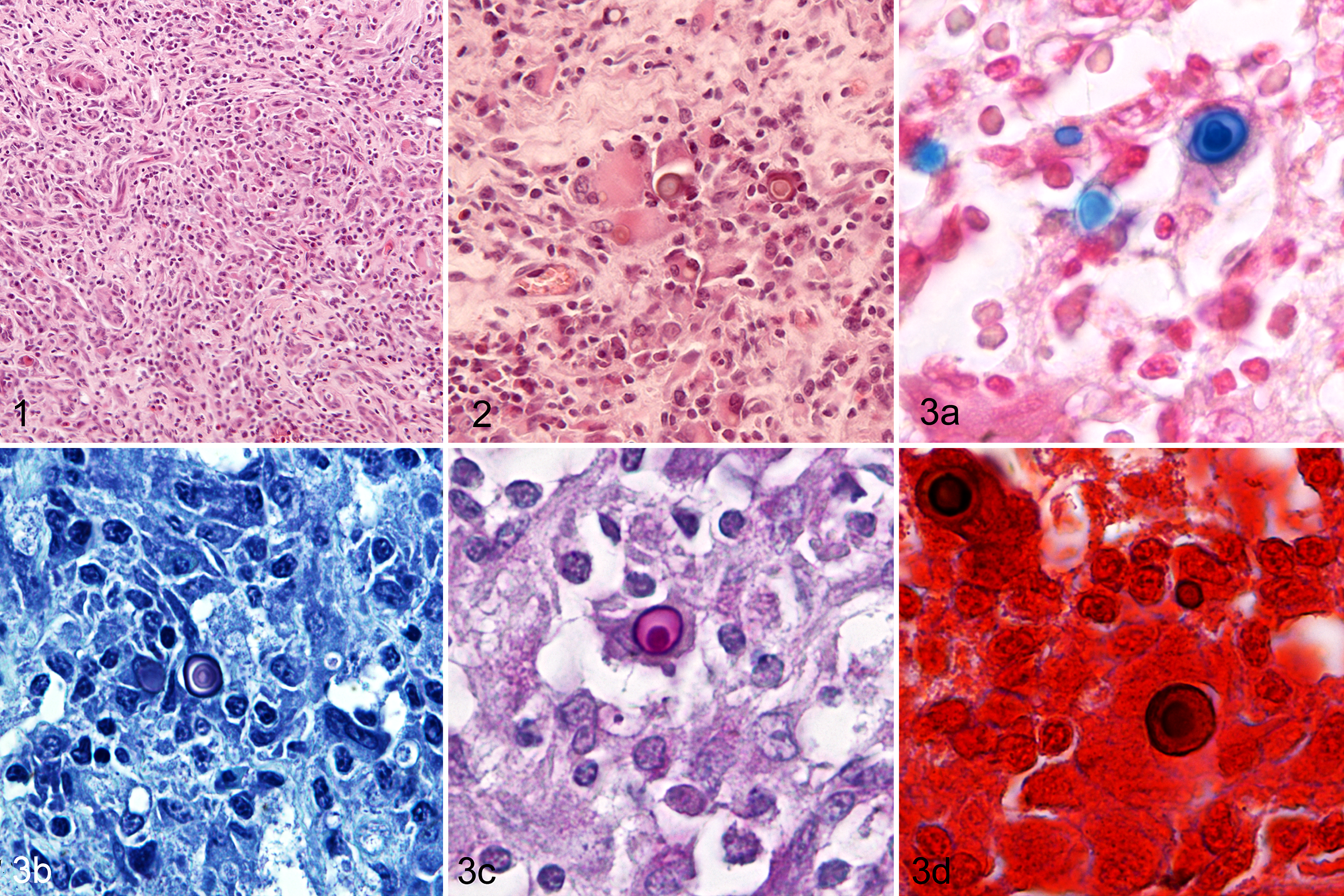
Pancreas; swine. Pleomorphic round cell infiltrate with tumor-like pattern. Hematoxylin and eosin (HE).
The infiltrating cells were organized in a typical granulomatous pattern with multiple aggregates of histiocytes and multinucleated giant cells intermingled with plasma cells, lymphocytes, and rare granulocytes. Histiocytes were large, polygonal, and noncohesive with moderate to abundant, granular, eosinophilic cytoplasm and oval to irregular nuclei with finely stippled chromatin. Round, refractile, basophilic, cytoplasmic inclusions, ranging from 2 to 20 μm and frequently with a concentric, target-like appearance, were observed in many histiocytes and multinucleated giant cells (Fig. 2). Finely granular PAS-positive granules were present in the cytoplasm of histiocytes. Microorganisms were not detected by the PAS reaction, Ziehl-Neelsen, Gram, or Warthin-Starry staining or by bacteriologic investigation.
By electron microscopy, most cells were identified as histiocytes with an uneven nuclear contour and moderate to abundant amounts of cytoplasm. The cells had numerous mitochondria, scattered profiles of rough and smooth endoplasmic reticulum, and lysosomes. The spherical inclusions varied in appearance. Most had irregularly round bodies composed of aggregates of needle-like crystals with a hydroxyapatite morphology set in a dense matrical core. Less frequently, the inclusions were large, annular, calcified structures with a central vacuole. Bacteria were not present in histiocytes containing Michaelis-Gutmann bodies (Fig. 4).
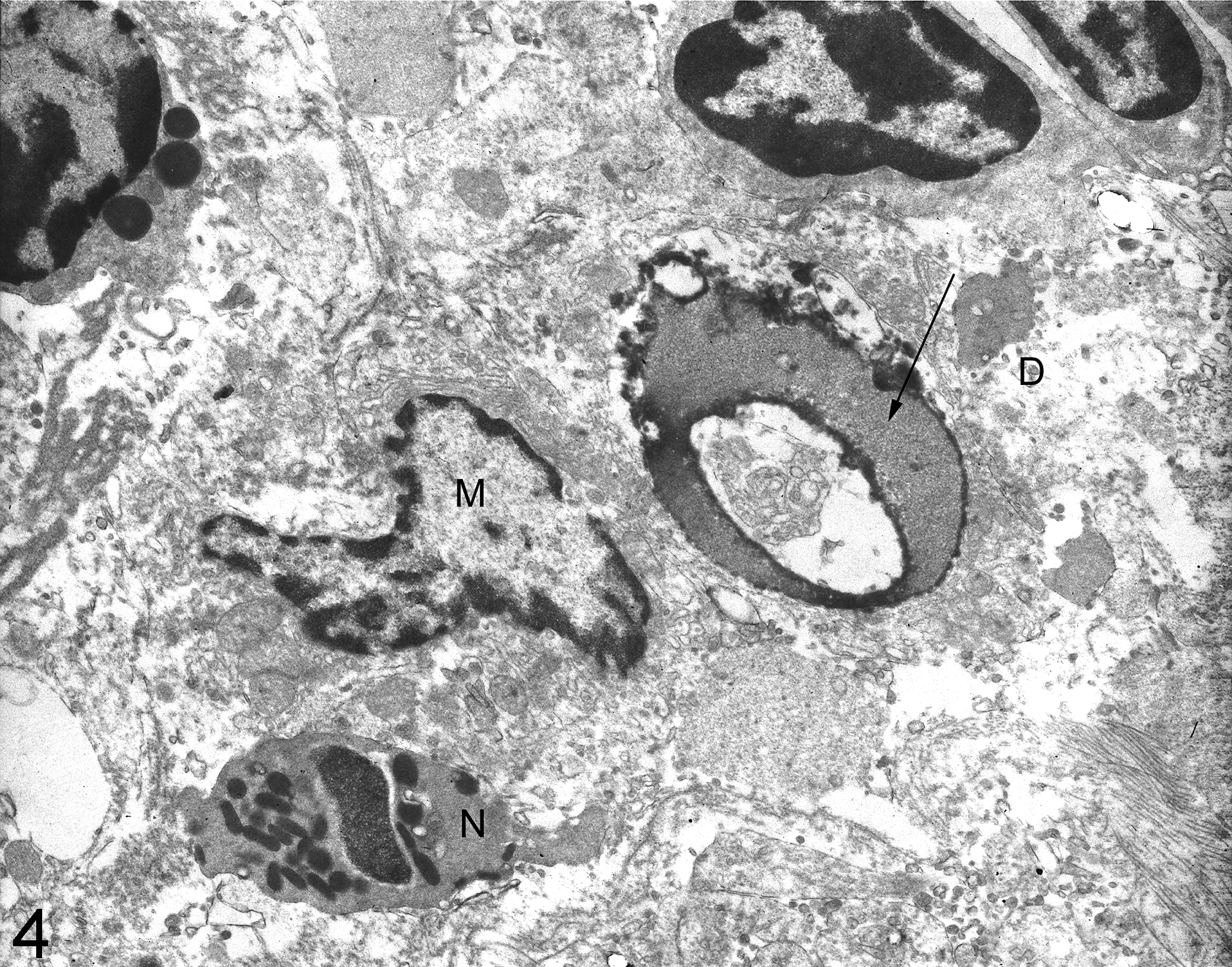
Lymph node, swine. Macrophage (M), neutrophil granulocyte (N), and cellular necrotic debris (D) in which a poorly calcified large phagolysosome (arrow) consistent with a Michaelis-Gutmann body is present. Transmission electron microscopy.
These results are consistent with a diagnosis of malakoplakia of the gastrointestinal tract. Malakoplakia can affect any organs or soft tissue, but the urinary and gastrointestinal tracts of humans and animals are affected most frequently. 1,10 Malakoplakia is an uncommon form of chronic inflammation related probably to inadequate bactericidal activity of monocyte-macrophage lineage cells due to defective phagolysosomal function. Lysosomes are unable to completely digest bacteria as a result of abnormal assembly of microtubules. 2 Glycolipid remnants accumulate in phagosomes and act as a nidus for the subsequent deposition of calcium and iron in the typical concentric laminations known as Michaelis-Gutmann bodies. 8
A wide variety of bacteria (E. coli, Staphylococcus aureus, Streptococcus spp, Mycobacterium spp, Rhodococcus equi) have been detected in malakoplakia-affected patients as a consequence of chronic infection, but malakoplakia has also been associated with a variety of tumors, both with or without a history of infection. 8
In pigs, malakoplakia has been associated with bacterial infection 3 and reproduced by subcutaneous inoculation of a R. equi strain of intermediate virulence. 6 In the case described herein, no bacteria were observed. Moreover, investigations for bacteria were negative and no microorganisms were detected in the cytoplasm of histiocytes containing Michaelis-Gutmann bodies. Taniyama et al 10 described a systemic malakoplakia in swine characterized by multiple disseminated lesions in several organs of the thoracic and abdominal cavities. The authors speculated that the presence of the granuloma-like lesions in perivascular areas is consistent with a hematogenous spread of the disease. In the current case, the stomach, pancreas, and peritoneum were severely infiltrated. Thus, a contiguous spread of the disease due to the proximity of the stomach, pancreas, omentum, and mesenteric lymph nodes might be more likely. The inciting event for the type of gastric malakoplakia we studied may be a chronic ulcer. This hypothesis suggests a correlation between gastric ulcers and gastric malakoplakia. However, the high frequency of gastric ulcers and the apparent rarity of malakoplakia in slaughtered swine do not support a relationship between these 2 conditions. In human medicine, gastric malakoplakia has been associated with H. pylori infection or, in its absence, to other bacteria. 4 Viral infections have never been associated with malakoplakia. The pathogenesis of malakoplakia is enigmatic, but our results suggest that the PCV2 and PRRS viruses and H. pylori are not causative. In 1981, McClure 8 suggested that the origin of Michaelis-Gutmann bodies’ core may be derived from endogenous cell membrane breakdown products. So far, the etiology of malakoplakia or the mechanism that leads to the formation of Michaelis-Gutmann bodies has not been determined.
From 1900 until the present, only 3 cases of malakoplakia have appeared in the veterinary literature. In veterinary medicine, slaughterhouses are an ideal venue for the monitoring of livestock disease. The use of histopathology is essential to achieve a correct diagnosis among lesions mimicking neoplasms. Awareness of malakoplakia and its characteristic histopathological features may increase the number of cases reported.
Footnotes
Declaration of Conflicting Interests
The author(s) declared no potential conflicts of interest with respect to the research, authorship, and/or publication of this article.
Funding
The author(s) received no financial support for the research, authorship, and/or publication of this article.