
Abstract
A die-off of passerine birds, mostly Eurasian siskins (Carduelis spinus), occurred in multiple areas of Switzerland between February and March 2010. Several of the dead birds were submitted for full necropsy. Bacteriological examination was carried out on multiple tissues of each bird. At gross examination, common findings were light-tan nodules, 1 to 4 mm in diameter, scattered through the esophagus/crop. Histologically, a necroulcerative transmural esophagitis/ingluvitis was observed. Bacterial cultures yielded Salmonella enterica subsp. enterica serovar Typhimurium. At the same time, 2 pet clinics reported an unusual increase of domestic cats presented with fever, anorexia, occasionally dolent abdomen, and history of presumed consumption of passerine birds. Analysis of rectal swabs revealed the presence of S. Typhimurium in all tested cats. PFGE (pulsed field electrophoresis) analysis was performed to characterize and compare the bacterial isolates, and it revealed an indistinguishable pattern between all the avian and all but 1 of the feline isolates. Cloacal swabs collected from clinically healthy migrating Eurasian siskins (during autumn 2010) did not yield S. Typhimurium. The histological and bacteriological findings were consistent with a systemic infection caused by S. Typhimurium. Isolation of the same serovar from the dead birds and ill cats, along with the overlapping results of the PFGE analysis for all the animal species, confirmed a spillover from birds to cats through predation. The sudden increase of the number of siskins over the Swiss territory and their persistency during the whole winter of 2009–2010 is considered the most likely predisposing factor for the onset of the epidemic.
Salmonella enterica subsp. enterica serovar Typhimurium is known to cause an infection in passerine birds, with pathognomonic lesions characteristically localized in the esophagus and crop. Finches (Fringillidae) and sparrows (Passeridae) seem to be particularly susceptible to Salmonella sp infection. 9,39 Infected birds may or may not develop clinical disease, and clinically healthy carriers are suspected to play a role in the transmission of the pathogen, representing an important source of infection for other birds. A healthy carrier state for S. Typhimurium has been shown in free-ranging sparrows (Passer domesticus), 36 as well as in few gull species (Laridae). 25,37 Additionally, S. Typhimurium has been detected both in sick and clinically healthy Norwegian passerine birds, including Eurasian siskins (Carduelis spinus). 27
Eurasian siskins are small finches that live in coniferous woodlands, where they feed on seeds, especially of spruce (Picea abies), alder (Alnus sp), and birch (Betula sp). Seasonal drop of food availability in winter contributes to frequent crowding of siskins at feeding places in private gardens. 31 This crowding is generally associated with the deposition of large amounts of feces in and around the feeding sites. When infected birds feed at these stations, significant bacterial load associated with fecal material may contribute to turn feeding stations into bacterial fomites with increased risk of Salmonella exposure for other birds. 15
Infected wild passerine birds represent also a potentially significant source of S. Typhimurium for nonavian susceptible species, including humans and cats, 24,29,34 and their occurrence in human settlements is also a matter of public health concern. Infection of humans has been suggested to occur either indirectly via infected cats 33 or directly by contact with wild birds and their droppings, respectively. 14
We report for the first time the complete picture of a salmonellosis outbreak in free-ranging passerine birds, which is apparently the largest ever recorded in Switzerland. This outbreak mostly affected Eurasian siskins, with the simultaneous occurrence of cases in domestic cats. Here (1) we describe in detail the associated pathology in birds with a proposed classification of the lesions; (2) we demonstrate the presence of the bacteria within the tissue lesions; (3) we provide epidemiological and molecular evidences via pulsed-field gel electrophoresis (PFGE) analyses of salmonellosis spillover from birds to cats; (4) we investigate the epidemiological context of the outbreak, considering different potential promoting factors, including environmental conditions, host–population frequency, and the role of Eurasian siskins migrating through Switzerland as potential carriers of S. Typhimurium.
Materials and Methods
Dead Passerine Birds Submitted for Pathological Examination
From February to March 2010, many passerine birds were observed in various areas of Switzerland, presenting apathic behavior and ruffled plumage or found dead in proximity of feeding places in private gardens (Fig. 1). These observations were reported to the Swiss Ornithological Institute (Schweizerische Vogelwarte Sempach), and carcasses of 16 birds were subsequently sent to the Centre for Fish and Wildlife Health (FIWI) for diagnostic workup. Postmortem examination was performed on 14 Eurasian siskins, 1 European goldfinch (C. carduelis), and 1 European greenfinch (C. chloris). Sex, weight, body condition, and gross pathological findings were recorded. Body condition was assessed according to the volume of the pectoral muscle and the amount of claviculocoracoid and substernal fat. 1 Representative specimens of the lung, heart, liver, esophagus/crop, brain, intestine, kidneys, spleen, and any additional tissue showing gross lesions were collected and fixed in 10% buffered formalin. Fixed specimens were processed routinely, embedded in paraffin, sectioned at 5 µm, and stained with hematoxylin and eosin for histological examination. 8 Special stains, including Gram (Brown and Brenn’s), Grocott, Ziehl-Neelsen, and periodic acid-Schiff, were performed as appropriate. Lesions within the crop and esophagus were classified in early, intermediate and late on the basis of (1) presence, distribution, and extent of the inflammatory infiltrate and (2) presence and extent of necrosis.
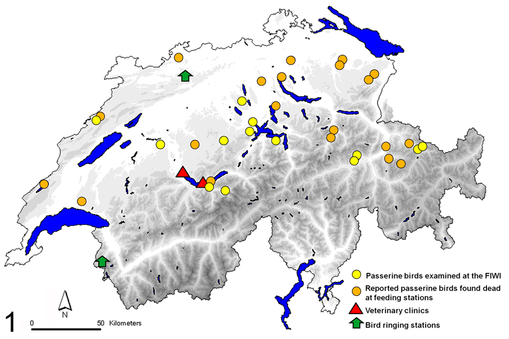
Map of Switzerland with main lakes (blue irregular surfaces) and relief (shades of grey: the darker, the higher the altitude) representing the distribution of the passerine birds found dead during the salmonellosis outbreak of 2010. Yellow circles = dead passerine birds examined at the FIWI. Orange circles = reported dead passerine birds to the Swiss Ornithological Institute. Red triangles = small animal clinics where the sick cats were treated. Green arrows = bird-ringing stations.
Diseased Cats Presented for Clinical Examination
In March 2010, 2 small animal clinics in Interlaken and Thun (Switzerland; Fig. 1) reported an increased number of domestic outdoor cats presenting fever, anorexia, occasionally dolent abdomen, vomiting, and history of hunting wild birds (presence of yellow feathers within the mouth). No evidence of diarrhea was reported by the cats’ owners. Eight cats were included in the study to assess if S. Typhimurium infection might have been the cause of the observed clinical signs and to establish a possible causal relationship with the predated birds. Rectal swabs (Transport swabs, Starstedt, Sevelen, Switzerland) were collected and kept in transport medium at room temperature until processing. An antibiogram was performed on 3 of the Salmonella isolates.
Environmental Conditions
Environmental conditions concerning the winter of 2009–2010 were obtained by the seasonal climate bulletin of the Swiss meteorological website (http://www.meteoschweiz.admin.ch/web/de/klima/klima_heute/saisonflash.html).
Siskin Population Size
Information concerning the siskin frequency (as a measure of the local–migrating siskin population size) in Switzerland during the winter of 2009–2010 and previous years was obtained from Müller and Volet 20 based on observations of birds by collaborators of the Swiss Ornithological Institute.
Clinically Healthy Eurasian Siskins Tested for Salmonella sp
Cloacal swabs (Transwabs, WW 172/P, Novoglas, Bern, Switzerland) were collected from 136 clinically healthy Eurasian siskins (71 males [45 adults, 2 subadults, 24 juveniles] and 65 females [39 adults, 26 juveniles]) caught at 2 bird-ringing stations in October 2010. These stations are both placed at locations suitable for capturing migrating birds and are located at the Col de Bretolet and Ulmet (Fig. 1), in the southwest and north of Switzerland, respectively. The swabs were kept in transport medium (Amies-Medium) at room temperature until examination.
Bacteriological Examination
General bacteriological examination of tissue samples from bird carcasses
Bacteriological analyses, including that specific for Salmonella sp, were performed on the intestine of 15, the esophagus/crop of 10, the lung of 7, and/or the subcutis of 1 bird, respectively, following accredited standard testing procedures performed at the Institute of Veterinary Bacteriology at the University of Bern. 28 Presumptive Salmonella colonies were confirmed by appropriate biochemical tests and serotyped by agglutination according to the White-Kauffmann-Le Minor scheme. 11
Bacteriological analyses of rectal swabs from diseased cats and cloacal swabs from healthy siskins
Rectal swabs from cats were inoculated in 10-ml of tetrathionate broth and incubated at 37°C for 24 hours. The tetrathionate broth was then subcultured onto brilliant green agar (Oxoid, Hampshire, England) and brilliant salmonella agar (Oxoid). Following 24 hours incubation at 37°C, presumptive Salmonella colonies were confirmed as described above. 11
Cloacal swabs collected from living birds were inoculated in 5 ml of buffered peptone water (Oxoid) and incubated at 37°C for 24 hours. Subsequently, 100 µl of the culture were transferred to 10 ml of tetrathionate broth (Oxoid), reincubated (37°C, 24 hours), and further processed as described for rectal swabs from the cats.
Antibiotic susceptibility testing for S. Typhimurium was carried out using ATB VET and ATB G-5 strips (Mini API BioMérieux; Geneva, Switzerland).
Genotypic Characterization of Bacterial Isolates
All isolates included in the present study were analyzed by pulsed-field gel electrophoresis (PFGE), which was performed according to the CDC PulseNet protocol (http://www.cdc.gov/pulsenet/protocols.htm) with minor modifications. Briefly, bacterial strains were grown on blood agar at 37°C overnight. Colonies from blood agar were resuspended in cell suspension buffer (OD600 = 1). The bacterial cell suspension was mixed with 400 μl of 1.4% BIO-RAD Agarose (BIO-RAD, Munich, Germany), and cells were lysed by proteinase K treatment overnight. After lysis, plugs were washed twice for 15 minutes in ultrapure water and 4 times for an hour in Tris-EDTA buffer. After washing with Tris-EDTA buffer, DNA agarose plugs were incubated overnight in the presence of XbaI (Roche, Mannheim, Germany) according to the manufacturer’s instructions. Restricted DNA in plug slices was separated in a 1% SeaKem Gold (BioConcept, Allschwil, Switzerland) agarose gel at 6 V/cm in 0.5 X Tris-Borate-EDTA buffer cooled to 14°C in a CHEF-DR III system (BIO-RAD, Munich, Germany). Pulse times were ramped from 5 to 50 seconds for 20 hours at an angle of 120°. Gels were stained with ethidium bromide and visualized under ultraviolet light transillumination using a Gel Doc system (BIO-RAD, Munich, Germany) and analyzed with BioNumerics software (Applied Maths, Sint-Martens-Latem, Belgium). Salmonella Braenderup strain H9812 (ATCC BAA 664) was used as a size standard.
Statistical Analysis
Sample size for healthy birds was calculated with Win Episcope 2.0. 6 A sample size of 124 birds was obtained for a population of 10 000 birds (estimated population of siskins in Switzerland during the breeding season 30 ), based on an expected prevalence of 8.8% 27 with a confidence interval (CI) of 95%. 35 CIs of prevalences in sampled healthy birds were calculated with the NCSS 2007 software (Hintze J. 2007; NCSS, Kaysville, Utah).
Results
Pathological and Bacteriological Findings in Dead Passerine Birds
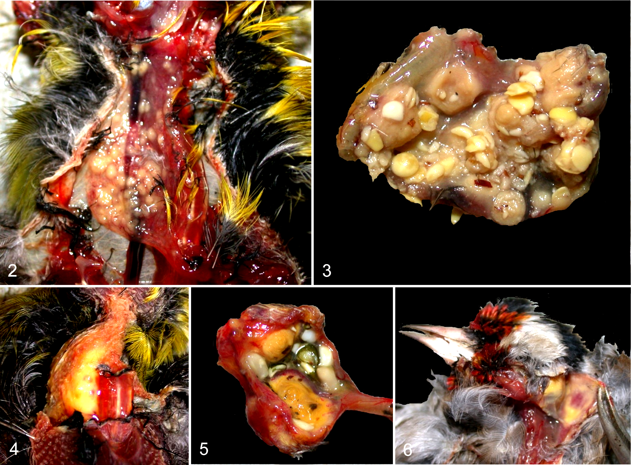
Ten of the 16 examined birds (63%) were in poor body condition, with markedly atrophic pectoral muscles and no visible fat, while the other 6 birds were in moderate body condition. At gross examination, the most common finding (10 of 14 Eurasian siskins, 71%) was the presence of multifocal to coalescent light tan nodules (1–4 mm in diameter) scattered throughout the esophagus/crop (Figs. 2, 3). A diffuse light tan discoloration and thickening of the crop wall were observed in 2 of 14 Eurasian siskins (Fig. 4). In the European greenfinch, nodules were fewer but larger than those found in the Eurasian siskins and ranged from 0.5 to 1 cm in diameter, often bulging into the lumen of the crop (Fig. 5). The European goldfinch presented nodules in the subcutis of the neck (4–5 mm in diameter) and in the coelomic cavity (3–4 mm in diameter; Fig. 6).
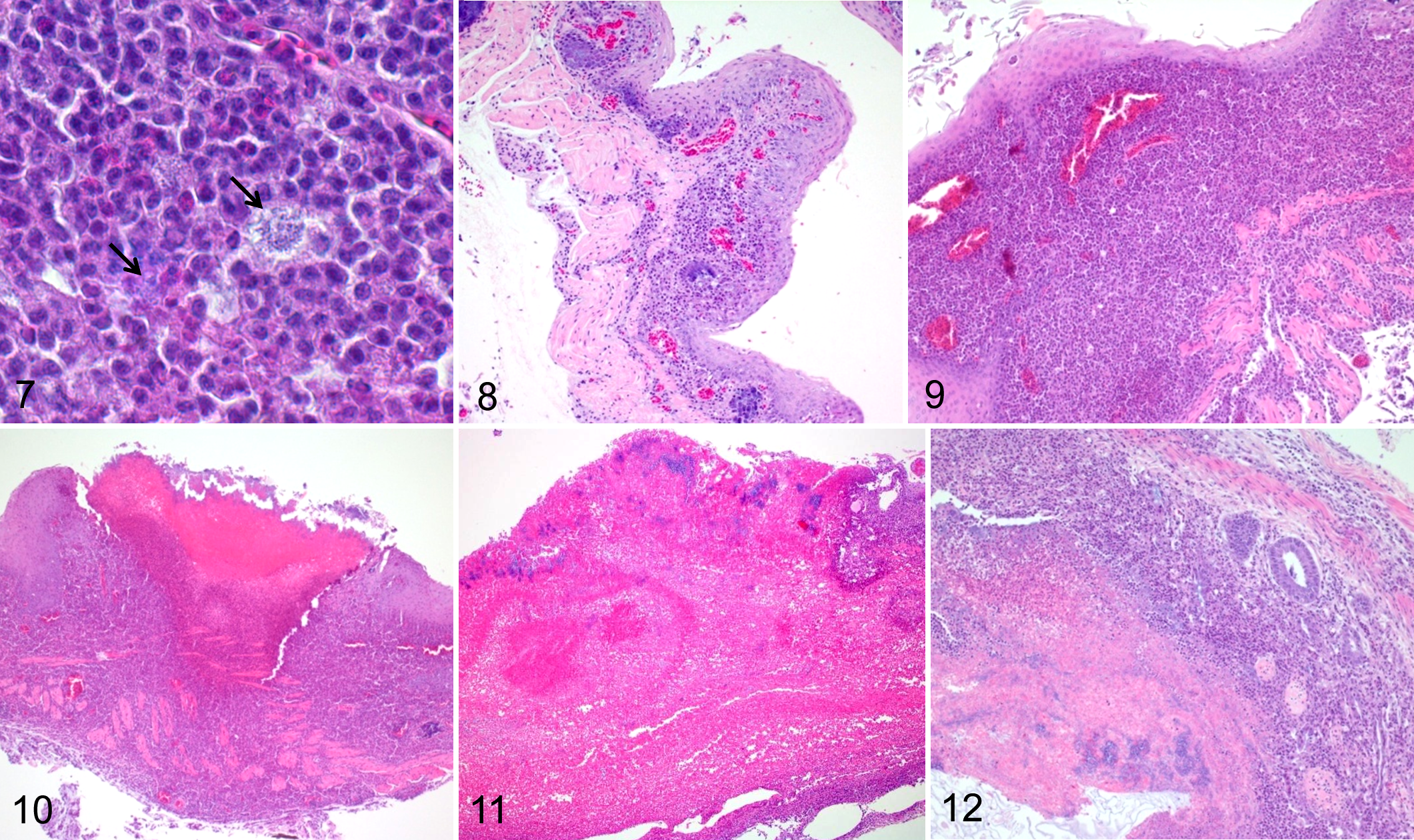
Histological examination of the esophagus/crop of siskins and greenfinch with macroscopical lesions revealed a severe transmural necrotizing and ulcerative esophagitis/ingluvitis (Figs. 7 –11). A severe necrotizing cellulitis was observed in the neck of the goldfinch. Macroscopic nodules observed in all species consisted in variably extensive necrotic areas, often ulcerated, characterized by collections of hypereosinophilic cellular and basophilic nuclear debris, respectively. An infiltration of viable and degenerating histiocytes and heterophils, with presence of intra- and extracellular bacterial rods, was observed surrounding the necrotic areas and expanding the esophageal wall (Fig. 7). In early lesions (8%), the submucosa and to some extent the tunica muscularis/serosa were mildly to moderately expanded by inflammatory cells, with occasional minute to small areas of necrosis affecting the basal layers of the mucosa (Fig. 8). In intermediate lesions (38%), the inflammation was more pronounced, and the submucosa, muscularis, and serosa were more diffusely infiltrated by large clusters of degenerating and necrotic inflammatory cells. The mucosa was moderately to markedly elevated, with multifocal areas of ulceration and more extensive necrosis (Fig. 9), occasionally forming buttonlike elements (Fig. 10). In late lesions (54%), diffuse areas of necrosis associated with massive inflammatory infiltrate extended across the esophagus/crop wall (transmural necrosis; Fig. 11). Necrotizing enteritis was also observed in 5 birds with esophageal lesions. The necrosis was mostly affecting the mucosa and then extending to the deeper layers of the organ (Fig. 12). The nature of the enteric inflammation (viable/degenerating histiocytes and heterophils) was similar to that observed within the esophagus/crop. Additional lesions observed in infected birds included necrotizing hepatitis (9 birds), interstitial pneumonia (4 birds), necrotizing splenitis (2 birds), necrotizing coelomitis (2 birds), epicarditis/myocarditis (2 birds), necrotizing nephritis (2 birds), necrotizing encephalitis (1 bird), and proventriculitis (1 bird; Table 1).
Histological and Bacteriological Findings in the 14 Passerine Birds Diagnosed With Salmonellosisa
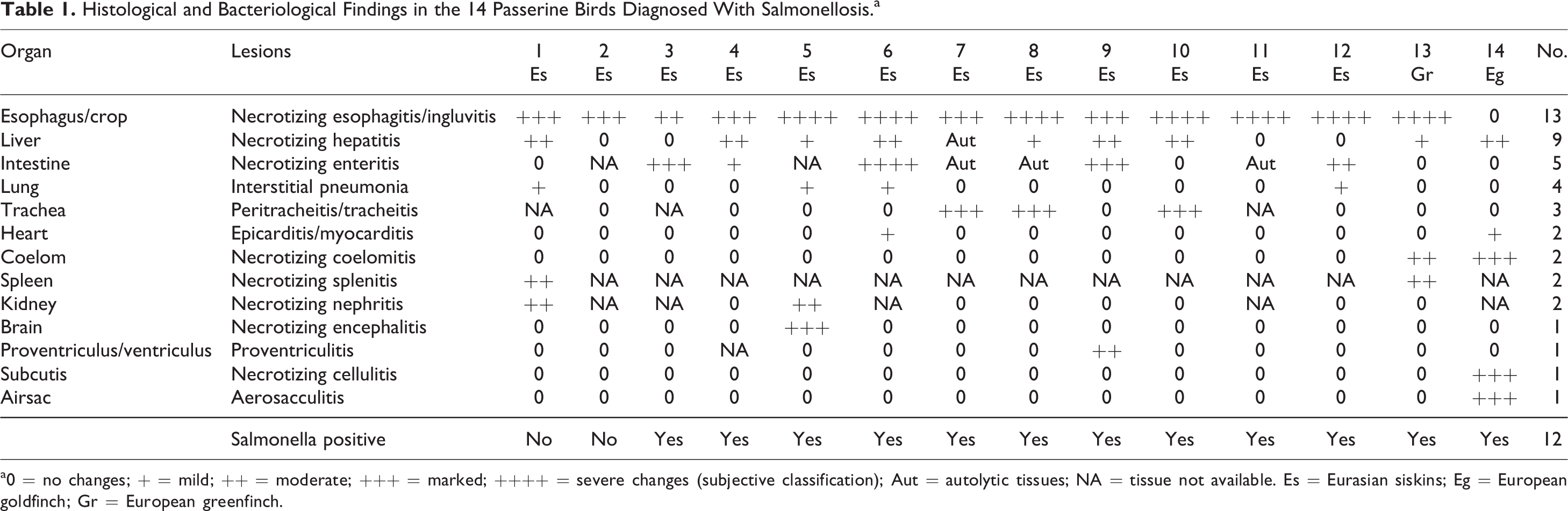
a0 = no changes; + = mild; ++ = moderate; +++ = marked; ++++ = severe changes (subjective classification); Aut = autolytic tissues; NA = tissue not available. Es = Eurasian siskins; Eg = European goldfinch; Gr = European greenfinch.
A pure culture of Salmonella enterica subsp. enterica serovar Typhimurium was obtained either from the intestine (11 birds), crop (10 birds), lung (3 birds), and/or subcutis (1 bird) of 12 of 14 (86%) birds presenting nodular lesions (Table 1). Detailed serotyping results can be summarized in the antigenic formula S. enterica subsp. enterica 4,12: i:1,2. Because of known antigenic variations within serovar Typhimurium, agglutinations with antisera against O1 and O5 were also carried out and were negative.
Of the 2 birds culturally negative for Salmonella sp and lacking nodular lesions (both Eurasian siskins), 1 had a severe necrotizing meningoencephalitis caused by Toxoplasma gondii, while severe fungal granulomatous coelomitis and enteritis were observed in the other bird.
Bacteriological Findings in Diseased Cats
Bacterial cultures of the rectal swabs from all tested cats yielded S. Typhimurium (for detailed serotyping results see corresponding section on birds).
The 3 selected isolates of S. Typhimurium were found to be sensitive to the following antimicrobial agents: β-lactams (amoxicillin, amoxicillin/clavulanic acid, ticarcillin, imipenem, cefalotin, cefoxitin, cefotaxime, ceftazidime), tetracyclines (tetracycline), aminoglycosides (gentamicin, tobramycin, amikacin), fluoroquinolones (ciprofloxacin), potentiated sulfonamides (cotrimoxazol), phenicols (chloramphenicol), and cyclic polypeptides (colistin).
All cats recovered following treatment with Clavaseptin (100 mg, twice per day, for 10 days).
Genotyping of S.Typhimurium Isolates
A single PFGE pattern was discernible among all the tested bird isolates, consistent with a single bacterial clone (outbreak strain; Fig. 13). Similarly, 7 of 8 cat isolates presented a PFGE pattern indistinguishable from that of the birds. The remaining cat presented a slightly different pattern characterized by the absence of the 490 kb band and the presence of a 740 kb band, respectively, when compared to the predominant genomic fingerprint (Fig. 13).
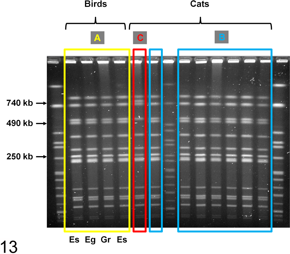
PFGE of serovar Typhimurium from avian and feline isolates. (A) The genomic fingerprint of 5 of the 12 avian bacterial isolates is shown in the figure, all displaying a unique pattern (Es = Eurasian siskins, Eg = European goldfinch, Gr = European greenfinch). (B) Seven feline isolates presented the same PFGE pattern of the birds isolates. (C) One of the cat isolates presented a slightly different PFGE pattern with 2 band differences (740 and 490 kb).
Environmental Conditions
According to the Swiss meteorological service, the winter of 2009–2010 (December 2009 to February 2010) was one of the coldest winters registered in the country since the beginning of the measurements in 1856. In mountain regions (Northern and Southern Alps, at approximately > 1000 m of altitude), the winter of 2009–2010 was the coldest of the past 29 years (since 1980–1981); in contrast, in the plain regions (Plateau, Rhône-, Rhine-, Reuss-Valley, Centre and South of Tessin) temperatures were close to the average seasonal values. During the winter of 2009–2010, a series of cold fronts and unusual persistence of snow cover were also registered all over Europe. 7
Siskin Population Size
During the winter of 2009–2010, Eurasian siskins were observed over the entire Swiss territory. From mid-December 2009 to mid-March 2010, Eurasian siskins were found to be 3 times more abundant than during the previous 2 years (mean values; Fig. 14; graphic from Müller et al, 2010, reproduced with the permission of the editor).
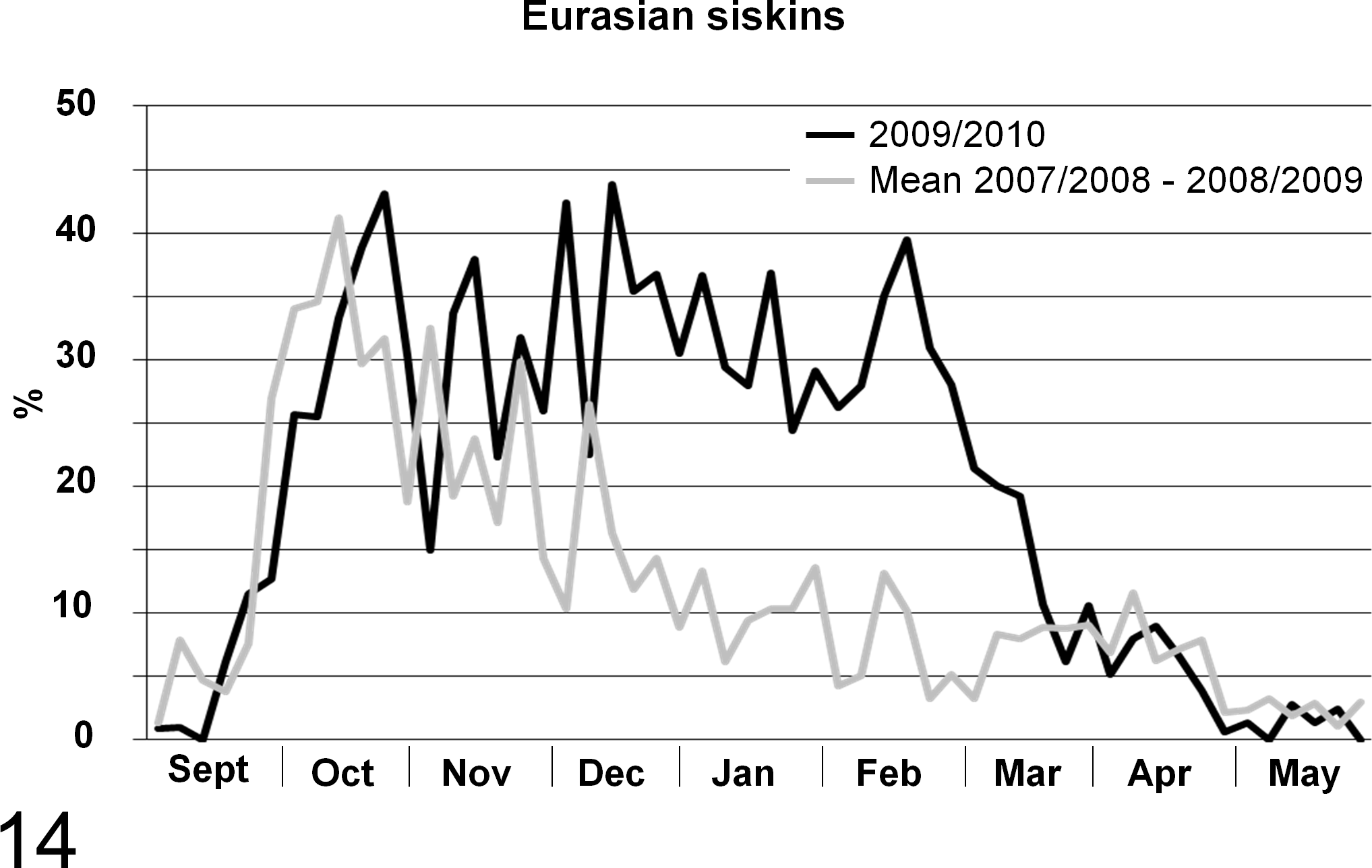
Eurasian siskin frequency assessment (sighting rate of Eurasian siskins in Switzerland as indicator of the population size). The value shown on y-axis corresponds to the actual proportion of positive siskin sightings over the total sightings attempts performed. Each sighting attempt was referred to an observation area of 1 km2 each during a period of 5 days (Pentad), performed by voluntary bird watchers. On the x-axis are displayed the months of the year when the sighting attempts were performed. Gray line = mean of observations in 2007–2008 and 2008–2009; black line = observations from September 3, 2009, to May 30, 2010.
Bacteriological Findings in Clinically Healthy Eurasian Siskins
Bacterial culture from cloacal swabs yielded S. enterica subsp. enterica serovar Agona in 1 of 136 samples (prevalence of 0.7%; 95% CI: 0.0%–4%). Salmonella Typhimurium was not isolated from any of the samples examined (prevalence of 0.0%; 95% CI: 0.0%–2.7%).
Discussion
Salmonellosis in passerine birds is a multifactorial disease with several recognized predisposing conditions, such as stress, crowding at feeding places, fecal contamination of food, and presence of healthy carriers. 36 Epidemics of salmonellosis in passerine birds are regularly observed in Northern Europe (Scandinavia and Great Britain), especially during the winter and spring. 13,22,27,32 In contrast, epidemics seem to be rare in Central/Southern Europe, where only occasional outbreaks have been described in Germany and Spain. 17,19 In Switzerland, no epidemic has been reported since the 1950s, when about 40 house sparrows (Passer domesticus) were found dead. 3 This event was believed to have occurred secondary to the introduction of S. Typhimurium to control the local rodent population. 3 Thus, the outbreak of 2010 is only the second reported epidemic of salmonellosis in passerine birds in Switzerland over the past 60 years, consistent with a rather unusual event, and the only reported epidemic in Switzerland affecting almost exclusively Eurasian siskins. Interestingly, a similar event with partially overlapping features was reported to have occurred during the same winter in neighboring Austria, affecting mostly Eurasian siskins, bullfinches (Pyrrhula pyrrhula), and greenfinches (Carduelis chloris). 4
Pathological Findings in Birds
Esophageal and crop nodules (ingluvitis and esophagitis) have long been recognized as pathognomonic lesions associated with salmonellosis in passerines. 38 Consistently, the most common gross findings (small light tan nodules scattered throughout the esophagus/crop) observed in the examined siskins in this study, their location, and histological features were overlapping with those reported in the literature and with those detected during a recent salmonellosis outbreak in Austria. 4 In few Eurasian siskins, diffuse crop lesions were observed, similar to what is more commonly seen in house sparrows. 16,23 Interestingly, the greenfinch presented nodules larger that those observed in the siskins, while in the European goldfinch, they were not found in the esophagus but surprisingly in the subcutis. We could not determine obvious reasons accounting for these differences, which might reflect species/individual specificities and/or predispositions.
There are no conclusive explanations concerning the pathogenesis of the crop lesions in songbirds infected with Salmonella sp. Lesions might start locally within the crop or affect the crop secondary. Daoust et al 9 proposed that the esophagus/crop might represent the sites where prolonged contact with contaminated food occurs, possibly enhancing the chance for bacterial colonization in situ. Alternatively, esophagus and crop have been proposed as preferred sites for bacterial growth following bacteremia originating from other tissues such as the intestine. 9 In our study we provided for the first time a proposed classification of the changes observed in the esophagus/crop in early, intermediate, and late lesions. This classification allowed us to better dissect the pathological changes in the attempt to understand the pathogenesis of the disease. Different from the late lesions, where the tissue changes were so severe to efface the details of the affected organs, in the early and intermediate lesions the architecture of the tissues was better preserved. The latter lesions were characterized by an inflammatory reaction expanding the submucosa and elevating a viable and often intact mucosa. In contrast, within the intestine the mucosa was invariably severely affected, and lesions were extending into deeper layers. Furthermore, S. Typhimurium was isolated from different organs. These findings are highly suggestive of a systemic infection originating from the intestines and spreading systemically to other organs, including the crop and finally leading to death. Florid proliferation of crop lesions and prominent inflammatory infiltrate observed in different affected organs are consistent with a subacute (to chronic) disease, which suggests that passerine birds are likely to have survived at least few days following the beginning of the septicemia.
Observations in Domestic Cats and Molecular Analysis
The treatment with large spectrum antibiotics (amoxicillin/clavulanic acid) administered to the cats was successful, and the antibiogram confirmed that S. Typhimurium was sensitive to Clavaseptin. These observations further support the diagnosis of salmonellosis in these cats.
Isolation of S. Typhimurium in all investigated ill cats indicated a possible association with the outbreak in siskins. All these cats had a history of yellow feathers within the mouth, strongly indicating consumption of siskins. Sick, weakened siskins searching for food in private gardens represented an easy prey for domestic cats. These observations strongly suggest that wild passerine birds were the source of infection of S. Typhimurium for outdoor cats, consistently with the conclusions of other reports performed in Sweden, Great Britain, and Germany. 12,33,34 Importantly, our PFGE analysis provided direct genetic evidence of the clonal relationship between feline and avian strains, in contrast to several previous studies, which documented only phenotypical similarities. 24,33,34 Recently, a study performed in Germany revealed the presence of Salmonella sp strains isolated from wild birds, humans, and cats with similar genotypic characteristics to the Swiss strains. However, a direct epidemiological link (such as the observation of bird consumption by the infected cats) was not reported in this investigation. 12 In our study, the predominant feline S. Typhimurium PFGE pattern was indistinguishable from that found in all examined bird isolates, irrespective of the bird species, suggesting the presence of a single bacterial clone. One cat presented a slightly different PFGE pattern; however, given our small sample size, we cannot exclude that other birds were infected with this second strain. Interestingly, the 2 isolates of S. Typhimurium observed among Swiss finches and cats were similar to the 2 isolates found in the Norwegian finches population, 26 where 42 out of 46 small passerines (91%, including Eurasian siskins), were infected by either one of them. These findings suggest that at least 2 clones might be endemic to different populations of small passerine birds in Europe and might have been carried along by the migrating siskins.
Epidemiology
From the end of September to mid-November, Eurasian siskins migrate southward and can be found as far as the south rim of the Mediterranean basin. From the end of February to the end of April, they return to more northern countries. Siskins are considered to be irruptive migrants because they can adjust their migration behavior according to food availability, which can strongly fluctuate from year to year. 21,32 Thus, in years of widespread food shortage or when migrating birds outsize food supplies, larger numbers of individuals migrate to lower latitudes. 2,10 Additionally, a population increase is generally observed in regions where Norway spruces (Picea abies) produce abundant fruits. 10,18 In 2009, particular abundant fruits of spruce, birch, and fir (Abies) were found in Switzerland. 5 The number of siskins that were observed in Switzerland during the fall (from September to November 2009) was comparable to that of the previous years. In contrast, from December to mid-March 2010, a higher number of siskins was observed, which might be explained by the presence of abundant food. Interestingly, this period corresponds to that when the described salmonellosis outbreak occurred. The winter of 2009–2010 was reported to be a very cold winter in Switzerland and all around Europe with unusual persistence of snow cover. 7 It is not known which role, if any, the environmental conditions might have played in influencing the occurrence of the outbreak. The snow cover should not have hampered the access of birds to the fruits of the preferred cones, as the cones are hanging on the tree and only the top of the cone should be covered with snow. The high number of birds might have favored crowding around the preferred wild food sources, enhancing the transmission of the pathogen through fecal material. The cold temperature might have led the birds to use more energy to maintain their body temperature, requiring more food intake and, consequently, more occasions for infection. High siskin abundance was observed not only within the mountain regions but also within the plain regions, where the temperature was more clement. The presence of feeding stations within private garden might have been an easier access to food for the birds. Consequently, higher abundance of siskins have caused particular crowding also at bird-feeding stations (observations reported to the Swiss Ornithological Institute by the bird watchers), with subsequent exposure to fecal bacteria, including salmonellae.
Asymptomatic Carriers
Of the cloacal swabs collected from the migrating Eurasian siskins tested during the autumn 2010 (migratory season), none yielded S. Typhimurium. Nevertheless, this result does not exclude that migrating Eurasian siskins might be carriers of S. Typhimurium. It might be that the number of individuals tested was too low for any carrier to be detected; alternatively, carriers might excrete the bacteria only intermittently. Nevertheless, our results suggest that the prevalence of possible carriers within the migrating siskin population is lower than 2.7%. Refsum et al 27 found a prevalence of possible carriers of 8.8% in Norwegian Eurasian siskins; however, the collected swabs were taken from birds that were captured in proximity of feeding places. Those birds might have had a higher probability of being exposed to the bacteria without being clinically ill at the time of the testing. Alternatively, other species of passerine birds might be the primary source of infection for Eurasian siskins. 27,36
Conclusion
The crop and esophagus lesions in the infected birds were the most consistent tissue changes, and their features were highly suggestive of a septicemic event and not of an “in situ” (local) bacterial seeding. At least 2 strains of S. Typhimurium are circulating in Northern and Central Europe, and given the circumstantial evidences, infected passerine birds might represent a significant source of Salmonella sp infection for outdoor cats. Finally, the exceptional high number of siskins during the winter of 2009–2010 appears to be the most likely priming factor for the salmonellosis outbreak described in this article.
Footnotes
Acknowledgements
We thank Bernhard Staehelin and Tobias Denzler from the Small Animal Clinic in Interlaken and Thun for providing the rectal swabs of the ill cats. Marco Thoma, Sarah Althaus, and Luzius Fischer collected cloacal swabs of migrating Eurasian siskins. We are grateful to Natacha Wu, Julien Casaubon, and Fabien Mavrot for their contributions to necropsies and statistical analyses. Grethe Sägesser performed the PFGE analyses; Bernard Volet provided data from the Swiss Ornithological Institute; and Caroline Frey performed a polymerase chain reaction to confirm toxoplasmosis in 1 bird. We would also like to thank the Swiss Federal Office of Public Health, Division Communicable Diseases, and the Swiss Federal Office of the Environment for their constant support of the diagnostic services.
Declaration of Conflicting Interests
The author(s) declared no potential conflicts of interest with respect to the research, authorship, and/or publication of this article.
Funding
The author(s) received no financial support for the research, authorship, and/or publication of this article.